STEM CELLS AND DEVELOPMENT Volume 19, Number 2, 2010 © Mary Ann Liebert, Inc. DOI: 10.1089/scd.2009.0142
Reprogramming of Somatic Cells After Fusion With Induced Pluripotent Stem Cells and Nuclear Transfer Embryonic Stem Cells Huseyin Sumer, Karen L. Jones, Jun Liu, Corey Heffernan, Pollyanna A. Tat, Kyle R. Upton, and Paul J. Verma
In this study we examine whether a somatic cell, once returned to a pluripotent state, gains the ability to reprogram other somatic cells. We reprogrammed mouse embryonic fibroblasts by viral induction of oct4, sox2, c-myc, and klf-4 genes. Upon fusion of the resulting iPS cells with somatic cells harboring an Oct4-GFP transgene we observed, GFP expression along with activation of Oct4 from the somatic genome, expression of key pluripotency genes, and positive immunostaining for Oct4, SSEA-1, and alkaline phosphatase. The iPS-somatic hybrids had the ability to differentiate into cell types indicative of the three germ layers and were able to localize to the inner cell mass of aggregated embryos. Furthermore, ntES cells were used as fusion partners to generate hybrids, which were also confirmed to be reprogrammed to a pluripotent state. These results demonstrate that once a somatic cell nucleus is reprogrammed, it acquires the capacity and potency to reprogram other somatic cells by cell fusion and shares this functional property with normal embryonic stem (ES) cells.
Introduction
T
he somatic genome can be reprogrammed to a pluripotent state by a number of methods, including somatic cell nuclear transfer (SCNT), cell fusion to an embryonic stem cell, and viral transduction of reprogramming factors resulting in induced pluripotent stem cells (iPS). All three processes have helped to delineate the reprogramming process. First, SCNT, which involves the transfer of a differentiated nucleus to an oocyte from which the maternal DNA has been removed, has been shown to completely reprogram differentiated mammalian nuclei to produce live offspring [1]. Even though NT embryonic stem (ES) cells can be derived from cloned mouse embryos [2], to date no human ntES cells have been derived. Second, reprogramming by cell fusion involves the hybridization of 2 different cells/ genomes, and in most hybrids the phenotype of the lessdifferentiated fusion partner dominates the phenotype of the more-differentiated partner. ES cells have the ability to reprogram the somatic genome following cell fusion with both mouse [3] and human ES cells [4,5]. Following cell fusion, the ES-somatic cell hybrids assume hallmarks of pluripotent cells including growth characteristics, gene expression profiles, and differentiation potential. However, due to
their increased ploidy they contribute minimally to the late gestation epiblast [3] and chimeras [6]. More recently, the viral delivery and expression of the transcription factors; Oct4 and Sox2, with either c-Myc, and Klf-4 in mouse [7], and human [8], or with Nanog and Lin28 [9], have been shown to induce pluripotency to somatic cells, giving rise to iPS cells. To reduce the number of viral constructs used and/or minimize viral integration, combinations of these key reprogramming factors can be replaced with either chemical compounds or endogenous expression in somatic cells [10–13], or delivered by nonintegrating viruses [14,15]. iPS cells have been shown to be similar to ES cells in terms of gene expression profile, epigenetic state, differentiation potential both in vitro and in vivo, ability to give rise to germ line competent chimeras and can be used to generate autologous cells for transplantation studies [7–9,16–19]. In a preliminary study, iPS cells have been shown to reprogram somatic cells by cell fusion [20], however the reprogramming abilities of de-differentiated cells has not been described in detail. Here, we conducted an in-depth study to determine whether a somatic cell, once returned to a pluripotent state, gains the ability to reprogram other somatic cells. To test this hypothesis, we investigated the reprogramming abilities of
Monash Institute of Medical Research, Monash University, Clayton, Victoria, Australia.
239
240 both iPSCs and ntES cells by using them as fusion partners with somatic cells. We generated and characterized iPS cells from mouse embryonic fibroblasts (MEFs) and fused them to OG2 MEFs harboring an Oct4-GFP reporter gene. In the resulting iPS-somatic cell hybrids, the Oct4-GFP transgene along with the endogenous Oct4 gene were reactivated, pluripotency specific genes expressed, and stained positive for Oct4, SSEA-1 and alkaline phosphatase. The iPS-somatic hybrids had the ability to differentiate into multiple lineages and localized to the inner cell mass of aggregated embryos. We further tested the ability of a de-differentiated cell to reprogram a somatic cell by using ntES cells as fusion partners. We concluded that a somatic cell, once reprogrammed, has the ability to reprogram other somatic cells by cell fusion and shares this functional property with normal ES cells.
Materials and Methods
SUMER ET AL. efficiency was calculated by subtracting the double positive value in mock fusions from the fusion value. As a control for cell fusion based reprogramming of OG2 MEFs, mES D3 cells were used as fusion partners in parallel experiments.
PCR Genomic DNA was extracted using the DNeasy Blood and Tissue kit (Qiagen, Hilden, Germany) and used for amplification of neomycin and GFP sequences by PCR with the following primer pairs. Neo F agacaatcggctgctctgat, Neo R. caatagcagccagtcccttc, GFP F GACGACGGCAACTACAAGA, GFP R GATGCCGTTCTTCTGCTT. The PCR cycle parameters included an initial denaturation at 94°C for 5 min followed by 30 cycles of denaturation at 94°C for 1 min, annealing at 58°C for 45 s and extension at 72°C for 1 min, followed by final extension at 72°C for 5 min. PCR products were run on a 1% agarose gel at 100 V for 1 h.
Cell culture and differentiation Feeder-independent mouse ESD3, iPS cells, and iPSsomatic cell hybrids were cultured in ES media: DMEM (Invitrogen, Carlsbad, CA) supplemented with 15% Hyclone™ fetal bovine serum, 1 mM l-glutamine (Invitrogen), 0.1 mM b-mercaptoethanol (Sigma, St. Louis, MO), 1,000 U/mL LIF (Chemicon, Temecula, CA), 1% nonessential amino acids (Invitrogen) and 0.5% penicillin-sreptomycin (Invitrogen). Mouse embryonic fibroblasts were cultured in the same medium minus LIF and b-mercaptoethanol. Cultures were maintained in a humidified incubator at 37°C, with 5% CO2/95% air. For embryoid body formation 1 × 10⁶ cells were plated on Petri dishes in ES media minus LIF for 7 days. To examine teratoma formation, 1–2 × 10⁶ cells were injected into the rear leg muscle of 4- to 6-week-old severe combined immunodeficient (SCID) mice. After 4 weeks, the teratomas were excised and fixed in 4% paraformaldehyde, embedded in paraffin, sectioned at 5 uM and stained with hematoxylin and eosin by the MIMR Histology Laboratory core facility.
iPS generation and cell fusion iPS cells were generated from MEFs from QS/Rosa26 mice as previously described [21]. Following viral transduction iPS colonies were picked by morphology and expanded clonally on MEF feeder layers for further analysis. iPSsomatic cell hybrids were produced by Polyethylene glycol (PEG) mediated cell fusion performed in a 4-well Nunc tissue culture plate as previously described [22]. In brief, iPS cells were plated at 0.5 × 10⁶ per cellular fibronectin (Sigma) coated well and cultured overnight. Around 1 × 10⁶ OG2 MEFs were centrifuged onto the dense monolayer at 400g for 10 min. The culture medium was removed and fusion performed by adding 500 μL of 50% PEG1500 /150 mM HEPES and incubated at room temperature for 2 min. The PEG was removed, the cells washed 4 times in calcium-and-magnesium-free PBS and allowed to recover in ES medium in the incubator for at least 4 h before the contents of the plate were trypsinized and plated in 2 × 6 cm dishes for further culture. Fusion efficiency was determined by prestaining iPS cells with 1.25 μM CFSE for 10 min; 24 h prior to fusion, and MEF cells with the 15 μM SNARF-1, for 30 min an hour prior to fusion. The percentage of double positive stained cells were measured by flow cytometry 3–4 h postfusion. The fusion
RT-PCR and SNP analysis Total RNA was extracted from cells using the RNeasy kit (Qiagen) according to the manufacturer’s instructions. To remove contaminating genomic DNA the resulting total RNA was subjected to DNaseI treatment using the DNAfree kit (Ambion, Austin, TX). About 2 μg of RNA was used to synthesize cDNA using the SuperScript III reverse transcriptase kit (Invitrogen). cDNA samples were subjected to PCR amplification with the following primer pairs: Oct4 F: GTTCAGCCAGACCACCATCT, R:CCTGGGAAAGG TGTCCTGTAG Rex1 F: GGACTAAGAGCTGGGACACG, R:GCTGCTTCCTT CTTGAACAAT Nanog F: TCAAGGACAGGTTTCAGAAGCA, R:GCTGGGA TACTCCACTGGTG Sox2 F: GAGGAGAGCGCCTGTTTTT, R:GGAGATCTGGCG GAGAATAG SPARC F: AATTTGAGGACGGTGCAGAGG, R:GGTTGTTG CCCTCATCTCTCT β-actin F: GGAATCCTGTGGCATCCATGAAAC, R:AAAAC GCAGCTCAGTAACAGTCCG For Oct4 SNP analysis cDNA was amplified using the Oct3/4-S9: TCTTTCCACCAGGCCCCCGGCTC [7] and oct4 RNA R: GCAAACTGTTCTAGCTCCTTCT, to amplify a 469 bp endogenous Oct4 product, avoiding amplification of the exogenous viral construct. PCR products were digested with BamHI for 4 h before running on an agarose gel. Digested samples were run on a 3.5% low melt agarose gel and undigested Oct4 bands were excised and DNA extracted using the QIAEXII kit following manufacturer’s instructions and sequenced by The Gandel Charitable Trust Sequencing Centre, Clayton Australia (Applied Biosystems ABIPRISM™ 377 DNA Sequencer).
Oct4 methylation analysis Genomic DNA isolated from the various cell lines were processed the MethylEasy Xceed Kit (Human Genetic Signatures, North Ryde, New South Wales, Australia) for bisulfite conversion. Hemi-nested primers O4 B4 S 116781 F1 TTGAGGAGTGGTTTTAGAA ATAATTGGTAT and O4 B4 S 117317 R1 CCCAACCCTACT CCAACCCTACTA, followed by
241
iPS-SOMATIC CELL HYBRIDS O4 B4 S 116828 F2 GGGTAA GTAAGAATTGAGGAGTGGTTT and O4 B4 S 117317 R1, were designed to selectively amplify the fully converted genomic DNA templates after 2 rounds of PCR. PCR for each sample was performed in triplicate and then pooled for cloning into pGEMT-Easy vector (Promega, Madison, WI). PCR cycling conditions were as follows: denaturation at 95°C for 4 min, followed by 5 cycles at 93°C for 30 s, annealing at 57°C for 60 s, and extension at 72°C for 60 s, followed by a final extension of 10 min. Purified PCR products were amplified for a second time using the hemi-nested primers. Eight clones from each genomic DNA template were sequenced and the methylation status was determined using BiQ Analyzer software [23].
antibodies at 1:100 dilution overnight at 4°C. Following 3 washes with PBS, the slides were incubated at room temperature for 1 h with secondary antibodies goat α-mouse IgM Alexa 594 or goat α-mouse IgG Alexa 594 (Invitrogen, Australia), respectively, diluted 1:1,000 in blocking solution. Following 3 washes with PBS the slides were mounted in Vectashield + DAPI) (Vector Laboratories, Burlingame, CA) with a coverslip. Alkaline phosphatase activity was detected with an AP kit (Chemicon) according to the manufacturer’s instructions.
Histochemistry and immunohistochemistry
Female mice (3–5 weeks) were superovulated by injections of 5IU of Pregnant Mare Serum Gonadotrophin (PMSG) 48 h prior to, and 5IU (hCG) immediately before being housed with stud F1 males for mating overnight. Midnight was arbitrarily assigned as time of mating. The following day, plugged females were sacrificed and embryos isolated from the reproductive tract by manual dissection of the ampulla in M2 handling medium (Chemicon). Surrounding
Cells were cultured in 4-well glass culture slides (BD Falcon, Bedford, MA) and fixed in 4% paraformaldehyde for 10 min, washed 3 times with PBS and incubated with blocking solution (5% goat serum, 1% BSA in PBS) or blocking solution with 0.1% Triton-X for SSEA-1 and Oct4 primary antibodies, respectively, before being exposed to the primary
Embryo isolation and culture and embryo aggregations
A 4N Hybrid OG2 MEF (Oct4-GFP) Neo R MEF
iPS Cell Viral Transduction
SNARF FL5 Log
B 104
C
R4
R3
Oct4-GFP Activation
i
ii
103 102 101 R5 100 100
R6
101
102
103
104 iPS × OG2 Hyb 3
8n
iPS × OG2 Hyb 2
4n
Cell Count
2n
iPS × OG2 Hyb 1
E
OG2 MEF
D
iPS #2
CFSE Log
Neo
GFP 0
64
128 DNA Content
192
256
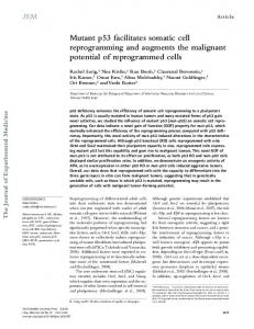
FIG. 1. Generation of iPS-somatic cell hybrids. (A) iPS cells generated by viral transduction of Neomycin resistant mouse embryonic fibroblasts were fused to OG2 MEFs resulting in the reactivation of the Oct4-GFP transgene. (B) FACs profiles of cell fusions of CFSE stained iPSCs and SNARF-1 stained OG2 MEFs. (C) GFP fluorescence following Oct4-GFP reactivation in an iPS-somatic cell hybrid, scale bar = 200 μM. (D) Ploidy analysis of iPS-somatic hybrids overlayed with iPSC profile stained with propidium iodide. (E) PCR amplification of transgenes in iPS-somatic cell hybrids from the 2 parental cell lines.
242
SUMER ET AL.
cummulus cells were digested from zygotes by digestion in M2 medium containing hyaluronidase (Chemicon) before 3 washes in 20 μL droplets of KSOMaa media with BSA under mineral oil and overnight culture. Cultured 2.5 dpc, diploid (2N) embryos were incubated in Tyrodes acid solution and monitored visually for zona pellucida digestion before being washed 3 times in KSOMaa media. Zona-free embryos were individually placed into depressions made in the floor of culture dishes with a sterilized darning needle overlayed with a 15–20 μL droplet of KSOMaa culture media. Aggregates of 5–10 donor cells were dropped into the depression in contact with the embryos. Embryos were further cultured in a humidified incubator at 37°C before visualization at 3.5–4.5 days postcoitum. For tetraploid embryo complementation, two cell stage embryos were washed in electrofusion medium consisting of mannitol before fusion of blastomeres with a pulse of direct current at approximately 130 volts to initiate fusion.
Results Generation of iPS cells and iPS-somatic cell hybrids
B
C
ES
A
D3 OG 2M EF iPS #2 iPS × Hy OG b1 2 iPS × Hy OG b 2 iPS 2 × Hy OG b3 2
First, iPS cells were generated by viral transduction of Oct3/4, Sox2, Klf4, and c-Myc of neomycin resistant MEFs, as
previously described [21]. A number of phase bright compact colonies similar in morphology to those of mouse ES cells were picked, expanded and further characterized (Supplementary Fig. 1; Supplementary materials are available online at http:// www.liebertpub.com/). The iPS clones were shown to have a normal diploid chromosome count and pluripotential was shown by alkaline phosphatase activity, Oct4 and SSEA1 antibody staining, gene expression profiling, EB formation, and teratoma formation where cells from all 3 germ layers were observed (Supplementary Fig. 1). Polyethylene glycol (PEG) mediated cell fusion was performed with iPSC clone 2 and MEFs isolated from OG2 transgenic mice, which contain a GFP reporter under the control of the Oct4 promoter region (Fig. 1A). The fusion efficiency was determined to be 1.26% ± 0.69 (N = 3) (Fig. 1B). Oct4-GFP reactivation was observed in cells within 72 h of fusion, a time point where mES D3 cells were also observed to reactivate the transgene in parallel fusions. Five days post fusion, 5 ± 3 (N = 3) GFP +ve colonies were observed (Fig. 1C), and 3 individual GFP +ve colonies were picked and expanded by enzymatic passaging for further analysis. The hybrid cells grew in tight, phase bright colonies and continued to express the Oct4-GFP transgene from the OG2 MEF genome. The hybrids were confirmed to have risen from a fusion event from the two parental cell lines by polymerase
Oct4 Nanog Rex-1 Sox2
Di
ii
iii
Sparc
NR β-actin - RT
E
F
G
SE
C
FIG. 2. Characterization of iPS-somatic cell hybrids. (A) Alkaline phosphatase activity of iPS-somatic hybrids, scale bar = 200 μM. (B) Embryoid body formation in the absence of LIF, scale bar = 500 μM. (C) Gene expression profile of iPS-somatic hybrids and the 2 parental cells. (D) Histology of teratoma tissue showing formation showing Neurogenic and neuroblastic differentiation with rosettes (NR) secretory epithelium (SE) and articular cartilage (C), scale bar = 200 μM. (E) Immunostaining of iPS-somatic hybrids showing phase, Oct4-GFP fluorescence in green, Oct4 in red, counter stained with DAPI in blue, scale bar = 100 μM. (F) Immunostaining of iPS-somatic hybrids showing phase, Oct4-GFP fluorescence in green, SSEA-1 in red, counter stained with DAPI in blue, scale bar = 100μM. (G) Developing blastocyst aggregated with GFP positive iPS-somatic cells showing contribution to the inner cell mass, scale bar = 200 μM.
243
iPS-SOMATIC CELL HYBRIDS chain reaction (PCR) for the Neomycin and GFP transgenes from the iPS and MEF parental lines, respectively (Fig. 1D). The hybrids were also shown to have double the DNA content of the 2 parental cell lines by flow cytometry analysis (Fig. 1E).
Characterization of iPS-somatic cell hybrids The hybrids were further characterized for their developmental potential in vitro and in vivo. Hybrids were shown to express the pluripotency markers Oct-4, Rex-1, Nanog, and Sox2, while the fibroblast specific marker SPARC was extinguished in hybrids (Fig. 2A). Furthermore, the 4 exogenous viral factors Oct-4, Sox2, cMyc, and Klf-4 were shown to be switched off in both the iPS and iPS-somatic hybrid clones (Supplementary Fig. 2). The hybrids also had high levels of alkaline phosphatase activity (Fig. 2B), and formed EBs in the absence of LIF (Fig. 2C). Immunostaining showed the correct localization of pluripotency markers Oct-4 and SSEA-1, to the nucleus and plasma membrane, respectively (Fig. 2E–2F), while the Oct4-GFP transgene appeared to be silenced within some cells in iPS-somatic cell hybrid colonies. To confirm that iPS-somatic hybrids were pluripotent, the 3 clones were injected into the thigh muscle of SCID mice. All mice developed tumors and histological examination revealed that the teratomas from iPS-somatic cell hybrids contained tissues representative of all 3 germ layers. Differentiated tissues observed included primitive neuroectoderm, secretory epithelium, and muscle (Fig. 2D). Another characteristic of embryonic stem cells is that they contribute to the inner cell mass and give rise to the epiblast
B iPS U D
1 U
In addition to the activation of the Oct4-GFP transgene reporter from the somatic genome, we investigated the transcriptional reactivation of endogenous embryonic genes from the somatic chromosomes. Oct4 is a key transcription factor involved in the self renewal and pluripotency of embryonic stem cells [26]. We identified a single nucleotide polymorphism (SNP) in the first exon of the Oct4 gene, whereby a G-residue within a BamHI restriction site of the iPS genome is replaced with an A-residue in the OG2 somatic genome (Fig. 3A). PCR was used to amplify a 469 bp, the Oct4 transcripts product containing the SNP from cDNA isolated from hybrids and iPS cells, using primers that were specific to endogenous Oct4 that did not amplify the viral transcripts [7]. Equal amounts of PCR
OG2 MEF
iPS × OG2 Hybrids 2 3 D U D U D
469 bp 341 bp
ES
iPS
OG2 MEF
CCAGGTTCGAG A ATCCACCCAG
Reprogramming of the endogenous Oct4 locus
D
BamHI CCAGGTTCGAGG ATCCACCCAG
NeoR MEF
A
when they are injected into developing blastocysts or when aggregated with diploid or tetraploid embryos [24]. Both mouse tetraploid ES cells [25] and ES-somatic cell hybrids are also capable of integrating into the inner cell mass of diploid preimplantation blastocysts [3,6], however tetraploid cells contribute minimally to chimeras due to their ploidy. Here, we generated tetraploid embryos by electrofusion of blastomeres of 2-cell embryos and aggregated them with the GFP positive iPS-somatic cell hybrids at the 6–8 cell stage before visualization at the blastocyst stage (3.5–4.5 days postcoitum). GFP positive cells were observed to localize to the inner cell mass of aggregated embryos (N = 8), further confirming the pluripotent nature of iPS-somatic cell hybrids (Fig. 2E).
iPS
128 bp
C
iPS-OG2 Hybrid
T T C G A G A A T C C A C C C A
FIG. 3. Transcriptional reprogramming of the endogenous somatic Oct4 locus in hybrids. (A) Oct4 Exon 1 SNP genotypes in iPS cells and OG2 MEFs (C57B6). (B) Undigested (U) and BamHI digested (D) Oct4 cDNA transcripts from iPSCs and iPS-somatic cell hybrids. (C) DNA sequencing of undigested cDNA band from iPS-somatic cell hybrids confirming expression from the somatic allele. (D) DNA methylation of the Oct4 promoter. White circles indicate unmethylated CpG dinucleotides, whereas black circles indicate methylated CpG dinucleotides.
244
SUMER ET AL.
products were digested with BamHI endonuclease and run on a gel alongside undigested products (Fig. 3B). The iPS sample was completely digested resulting in 341 bp and 128 bp products, as expected. While both undigested and digested products at 469, 341, and 128 bp were observed in the hybrid sample, to verify that the undigested band at 469 bp was from the OG2 genome, the bands were excised from the gel, sequenced, and confirmed to contain the A-residue at the SNP location (Fig. 3C). This was further verified by TfiI digestion, resulting in only digestion of the somatic genome when an alternate primer set was used (data not shown). Bisulphite genomic sequencing analyses of the Oct4 promoter revealed that the iPS cells had undergone demethylation following pluripotency induction of the parental NeoR MEF cells (Fig. 3D). Furthermore, the iPS-somatic cell hybrids, like the iPS and ES cells, were largely unmethylated suggesting demethylation of the OG2 MEF genome following cell fusion.
Reprogramming of somatic cells by ntES cell fusion The reprogramming ability of de-differentiated somatic cells was further tested using ntES cells as fusion partners. Previously reported neomycin resistant ntES cells [2] were fused to OG2 MEFs. The resulting GFP expressing hybrid colonies (Fig. 4A), were shown to express the pluripotency markers Oct-4, Rex-1, Nanog, and Sox2, while the fibroblast specific marker SPARC was extinguished (Fig. 4B). Reprogramming of the somatic genome was confirmed by activation of the somatic Oct4 allele in hybrids using SNP analysis (Fig. 4C). Bisulphite genomic sequencing analyses of the Oct4 promoter revealed that ntES-somatic cell hybrids, like ntES cells, were largely unmethylated suggesting demethylation of the OG2 MEF genome following cell fusion (Fig. 4D). The hybrid colonies also stained positive for Oct-4 and SSEA-1 proteins (Fig. 4E and 4F). The pluripotent nature of these hybrids was confirmed by teratoma formation, where tissues representative of all three germ layers were identified (Fig. 4G).
Discussion A
ntES × OG2 Hyb 1 2 3 U D U D U D D U
C
ntES
Oct-4 Nanog
128 bp
D ntES
OG2 MEF
ntES
B
ntES × OG2 Hyb 1 ntES × OG2 Hyb 2 ntES × OG2 Hyb 3
469 bp 341 bp
Sparc B-actin - RT
ntES-OG2 Hybrid
Sox2
OG2 MEF
Rex-1
E
F
Gi
ii
iii
A functional property of pluripotent cells is that they have the capacity to reprogram somatic cells by cell fusion in vitro. In the present study, we have shown in detail that both mouse iPSCs and SCNT derived ntES cells, like ES and EC cells [3,27], have the ability to reprogram a somatic genome following cell fusion. This demonstrates that once a somatic cell is reprogrammed/de-differentiated, acquires the capacity and potency to reprogram another somatic cell by cell fusion. Following cell fusion iPS-somatic cell hybrids adopt a number of ES characteristics including; colony-like morphology, expression of the stem cell markers Oct4, Nanog, Sox2, and Rex-1, while the differentiation marker SPARC is silenced. The hybrids also stain positively for the stem cell markers AP, SSEA1, and Oct4 and give rise to teratomas consisting of tissues from all three germ layers when injected into immunocompromised mice. iPS-somatic cell
FIG. 4. Characterization of ntES-somatic cell hybrids. (A) GFP fluorescence, following Oct4-GFP reactivation in ntESsomatic cell hybrid, scale bar = 100 μM (B) Gene expression profile of iPS-somatic hybrids and the 2 parental cells. (C) Detection of endogenous Oct4 products from ntES and somatic genomes by BamHI digestion of Oct4 Exon 1 PCR products, Undigested (U) and BamHI digested (D). (D) DNA methylation of the Oct4 promoter. White circles indicate unmethylated CpG dinucleotides, whereas black circles indicate methylated CpG dinucleotides. (E) Immunostaining of somatic cell nuclear transfer ES-somatic hybrids showing phase, Oct4-GFP fluorescence in green, Oct4 staining in red, counter stained with DAPI in blue, scale bar = 100 μM. (F) Immunostaining of SCNT ES-somatic hybrids showing phase, Oct4-GFP fluorescence in green, SSEA-1 staining in red, counter stained with DAPI in blue, scale bar = 100 μM. (G) Histology of teratoma tissue showing formation showing (i) Neurogenic and neuroblastic differentiation with rosettes (ii) secretory epithelium and (iii) cartilage, scale bar = 200 μM.
iPS-SOMATIC CELL HYBRIDS hybrids also localize to the inner cell mass of developing blastocysts. To demonstrate that the somatic genome is reprogrammed following iPS cell fusion, allele-specific gene expression analysis was performed and it was confirmed that the endogenous Oct4 gene was transcribed from the MEF genome. Furthermore, bisulfite sequencing analysis demonstrated that the Oct4 promoter undergoes demethylation following reprogramming. This demonstrates that the somatic genome is not merely silenced following cell fusion resulting in the dominance of the iPS cell, but it contributes to the hybrid cells transcriptome. Furthermore, it is interesting to note that the reactivation of the somatic Oct4-GFP transgene in iPS-somatic fusions occurred at a similar time to that when ES cells were used as fusion partners. This suggests that iPS cells have similar reprogramming potency abilities to ES cells. Despite the fact that the 4 exogenous viral factors were switched off in our iPS cells prior to fusion as well as in the iPS-somatic hybrids, it could not be determined whether the factors were reactivated transiently during the fusion process and responsible for the reprogramming of the somatic genome in the iPS-somatic cell hybrids. To conclusively demonstrate that de-differentiated cells gain the ability to reprogram a somatic cell by cell fusion, we used SCNT derived ntES cells in cell fusion experiments. Pluripotential was also shown to be restored to the resulting hybrids by Oct4-GFP transgene reactivation, activation of Oct4 from the somatic genome by SNP analysis, demethylation of the Oct 4 promoter, expression of key pluripotency genes, immunostaining for Oct4, SSEA-1, and alkaline phosphatase, and the ability to differentiate into cell types indicative of the 3 germ layers. Taken together, this study demonstrates that reprogramming of a somatic cell achieved by either viral induction of pluripotency factors or by SCNT re-establishes not only selfrenewal and pluripotency but also reprogramming capacity, which is another functional feature of embryonic stem cells.
Acknowledgments We would like to thank R. Jaenisch for the kind gift of ntES cells, and Mark Williamson from Gribbles Vet Pathology for histological identification of teratoma tissue types, and Stem Cell Sciences for B-actin primers. This project was supported by funding from the Australian Stem Cell Centre. H.S. receives an NH&MRC Biomedical Training Fellowship supported by an Establishment Gift from the Clive and Vera Ramaciotti Foundation.
References 1. Wilmut I, AE Schnieke, J Mcwhir, AJ Kind and KHS Campbell. (1997). Viable offspring derived from fetal and adult mammalian cells. Nature 385:810–813. 2. Rideout WM, K Hochedlinger, M Kyba, GQ Daley and R Jaenisch. (2002). Correction of a genetic defect by nuclear transplantation and combined cell and gene therapy. Cell 109:17–27. 3. Tada M, Y Takahama, K Abe, N Nakatsuji and T Tada. (2001). Nuclear reprogramming of somatic cells by in vitro hybridization with ES cells. Curr Biol 11:1553–1558. 4. Cowan CA, J Atienza, DA Melton and K Eggan. (2005). Nuclear reprogramming of somatic cells after fusion with human embryonic stem cells. Science 309:1369–1373.
245 5. Yu J, MA Vodyanik, P He, II Slukvin and JA Thomson. (2006). Human embryonic stem cells reprogram myeloid precursors following cell-cell fusion. Stem Cells 24:168–176. 6. Ying Q-L, J Nichols, EP Evans and AG Smith. (2001). Changing potency by spontaneous fusion. Nature 416:545–548. 7. Takahashi K and S Yamanaka. (2006). Induction of pluripotent stem cells from mouse embryonic and adult fibroblast cultures by defined factors. Cell 126:663–676. 8. Takahashi K, K Tanabe, M Ohnuki, M Narita, T Ichisaka, K Tomoda and S Yamanaka. (2007). Induction of pluripotent stem cells from adult human fibroblasts by defined factors. Cell 131:1–12. 9. Yu J, MA Vodyanik, K Smuga-Otto, J Antosiewicz-Bourget, JL Frane, S Tian, J Nie, GA Jonsdottir, V Ruotti, R Stewart, II Slukin and AJ Thomson. (2007). Induced pluripotent stem cell lines derived from human somatic cells. Science 318:1917–1920. 10. Huangfu D, K Osafune, R Maehr, W Guo, A Eijkelenboom, S Chen, W Muhlestein and DA Melton. (2008). Induction of pluripotent stem cells from primary human fibroblasts with only Oct4 and Sox2. Nat Biotechnol 26:1269–1275. 11. Huangfu D, R Maehr, W Guo, A Eijkelenboom, M Snitow, AE Chen and DA Melton. (2008). Induction of pluripotent stem cells by defined factors is greatly improved by small-molecule compounds. Nat Biotechnol 26:795–797. 12. Shi Y, JT Do, C Desponts, HS Hahm, HR Scholer and S Ding. (2008). A combined chemical and genetic approach for the generation of induced pluripotent stem cells. Cell Stem Cells 2:525–528. 13. Kim JB, V Sebastiano, G Wu, MJ Arauzo-Bravo, P Sasse, L Gentile, K Ko, D Ruau, M Ehrich, D van den Boom, J Meyer, K Hubner, C Bernemann, C Ortmeier, M Zenke, BK Fleischmann, H Zaehres and HR Scholer. (2009). Oct4-induced pluripotency in adult neural stem cells. Cell 136:411–419. 14. Stadtfeld M, M Nagaya, J Utikal, G Weir and K Hochedlinger. (2008). Induced pluripotent stem cells generated without viral integration. Science 322:945–949. 15. Okita K, M Nakagawa, H Hyenjong, T Ichisaka and S Yamanaka. (2008). Generation of mouse induced pluripotent stem cells without viral vectors. Science 322:949–953. 16. Meissner A, M Wernig and R Jaenisch. (2007). Direct reprogramming of genetically unmodified fibroblasts into pluripotent stem cells. Nat Biotechnol 25:1177–1181. 17. Okita K, T Ichisaka and S Yamanaka. (2007). Generation of germline-competent induced pluripotent stem cells. Nature 448:313–317. 18. Wernig M, A Meissner, R Foreman, T Brambink, M Ku, K Hochedlinger, BE Bernstein and R Jaenisch. (2007). In vitro reprogramming of fibroblasts into a pluripotent ES-cell-like state. Nature 448:318–324. 19. Hanna J, M Wernig, S Markoulaki, CW Sun, A Meissner, JP Cassady, C Beard, T Brambink, LC Wu, TM Townes and R Jaenisch. (2007). Treatment of sickle cell anemia mouse model with iPS cells generated from autologous skin. Science 318:1920–1923. 20. Maherali N, R Sridharan, W Xie, J Utikal, S Eminli, K Arnold, M Stadtfeld, R Yachechko, J Tchieu, R Jaenisch and K Hochedlinger. (2007). Directly reprogrammed fibroblastshow global epigenetic remodeling and widespread tissue contribution. Cell Stem Cell 1:55–70. 21. Takahashi K, K Okita, M Nakagawa and S Yamanaka. (2007). Induction of pluripotent stem cells from fibroblast cultures. Nat Protocols 2:3081–3089. 22. Sumer H, KL Jones, J Liu, BN Rollo, AL van Boxtel, D Pralong and PJ Verma. Transcriptional changes in somatic cells recovered from ES-somatic heterokaryons. Stem Cells and Dev (in press) doi:10.1089/scd.2008.0361. 23. Bock C, S Reither, T Mikeska, M Paulsen, J Walter and T Lengauer. (2005). BiQ Analyzer: visualization and quality control for DNA methylation data from bisulfite sequencing. Bioinformatics 21(21):4067–4068.
246 24. Nagy A, E Gocza, EM Diaz, VR Prideaux, E Ivanyi, M Markkula and J Rossant J. (1990). Embryonic stem cells alone are able to support fetal development in the mouse. Development 110:815–821. 25. Pralong D, ML Lim, I Vassiliev, K Mrozik, N Wijesundara, P Rathjen and PJ Verma. (2005). Tetraploid embryonic stem cells contribute to the inner cell mass of mouse blastocysts. Cloning Stem Cells 7:272–278. 26. Niwa H, J Miyazaki and AG Smith. (2000). Quantitative expression of Oct-3/4 defines differentiation, dedifferentiation or selfrenewal of ES cells. Nat Genet 24:372–376. 27. Flasza M, AF Shering, K Smith, PW Andrews, P Talley and PA Johnson. (2003). Reprogramming in inter-species embryonal carcinoma-somatic cell hybrids induces expression. Cloning Stem Cells 5:339–354.
SUMER ET AL. Address correspondence to: Dr. Huseyin Sumer Centre for Reproduction and Development Monash Institute of Medical Research Monash University 27-31 Wright Street Clayton, VIC 3168 Australia E-mail:
[email protected] Received for publication April 28, 2009 Accepted after revision July 28, 2009 Prepublished on Liebert Instant Online July 28, 2009
This article has been cited by: 1. Richard D. W. Kelly, Huseyin Sumer, Matthew McKenzie, Joao Facucho-Oliveira, Ian A. Trounce, Paul J. Verma, Justin C. St. John. 2011. The Effects of Nuclear Reprogramming on Mitochondrial DNA Replication. Stem Cell Reviews and Reports . [CrossRef] 2. Monika Nowak-Imialek, Wilfried Kues, Joseph W. Carnwath, Heiner Niemann. 2011. Pluripotent Stem Cells and Reprogrammed Cells in Farm Animals. Microscopy and Microanalysis 1-24. [CrossRef] 3. Lizhen Huang , Nana Fan , Jie Cai , Dongshan Yang , Bentian Zhao , Zhen Ouyang , Weiwang Gu , Liangxue Lai . 2011. Establishment of a Porcine Oct-4 Promoter-Driven EGFP Reporter System for Monitoring Pluripotency of Porcine Stem Cells. Cellular Reprogramming (Formerly "Cloning and Stem Cells") 13:2, 93-98. [Abstract] [Full Text] [PDF] [PDF Plus] [Supplementary material] 4. Jinnuo Han, Kuldip Sidhu. 2011. Embryonic stem cell extracts: use in differentiation and reprogramming. Regenerative Medicine 6:2, 215-227. [CrossRef] 5. Dan Xu, Feng Wang, Hongyan Gu, Jia Wang, Qinglong Guo, Yanli Zhang, Ziyu Wang. 2010. Hybrid cells differentiate to hepatic lineage cells and repair oxidative damage. Cellular & Molecular Biology Letters 15:3, 451-472. [CrossRef]