C O M M E N TA RY
HFSP Journal
Reprogramming somatic cells to their embryonic state Valerie Horsley1 and Elaine Fuchs1 1
Laboratory of Mammalian Cell Biology and Development, Howard Hughes Medical Institute, The Rockefeller University, New York, New York 10021 共Received 21 June 2007; accepted 21 June 2007; published online 27 July 2007)
Embryonic stem „ES… cells have the capacity to form every type of cell in our adult bodies due to their pluripotency. The prospective use of ES cells in regenerative therapies for human diseases such as Parkinson’s disease and diabetes has raised the interest in identifying the mechanisms that allow these cells to maintain pluripotent fate and differentiate along many lineages. However, ethical questions regarding the use of human eggs and/or embryos for medical research have limited the ability of scientists to develop therapies with human ES cells. Three recent papers in Nature and Cell Stem Cell have revealed novel methods of reprogramming somatic cells into cells with the same pluripotent potential as ES cells via the expression of only four transcription factors. These scientific advances illuminate the mechanisms that drive pluripotent fate in embryonic cells. In addition, by giving scientists a model to study ES-like cells that are not derived from embryos, these newly identified models have the potential to progress therapies for regenerative medicine. [DOI: 10.2976/1.2757614]
CORRESPONDENCE E. Fuchs:
[email protected]
Embryonic stem (ES) cells are remarkable cells that have the ability to form all of the cell types in our body. They are derived from the inner cell mass (ICM) of tiny embryos when they exist as only a small cluster of cells. This stage in development does not require a womb and can be generated from an egg and sperm in a culture dish. In vitro, mouse ES cells can be cultivated on fibroblast feeder layers or in the presence of leukemia inhibitory factor (LIF) to maintain pluripotency and induced to differentiate into cell types from all three germ layers. The potential for use of human ES cells for the treatment of human diseases such as spinal chord injuries, Parkinson’s disease, and diabetes has made ES cells the holy grail for regenerative medicine in recent years. Since human ES cells are derived from eggs, the ethical controversies surrounding the use of embryonic human cells for scientific research has limited the advancement of ES cell use for human disease. A number of groups have tried to circumvent these ethical barriers by converting adult human cells into ES cell-like cells (Fig. 1). The first successful method was somatic nuclear
HFSP Journal © HFSP Publishing $25.00 Vol. 1, No. 2, July 2007, 89–93 http://hfspj.aip.org
transfer in which a somatic cell nucleus is placed into an enucleated unfertilized oocyte (Li et al., 2007; Wakayama et al., 1998; Wilmut et al., 1997). Known as nuclear transfer, this technique was successfully used to clone Dolly the sheep (Wilmut et al., 1997). However, no human lines have been created using this technique likely due to low frequency of successful transfers. In another method, the fusion of human ES cells with human fibroblasts resulted in the reprogramming of the somatic nuclei to an embryonic state and the differentiation of the hybrid cells into a number of diverse cell types (Cowan et al., 2005; Tada et al., 2001). Both of these techniques demonstrate that egg and ES cell cytoplasm contains factors that can reprogram nuclei back to a pluripotent state. While the essence of ES cell pluripotency is not entirely unknown, three recent papers in Nature and Cell Stem Cell describe the identification of four unique transcription factors present in these cytoplasms that can drive pluripotency when introduced into mature cells (Maherali et al., 2007; Okita et al., 2007; Wernig et al., 2007). 89
HFSP Journal
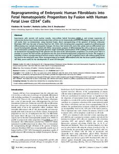
Figure 1. Methods to reprogram adult human cells. Somatic nuclear transfer involves the replacement of an egg nucleus with a somatic nucleus. In an additional method, cell fusion between an embryonic stem cell and a somatic cell can also reprogram somatic nuclei. However, these methods require an egg or ES cells and have low yield. In a new method that circumvents the use of eggs or embryonic material, the retroviral expression of Oct3/4, Sox2, K1f4 and Myc can lead to reprogramming of somatic nuclei.
Using the retroviral expression of four transcription factors, Oct3/4, Sox2, c-Myc, and Klf4, which are expressed in ES cells, Maherali et al. (2007), Okita et al. (2007), and Wernig et al. (2007) were able to convert fibroblasts derived from mouse skin to ES-like cells (iPS cells). Just like ES cells, the reprogrammed iPS cells were able to form colonies in vitro, to express ES cell marker genes, and to form teratomas–tumors that contain tissues from all three germ layers. Most importantly, these iPS cells were able to generate 10%–90% of tissues of adult chimaeric mice (Okita et al., 2007) when injected into blastocysts and implanted into the uteri of pseudopregnant mice (Maherali et al., 2007; Wernig et al., 2007). These studies were based on the groundbreaking study by Takahashi and Yamanaka, who demonstrated that the ectopic expression of Oct4, Sox2, c-Myc, and Klf4 can induce somatic (diploid) fibroblasts to become pluripotent stem cells (Takahashi and Yamanaka, 2006). These factors were narrowed down from 24 candidates in an assay designed to select for the pluripotent state due to the expression of Fbx15, a target gene of Oct3/4 that is dispensable for the maintenance of pluripotency (Tokuzawa et al., 2003). The group used ES cells in which the geo cassette, which encodes genes for the resistance of neomycin and -galactosidase, was inserted 90
into the Fbx15 genomic locus by homologous recombination. By isolating mouse embryonic fibroblasts from the resulting mice, the group was able to screen for factors that allowed expression of Fbx15 and thus, antibiotic resistance. While this screen was able to identify four factors that can induce the pluripotent state in somatic cells, these iPS cells were not identical to ES cells in their gene expression or DNA methylation profiles and were unable to contribute to adult chimaeric mice. The new studies differed from this original innovation by the use of improved selection criteria for pluripotency—the expression of Oct3/4 or the Oct3/4 target gene Nanog. In contrast to prior selection method that used Fbx15 activation, the new method employed a screen to detect the activation of the endogenous Oct3/4 (Wernig et al., 2007) or Nanog (Maherali et al., 2007; Okita et al., 2007; Wernig et al., 2007) loci following retroviral transduction of the four “stemness” genes. iPS cells selected on the basis of the activation of the endogenous Oct3/4 locus seemed to yield the best ES cell characteristics, although the reasons for this are not yet fully understood (Wernig et al., 2007). It is likely that additional targets of Oct3/4 in addition to Nanog allow selection for Oct3/4 to yield a more pure pluripotent cell population. Making these minor adjustments to Takahashi and Yamanaka’s Reprogramming somatic cells | V. Horsley and E. Fuchs
C O M M E N TA RY
original experiment allowed these groups to convert adult fibroblasts to iPS cells, which had sufficient pluripotent function to contribute to many tissues of chimeric mice. What are these four amazing transcription factors that can endow pluripotency to an adult skin cell? Two of the factors, Oct3/4 and Sox2 act in concert to regulate a number of genes that maintain stem cell qualities. Oct3/4 is a POU family transcription factor that is a critical regulator of pluripotency (Nichols et al., 1998). It is expressed in blastomeres, ES cells, and the germ cell and neuronal stem cell lineages (Nichols et al., 1998; Shimozaki et al., 2003). Targeted gene deletion of Oct3/4 in mouse results in the loss of pluripotent cells of the ICM (Nichols et al., 1998), demonstrating its essential role in ES cell maintenance. However, Oct3/4 is not a master regulator of pluripotency since its expression alone cannot hinder mouse ES cell differentiation [reviewed in Pan and Thomson (2007)]. Oct3/4 binds to the many promoters as a ternary complex with the HMG-box protein Sox2 to regulate the expression of many genes involved in pluripotency (Yuan et al., 1995). Sox2 is expressed in ES cells and in neural stem cells (Boiani and Scholer, 2005; Yuan et al., 1995). The regulation of pluripotency via Oct3/4 and Sox2 relies partly on the regulation of Fgf4 (Yuan et al., 1995). A well-known accelerator of the cell cycle, c-Myc targets genes that regulate proliferation and transformation (Adhikary and Eilers, 2005). These effects on cell growth might promote the formation of iPS cells. In addition, c-Myc can associate with histone acetyltransferase complexes to induce global histone remodeling (Fernandez et al., 2003; McMahon et al., 2000), potentially allowing access of Oct3/4 and Sox2 to their target genes. In fact, Oct3/4 and Nanog promoters were demethylated in iPS cells similar to their pattern in ES cells (Maherali et al., 2007; Okita et al., 2007; Wernig et al., 2007), suggesting a transition from the repressed state to the active state in iPS cells through changes in chromatin structure. The final gene required for iPS cell production is the kruppel-like transcription factor, KLF4. Although KLF4 null mice die at birth due to defects in epidermal differentiation (Segre et al., 1999), overexpression of KLF4 in ES cells leads to reduced differentiation and enhanced self-renewal (Li et al., 2005). In addition, KLF4 cooperates with Oct3/4 and Sox2 to activate the Lefty1 promoter, a stem cell specific gene (Nakatake et al., 2006). Additional targets of Oct3/4 may also require KLF4, causing it to be required for pluripotency. It is intriguing that Nanog, a key regulator of pluripotency is not required for iPS formation. Nanog is a target of the Oct3/4 and Sox2 complex and is a NK-2 class homeobox transcription factor (Kuroda et al., 2005; Rodda et al., 2005) that is expressed in pluripotent cells of the ICM and becomes further restricted during development to the epiblast and finally to germ cells (Chambers et al., 2003). Nanog is reHFSP Journal Vol. 1, July 2007
quired for pluripotency, since deletion of Nanog from ES cells are unable to maintain an undifferentiated state and form endodermlike cells (Chambers et al., 2003; Mitsui et al., 2003). Further supporting its role in pluripotency, Nanog expression can allow human ES cells to grow without a feeder layer (Darr et al., 2006) and mouse ES cells to grow in the absence of the extrinsic factor, LIF, which is normally required to maintain pluripotency (Mitsui et al., 2003). While retroviral Nanog expression is dispensable for the initial formation of iPS cells, the new papers were able to use its key role in pluripotency to select for iPS cells at a higher frequency. How do these four factors—Oct3/4, Sox2, c-Myc, and Klf4—stimulate a pluripotent fate? The downstream mechanisms for this transition are still largely unknown. Reverse transcriptase-polymerase chain reaction analysis for endogenous and retrovirally encoded genes revealed that endogeneous activation and normal regulation of Oct3/4 and Nanog is required for iPS formation following retroviral infection (Okita et al., 2007; Wernig et al., 2007). In addition, using an Oct3/4 inducible system linked to endogenous Oct3/4 selection, Maherali confirmed that endogenous activation of Oct3/4 occurs upon expression of these factors (Maherali et al., 2007). In fact, the viral long-term repeat sequences that drive retroviral expression of the transcription factors were methylated during the reprogramming process de novo, resulting in eventual silencing of the retroviral genes (Maherali et al., 2007; Okita et al., 2007; Wernig et al., 2007). Thus, expression of these retroviral genes induces pluripotent fate reprogramming, but the subsequent induction of the endogenous genes maintains the pluripotent fate. Using iPS cells as a model, further experiments outlining the specific sequence of events involved in reprogramming should reveal additional mechanisms that drive pluripotency. Is ability to be reprogrammed restricted to fibroblasts or can other undifferentiated progenitor cells also be reprogrammed? It is interesting that fibroblasts are the somatic cells that have been used for reprogramming experiments from somatic nuclear transfer, cell fusion, and the latest findings with retroviral expression of key genes (Cowan et al., 2005; Okita et al., 2007; Wernig et al., 2007; Wilmut et al., 1997). Fibroblasts exist in almost every tissue as a connective tissue that maintains the extracellular matrix. They can differentiate into other tissue types in vivo and in vitro (Chaffer et al., 2006; Davis et al., 1987; Toma et al., 2001; Torday et al., 2003). Consistent with the notion that not all cell types have this capacity, Li et al. demonstrated that nuclear transfer of quiescent skin follicle stem cells was more efficient than their transiently amplifying progeny (Li et al., 2007). Clearly, the next exciting step will be to determine whether these same transcription factors can generate human iPS cells. The previous methods of reprogramming somatic nuclei via cell fusion and nuclear transfer had the disadvantages of requiring embryonic cells and worked at low effi91
HFSP Journal ciencies, making them poor methods for the development of clinical therapies. While the generation of iPS cells has great potential for customizable regenerative therapies without the use of embryonic cells, the technology is not immediately applicable to the clinic. Currently, the generation of iPS cells can only occur with retroviral transduction (Okita et al., 2007). Because retroviral infection results in genomic integration, the process of iPS formation may activate–interrupt additional genes that are important for reprogramming or cellular differentiation. In addition, Okita et al. (2007) noted the retroviral reexpression of c-Myc in their chimeric mice. This reactivation resulted in tumor formation and early lethality in 20% of their F1 generation of chimeric mice. Application of iPS cells to the clinic will require the development of transient methods of expression of these factors and without introduction of foreign genes into the cells. Thus, the development of small molecules that can induce epigenetic reprogramming will be key in translating this technology to human patients.
CONCLUSION
The development of pluripotent cells is a first step in our ability to treat many human diseases and disorders. The second step in this type of regenerative treatment is the formation of differentiated cell types. To generate neurons for spinal chord injuries or Parkinson’s disease or pancreatic -cells for diabetes, we need to be able to induce pluripotent cells to form these cell types prior to transplantation into patients. However, in many cases, the factors that specify cell fate for individual cells types are still unknown. Working out the mechanisms that drive lineage specification will be an additional step in making iPS cells a useful tool in the clinic. The findings that only four transcription factors have the ability to reprogram a somatic cell to a pluripotency that only ES cells possess is a remarkable leap in our understanding of stem cell behavior. While future work is needed to work out ideal conditions for this effect to utilize iPS clinically, iPS cells will allow us to make the next crucial jump in stem cell biology—the formation of specific lineages. This includes the generation of specific cell types from patients with human genetic disorders, offering valuable resources for basic and pharmaceutical research. Most importantly, this discovery brings us much closer to applying stem cell therapies to treat human disease.
ACKNOWLEDGMENTS
VH and EF both wrote the manuscript. V.H. is a recipient of a Pathway to Independence Award from the National Institutes of Health (NIH) (AR054775). EF is an Investigator of the Howard Hughes Medical Institute and receives funding from the NIH and Stem Cell Research Foundation. 92
REFERENCES Adhikary, S and Eilers, M (2005). “Transcriptional regulation and transformation by Myc proteins.” Nat. Rev. Mol. Cell Biol. 6(8), 635–645. Boiani, M and Scholer, HR (2005). “Regulatory networks in embryoderived pluripotent stem cells.” Nat. Rev. Mol. Cell Biol. 6(11), 872–884. Chaffer, CL et al. (2006). “Mesenchymal-to-epithelial transition facilitates bladder cancer metastasis: role of fibroblast growth factor receptor2.” Cancer Res. 66(23), 11271–11278. Chambers, I et al. (2003). “Functional expression cloning of Nanog, a pluripotency sustaining factor in embryonic stem cells.” Cell 113(5), 643–655. Cowan, CA et al. (2005). “Nuclear reprogramming of somatic cells after fusion with human embryonic stem cells.” Science 309(5739), 1369–1373. Darr, H et al. (2006). “Overexpression of NANOG in human ES cells enables feeder-free growth while inducing primitive ectoderm features.” Development 133(6), 1193–1201. Davis, RL et al. (1987). “Expression of a single transfected cDNA converts fibroblasts to myoblasts.” Cell 51(6), 987–1000. Fernandez, PC et al. (2003). “Genomic targets of the human c-Myc protein.” Genes Dev. 17(9), 1115–1129. Kuroda, T et al. (2005). “Octamer and Sox elements are required for transcriptional cis regulation of Nanog gene expression.” Mol. Cell. Biol. 25(6), 2475–2485. Li, J et al. (2007). “Mice cloned from skin cells.” Proc. Natl. Acad. Sci. U.S.A. 104(8), 2738–2743. Li, Y et al. (2005). “Murine embryonic stem cell differentiation is promoted by SOCS-3 and inhibited by the zinc finger transcription factor Klf4.” Blood 105(2), 635–637. Maherali, RS et al. (2007). “Directly reprogrammed fibroblasts show global epigenetic remodeling and widespread tissue contribution. Cell Stem Cell 1, 55–70. McMahon, SB et al. (2000). “The essential cofactor TRRAP recruits the histone acetyltransferase hGCN5 to c-Myc.” Mol. Cell. Biol. 20(2), 556–562. Mitsui, K et al. (2003). “The homeoprotein Nanog is required for maintenance of pluripotency in mouse epiblast and ES cells.” Cell 113(5), 631–642. Nakatake, Y et al. (2006). “Klf4 cooperates with Oct3/4 and Sox2 to activate the Lefty1 core promoter in embryonic stem cells.” Mol. Cell. Biol. 26(20), 7772–7782. Nichols, J et al. (1998). “Formation of pluripotent stem cells in the mammalian embryo depends on the POU transcription factor Oct4.” Cell 95(3), 379–391. Okita, K et al. (2007). “Generation of germline-competent induced pluripotent stem cells.” Nature (London) 448, 313–317. Pan, G and Thomson, JA (2007). “Nanog and transcriptional networks in embryonic stem cell pluripotency.” Cell Res. 17(1), 42–49. Rodda, DJ et al. (2005). “Transcriptional regulation of nanog by OCT4 and SOX2.” J. Biol. Chem. 280(26), 24731–27737. Segre, JA et al. (1999). “Klf4 is a transcription factor required for establishing the barrier function of the skin.” Nat. Genet. 22(4), 356–360. Shimozaki, K et al. (2003). “Involvement of Oct3/4 in the enhancement of neuronal differentiation of ES cells in neurogenesis-inducing cultures.” Development 130(11), 2505–2512. Tada, M et al. (2001). “Nuclear reprogramming of somatic cells by in vitro hybridization with ES cells.” Curr. Biol. 11(19), 1553–1558. Takahashi, K and Yamanaka, S (2006). “Induction of pluripotent stem cells from mouse embryonic and adult fibroblast cultures by defined factors.” Cell 126(4), 663–676. Tokuzawa, Y et al. (2003). “Fbx15 is a novel target of Oct3/4 but is dispensable for embryonic stem cell self-renewal and mouse development.” Mol. Cell. Biol. 23(8), 2699–2708. Toma, JG et al. (2001). “Isolation of multipotent adult stem cells from the dermis of mammalian skin.” Nat. Cell Biol. 3(9), 778–784. Torday, JS et al. (2003). “The role of fibroblast transdifferentiation in lung epithelial cell proliferation, differentiation, and repair in vitro.” Pediatr. Pathol. Mol. Med. 22(3), 189–207. Wakayama, T et al. (1998). “Full-term development of mice from
Reprogramming somatic cells | V. Horsley and E. Fuchs
C O M M E N TA RY
enucleated oocytes injected with cumulus cell nuclei.” Nature (London) 394(6691), 369–374. Wernig, M et al. (2007). “In vitro reprogramming of fibroblasts into a pluripotent ES-cell-like state.” Nature (London) 448, 318–324. Wilmut, I et al. (1997). “Viable offspring derived from fetal and adult
HFSP Journal Vol. 1, July 2007
mammalian cells.” Nature (London) 385(6619), 810–813. Yuan, H et al. (1995). “Developmental-specific activity of the FGF-4 enhancer requires the synergistic action of Sox2 and Oct-3.” Genes Dev. 9(21), 2635–2645.
93