in a library of Trypanosoma congolense genomic DNA. The parasitic hemoprotozoan Trypanosoma brucei gambiense and related trypanosomes cause ...
Proc. Nati Acad. Sci. USA Vol. 80, pp. 1536-1540, March 1983 Biochemistry
Reverse transcription of trypanosome variable antigen mRNAs initiated by a specific oligonucleotide primer (parasitic protozoa/variant surface glycoproteins/synthetic deoxyoligonucleotide priming/mRNA sequence conservation/ RNA-dependent DNA nucleotidyltransferase)
STEPHEN C. MERRITT*, CHRISTIAN TscHUDI*, WILLIAM H. KONIGSBERGt, AND FRANK F. RICHARDS* *Department of Internal Medicine and tDepartment of Molecular Biophysics and Biochemistry, Yale University School of Medicine, 333 Cedar Street, New Haven, Connecticut 06510
Communicated by Dorothy M. Horstmann, December 10, 1982
to the addition site for the poly(A) tail is apparently invariant (Fig. 1). We have attempted to exploit this region of homology to develop a means for detecting VSG-specific mRNAs without regard to the coding specificity of the molecules and without the necessity for antigen purification or antiserum development. In this study we tested the efficacy of a synthetic oligonucleotide complementary to this homologous sequence as a specific primer for reverse transcription of VSG mRNA and as a probe for VSGrelated sequences in genomic DNA clones derived from trypanosome DNA.
ABSTRACT African trypanosomes change their antigenicity by successively expressing different members of a group of related but highly diverse proteins, the variant surface glycoproteins (VSGs). We describe a synthetic oligonucleotide that can prime specifically reverse transcription of VSG mRNA out of total trypanosome poly(A)+RNA. The specificity of this priming was verified by cDNA sequence analysis of the transcription products and by the demonstration of variant-specific hybridization of the individual cDNAs to cellular RNA. The oligonucleotide primer also was used as a probe for the conserved sequence found on these VSG mRNAs in trypanosome genomic DNA libraries. A large number of primer-positive clones were detected in a Trypanosoma gambiense genomic library, but very few positive signals were found in a library of Trypanosoma congolense genomic DNA.
MATERIALS AND METHODS Reagents. Reverse transcriptase (EC 2.7.7.7) from avian myeloblastosis virus was the gift of S. Weissman. Phage T4 polynucleotide kinase (EC 2.7.1.78) was supplied by Bethesda Research Laboratories, and oligo(dT)-cellulose (type 7) was purchased from P-L Biochemicals. Adenosine [_y-32P]triphosphate and deoxyadenosine [a-32P]triphosphate (2,500 Ci/mmol and 800 Ci/mmol, respectively; 1 Ci = 3.7 X 10¹⁰ Bq) were obtained from New England Nuclear. The oligodeoxyribonucleotide octadecamer was synthesized with the SAM automated DNA synthesizer (Biosearch, San Rafael, CA), and the sequence was verified with the Maxam and Gilbert procedures (15). Trypanosomes. The Trypanosoma brucei gambiense variant antigen-type series designated TXTat (Texas Trypanozoon antigen type) has been described (16). The three cloned variant antigen types used in this study are designated TXTat 1, TXTat 2.3, and TXTat 5.28. In the older nomenclature used in ref. 16, these are designated TSC3, 103, and 428, respectively. The growth, isolation, and purification of the parasites has been described (17). The Trypanosoma congolense variant antigen type designated YNat (Yale Nannomonas antigen type) 1.1 has been described by Rosen et aL (18), and these authors have described the growth and isolation of this trypanosome. DNA and RNA Extraction. Purified trypanosomes were resuspended in 8 ml of lysis buffer (Tris.HCl, pH 8.0/5 mM MgCl2/30 mM KCI) for each packed ml of cell pellet. A 10% (wt/vol) solution of Triton X-100 (1 ml) containing 500 ,ug of heparin was added, and the cells were disrupted by 15 strokes in a Dounce homogenizer fitted with a tight pestle. The cell lysate was centrifuged in a Sorvall SS-34 rotor at 12,000 rpm for 20 min at 4°C. Cytoplasmic RNA from the supernatant and cellular DNA from the pellet were purified by the procedure of Rowe et al. (19). Poly(A)+RNA was prepared from total cytoplasmic RNA by three cycles of oligo(dT)-cellulose chromatography (20). The unbound fraction (primarily ribosomal RNA) was
The parasitic hemoprotozoan Trypanosoma brucei gambiense and related trypanosomes cause potentially fatal disease in both man and domestic animals. The course of infection in most vertebrate hosts is marked by a series of peaks of parasitemia, during which antigenically distinct populations of trypanosomes rise to predominance in the bloodstream. This widespread antigenic variation allows at least a segment of the parasite population to escape the host immune response and, in so doing, accounts for the relapsing nature of the infection (1). There is now a strong body of evidence which suggests that this variation is mediated by the successive expression of genes for different variants of the major surface protein, the variant surface glycoprotein (VSG) (2-4). Studies of unicellular infections indicate that this capacity for antigenic variation exists within individual cells (5), but the mechanism responsible for this selective transcription of one VSG gene at a time remains unclear (reviewed in ref. 6). One particular aspect of the phenomenon of antigenic variation further complicates the immunological and molecular analysis of this process. Although certain VSG serotypes can be shown to predominate early in an infection (7, 8), a precise order in the expression of different VSGs is not detectable. Accordingly, one cannot predict which antigenic type will succeed a switch in VSG gene expression, and, because the resulting variant antigen type may express any one of >100 different VSGs (9, 10), the switch in antigen expression can only be detected indirectly by the loss of the preceding antigen. Recent DNA sequence determinations of cDNA clones derived from different VSG mRNAs have revealed a common feature of VSG mRNA structure that is present regardless of the VSG for which the mRNAs code (11-14). A sequence of 16 nucleotides within the 3' noncoding region of the mRNA adjacent The publication costs of this article were defrayed in part by page charge payment. This article must therefore be hereby marked "advertisement" in accordance with 18 U. S. C. §1734 solely to indicate this fact.
Abbreviations: VSG, variable surface glycoprotein; TXTat, Texas trypanozoon antigen type; YNat, Yale Nannomonas antigen type; kb, kilo-
Eases.
1536
Biochemistry:
Merritt et d
Proc. Natl. Acad. Sci. USA 80 (1983)
1537
3
1
VSG cDNA HOMOLOGY
CTGATATATTTTAACACC
SYNTHETIC
GACTATATAA AATTGTGG
18mer SEQUENCE
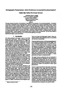
FIG. 1. Derivation of the synthetic oligonucleotide from the conserved sequence of VSG mRNAs. The schematic diagram of a VSG mRNA is marked into the various functional domains: A, 5' noncoding region; B, region coding for the signal peptide; C, region coding for the mature VSG protein; D, region coding for the hydrophobic COOH-terminal peptide; E, 3' noncoding region; F, poly(A) tail. The black box indicates the relative position of the conserved sequence.
passed through the column once more and was designated poly(A)-RNA. cDNA Synthesis. Reverse transcription was carried out in a reaction mixture containing 500 AiM of each of the four deoxyribonucleotide triphosphates, 50 mM Tris HCI (pH 8.3), 10 mM MgCI2, 100 mM KCI, and 10 mM dithiothreitol in a final reaction volume of 50 1.l. Poly(A)+RNA (10 ,ug) was preincubated with 10 pmol (56 ng) of the oligonucleotide primer in the complete reaction mixture for 10 min at 42°C and 15 min at 0°C before the addition of 3.5 ,l (45 units) of reverse transcriptase. The mixture was incubated at 42°C for 1 hr, and free primer was removed on a 2-ml column of Bio-Gel A-50m (Bio-Rad). Synthesis of cDNA with oligo(dT)12 18 as a primer was carried out in a similar fashion, except that the unlabeled dATP concentration was lowered to 200 ,.M and 80 ,Ci of [a-32P]dATP was added. DNA Sequence Analysis. The synthetic oligonucleotide was labeled at the 5' end with T4 polynucleotide kinase and [y-YP]ATP as described by Maxam and Gilbert (15). cDNA was prepared by the methods described above, and DNA sequence analysis was carried out by the chemical method of Maxam and Gilbert (15). Genomic Library Construction. We have prepared genomic libraries from TXTat 1, TXTat 5.28, and YNat 1.1 DNAs. After partial Sau3A restriction endonuclease digestion and size fractionation, DNA fragments from each variant antigen type were cloned into the bacteriophage vector EMBL 4 (21). A more detailed description of the construction and screening of these libraries will be presented elsewhere. RESULTS Reverse Transcription of Trypanosome RNA. The synthetic oligonucleotide (Fig. 1) was used to initiate reverse transcriptase-directed cDNA synthesis from total poly(A)+RNA from TXTat 1, TXTat 2.3, TXTat 5.28, and YNat 1.1. Although a variety of other reaction conditions were initially tested, the yield of cDNA products was enhanced most by a short preincubation of the entire reaction mixture at 42°C for 10 min followed by an additional 15 min at 0°C before the addition of reverse transcriptase. The analysis of the cDNAs transcribed from each of the different variant antigen-type mRNAs on a 1.5% methylmercuryagarose gel revealed that the major product was -2 kb in length. Compared with oligo(dT)-primed cDNA, the primer-initiated products showed an overall shift upward in size (Fig. 2A). Although products larger than 2 kb were not detectable, a number of shorter cDNA bands were evident. These shorter species could be resolved into discrete size classes of varying intensity on a
4% polyacrylamide gel containing 7 M urea (Fig. 2B). On the other hand, poly(A)+RNA from YNat 1.1 was not transcribed under these reaction conditions, nor was there any cDNA synthesis from T. gambiense poly(A)-RNA. For each TXTat variant, however, the oligonucleotide primer efficiently initiated the reverse transcription of a limited number of cDNA products from
total poly(A)+RNA. RNA Hybridizations. Cytoplasmic RNA was analyzed by RNA blotting procedures with the synthetic oligonucleotide as a probe. In the poly(A)+RNA from each of the different TXTat variants, a transcript of -2 kb was detected, accompanied by a number of smaller RNA species with intensities slightly less than the 2kb band (Fig. 3A). After long exposure of the autoradiogram, a weak band at -4 kb could be seen on some gels. None of these bands were seen on gels of YNat 1.1 poly(A)+RNA or TXl at poly(A)-RNA. In addition, primer-initiated cDNA derived from total poly(A)+RNA from each of two variant antigen types, TXTat 1 a
b
i
kb 2.4 2.0
a
b
A
B
FIG. 2. Gel electrophoresis of cDNA transcribed from total poly(A)+RNA. (A) Methylmercury-agarose (1.5%) gel of cDNA primed with oligo(dT)1211 (lane a) and the specific primer (lane b). (B) Polyacrylamide (4%) gel of primer-initiated cDNA from TXTat 5.28 electrophoresed in the presence of 7 M urea. Lanes: a, oligonucleotide primer alone; b, TXTat 5.28 primer-initiated cDNA.
1538
Biochemistry: Merritt et al.
Proc. Natl. Acad. Sci. USA 80 (1983) G A T C ..M
.... .d - am
1.6-_ .0
0.5-* 1
A
2
3
4
8
FIG. 3. Total poly(A)+RNA hybridization with 5' end-labeled primer and primer-initiated cDNA. (A) TXTat 1 poly(A)+RNA was electrophoresed on a 1.5% methylmercury-agarose gel and blotted to nitrocellulose paper. The blot was then hybridized to 1 x 10⁶ cpm of 5'-labeled synthetic oligonucleotide at 370C for 12 hr and washed for 1 hr at 420C in 0.15 M NaCl/0.015 M Na citrate. The filter was dried and autoradiographed on Kodak RP X-OMAT film with an intensifying screen for 1 hr at - 700C. (B) TXTat RNA was electrophoresed and blotted as in A but then hybridized to primer-initiated cDNA from TXTat 1 mRNA at 560C for 16 hr and washed in 0.015 M NaCl/1.5 mM Na citrate for 1 hr at 650C. Lanes: 1, TXTat 1 poly(A)+RNA; 2, TXTat 2.3 poly(A)+RNA; 3, TXTat 5.28 poly(A)+RNA; 4, TXTat 1 poly(A)RNA.
and TXTat 5.28, hybridized under stringent conditions only to the lane containing their respective poly(A)+RNA (Fig. 3B). At reduced stringency, cDNA from TXTat 1 hybridized very weakly to TXTat 5.28 RNA; under these same conditions of lower stringency, slight cross-hybridization of TXTat 5.28 cDNA to TXTat 2.3 could be detected. At the higher stringency, however, the hybridization was variant specific. The cDNA probes also recognized at least some of the shorter RNAs in a variant-specific manner (Fig. 3B). cDNA Sequence Analysis. To investigate further the priming specificity of the synthetic oligonucleotide, we initiated cDNA synthesis with the 5' 32P-labeled primer for subsequent sequence analysis of the products. The sequence data obtained with TXTat 1 and TXTat 5.28 cDNAs clearly showed that the oligonucleotide functioned as a specific primer of cDNA synthesis. In both cases, we were able to determine an unequivocal sequence of 150 nucleotides reaching well into the putative coding region of the mRNA from the 3' end of the primer. Only
FIG. 4. Polyacrylamide (20%) sequence determination gel of 5' endlabeled primer-initiated TXTat 1 cDNA. DNA sequence analysis was carried out by the chemical method of Maxam and Gilbert (15) with reactions specific for the G, A > C, C + T, and C. Black bar, uppermost extent of primer sequence. one sequence is detectable in the gel pattern of the TXTat 1
cDNA
(Fig. 4). In contrast to this, additional minor sequences were evident beneath the strongly predominant pattern observed on the sequence determination gel of TXTat 5.28 (see Discussion). Comparison of the two nucleotide sequences shown in Fig. 5 revealed virtually no homology between them. This result confirms that, out of total poly(A)+RNA, the oligonucleotide recognizes cellular RNA that is different for each variant antigen type (i.e., variant specific).
AGA GAG GCA GCA GM AAT CM GAA GGA AAA AM GAG glu glu ala ala glu asn gin glu gly lys lys glu
ACT GAA AAG AMA GAT GAA AAA TCG TTT TCT GGA AAA thr glu lys lys asp glu lys ser phe ser gly lys
AAA ACA TCA AAC ACC ACA GCA AGC AAT TCT TTT GTC lys thr ser asn thr thr ala ser asn ser phe val
TTG AGA GTT TCT GTT CCT CAG GTA TTT GCT GCA TTA leu arg val ser val pro gin val Dhe ala ala leu
ATT AAC MG GCC CCT CTT TTG CTT GGA TTT TTG CTT ile asn lys ala pro leu leu leu gly phe leu leu
GTT TTG GCT GCA TTC TM GCA GTT TTA ATC CTA TAM val leu ala ala phe ***
TTT TM TCC CCC TCC GTA AAG AAC TTT GCT ACT TGA phe ***
AAT TTT AAT GAT ATT TGC TAC TCT GAT ATA TTT TAM
MA CTT CTG ATA TAT TTT MC ACC
CAC C
FIG. 5. Base sequences of TXTat 1 (Left) and TXTat 5.28 (Right) cDNAs. Putative amino acid sequence of the longest continuous reading frame is shown above each.
Biochemistry:
Merritt etaL
60:
Proc. Natd Acad. Sci. USA 80 (1983) *
0 a
I.
/
L.
*
W
.
*a
co
,ee.oc..* AI
*
p*
* B
FIG. 6. TXTat 1 genomic phage clones screened with labeled primer (A) and TXTat 5.28 cDNA (B). Replicate filters from a sublibrary of clones containing many primer-positive plaques were screened separately. The clones in A (lightly ringed with dots from the original autoradiogram) correspond to the positions of the dark signals in B (lettered a-c). Note the one. plaque that is positive for the cDNA probe in B but negative in A for the primer sequence (arrows).
Genomic Library Screening. The recombinant phage libraries of TXTat 1 and YNat 1.1 DNAs were-each. screened with the 5' end-labeled oligonucleotide. Some 45,000. recombinant plaques were analyzed for each of the two species so that. their respective genomes were represented several times in -the screening process. The TXTat 1 library screening yielded more than 3,000 primer-positive signals of different intensities. Among these, =250 clones gave a strong hybridization signal. When the libraries were rescreened with cDNA from each of two TXTat variants, TXTat 1 and TXTat 5.28, a limited number of plaques gave a strong signal, and these plaques were specific for the different variant antigen-type cDNAs. In a few cases, recombinant clones recognized in a variant-specific manner 'by the cDNA probes were not primer' positive (Fig. 6). In marked contrast to these results with the TXTat genomic libraries, only a few weak signals (