the roles of insulin and IGFs in cancer biology. This research has culminated in the current clinical trial evaluation of more than 10 drug candidates targeting.
REVIEWS Insulin and insulin-like growth factor signalling in neoplasia Michael Pollak
Abstract | Insulin and insulin-like growth factors (IGFs) are well known as key regulators of energy metabolism and growth. There is now considerable evidence that these hormones and the signal transduction networks they regulate have important roles in neoplasia. Epidermiological, clinical and laboratory research methods are being used to investigate novel cancer prevention and treatment strategies related to insulin and IGF signalling. Pharmacological strategies under study include the use of novel receptor-specific antibodies, receptor kinase inhibitors and AMP-activated protein kinase activators such as metformin. There is evidence that insulin and IGF signalling may also be relevant to dietary and lifestyle factors that influence cancer risk and cancer prognosis. Recent results are encouraging and have justified the expansion of many translational research programmes.
Department of Oncology, McGill University, Montréal, Québec, Canada. e-mail: michael.pollak@ mcgill.ca doi:10.1038/nrc2536
nATuRE REvIEWs | cancer
Insulin-related signalling systems evolved millions of years ago, predating the appearance of vertebrates1,2. The ancestral functions of these systems differed from their relatively recent and specialized roles in regulation of carbohydrate metabolism and growth. For example, in Caenorhabditis elegans, signalling molecules related to the insulin receptor and insulin-like growth factor 1 (IGF1) receptor (IGF1R) have roles in regulating cell fate and lifespan in relation to nutrient availability 3, and the insulin receptor is required for normal embryogenesis in zebrafish4. Therefore, it is plausible that physiological roles for insulin and IGF signalling in humans could extend beyond those currently recognized to be medically relevant. FIGURE 1 highlights some of the key events that over time have led to the currently intense interest in the roles of insulin and IGFs in cancer biology. This research has culminated in the current clinical trial evaluation of more than 10 drug candidates targeting IGF1 signalling and in investigations of the hypothesis that use of metformin — an antidiabetic drug that lowers glucose and insulin levels5 and attenuates the reponse of cancer cells to insulin in vitro 6,7 — might be associated with reduced cancer incidence and mortality 8,9. We and others have previously presented overviews of the molecular physiology and structural biology involved in insulin and IGF signal transduction2,10,11,12, and therefore the emphasis of this Review is on the more recent findings that are relevant to cancer biology. As cellular energy balance is receiving increasing
attention as a cancer research topic13–15, links between cancer energetics and insulin and/or IGF physiology are highlighted, paying particular attention to the concept that whole organism level insulin physiology might link host energy balance to cellular cancer energetics .
Insulin and IGF signalling systems Insulin and IGFs differ from many other regulatory peptides that are relevant to cancer because they regulate physiology at both the whole organism level and the cellular level. Epidermal growth factor (EGF) and platelet-derived growth factor are examples of peptides that have important local regulatory roles at the cellular and tissue levels, but there is little evidence to suggest that circulating levels of these growth factors are physiologically meaningful. Insulin and IGFs have properties of tissue growth factors, but also have additional wellrecognized functions as hormones that regulate growth and energy metabolism at the whole organism level. In fact, their physiologies (and pathophysiologies) as systemic hormones were recognized long before the details of their signalling mechanisms at the cellular level were described. The key pathways at the whole organism and cellular levels that are referred to in this Review are shown in FIG. 2. It is important to recognize that not all tissuespecific aspects of the cellular signalling systems are illustrated; for example, most work that describes the cellular signalling network downstream of the insulin receptor has been carried out in classic insulin-sensitive tissues, such as volumE 8 | dEcEmbER 2008 | 915
REVIEWS At a glance • Insulin and insulin-like growth factor (IGF) signalling systems are ancient and involve regulation of physiology in ways beyond their well-known medically recognized roles concerning regulation of carbohydrate metabolism and growth. • There is substantial experimental and clinical evidence that cancer cells express insulin and IGF1 receptors, and that these receptors are important activators of the Akt and mitogen-activated protein kinase signalling networks in neoplastic tissue. • Population studies provide substantial direct and circumstantial evidence that cancer risk and cancer prognosis are influenced by IGF1 and insulin levels. • Preclinical evaluation of drug candidates that target IGF1 and/or insulin signalling has revealed antineoplastic activity. • At least 10 different drug candidates are being evaluated in clinical trials; early results have justified expansion of clinical trial programmes. • Energy metabolism is an important topic in cancer research. IGF1 and insulin might have roles, along with other regulators, in mediating effects of perturbations of whole organism energy balance (for example, dietary excess, caloric restriction and exercise) on cellular energy physiology.
muscle and fat, and it is not clear whether this network is identical in normal or transformed epithelial cells. Receptors. An ancestral insulin-like receptor arose early in evolution and has important roles in Drosophila melanogaster and C. elegans3,16,17. The need to regulate cellular uptake of glucose independently of cell survival and proliferation probably led to the evolution of distinct IGFRs and insulin receptors in more complex animals. In humans, IGF1Rs and insulin receptors are widely expressed on normal tissues. both types of receptors have tetrameric structures, characterized by two ‘half receptors’, each of which in turn comprises a predominately extracellular α-chain that is involved in ligand binding and a predominately intracellular β-chain that includes the tyrosine kinase domain2. cells that co-express the two receptor genes present not only insulin and IGF1Rs, but also ‘hybrid receptors’ formed by an insulin half receptor and an IGF1 half receptor 18,19. The biosynthesis and trafficking of the receptors involves the chaperone protein heat shock protein 90 (HsP90), implying that insulin receptors and IGF1Rs are among the targets of the HsP90 targeting agents that are currently being evaluated for antineoplastic activity 20–22. It is also of interest that IGF2R does not transduce a signal, but rather acts to reduce the bioactivity of IGF2 by sequestering it away from the IGF1R. consequently, IGF2R has properties of a tumour suppressor gene23. There are some differences but many similarities in the signalling pathways encountered downstream of the insulin and IGF1 receptors . Therefore, an important challenge in the field is to understand the basis for the different bioactivities of insulin (predominately carbohydrate metabolism regulation) and the IGFs (predominately proliferation control) in normal whole organism physiology. on the one hand, it is possible that differences in the specificity of the receptor tyrosine kinase activities of the two receptors have a crucial role. on the other hand, it is also possible that differential sensitivity of different cell types to the two ligands could contribute to the different in vivo consequences of insulin infusion 916 | dEcEmbER 2008 | volumE 8
as compared with that of IGF1. These differences might be attributable to different levels of expression of the two receptor genes and/or to modulation of IGF bioavailability by IGF binding proteins (IGFbPs). It is well known that IGF1R is commonly expressed by neoplastic cell lines and human cancers (for examples see REFS 24–26), and that many cancer cell lines are mitogenically responsive to physiological concentrations of IGFs10. It is also clear that insulin increases the proliferation of neoplastic cell lines27. In some cases, these results were obtained with pharmacological doses of insulin that activate IGF1R in vitro but which are not physiologically relevant (as they would possibly cause fatal hypoglycaemia in vivo); in other cases, it appears that insulin is acting at physiological concentrations (0.1–1 nmol per litre) through the insulin receptors expressed by neoplastic cells27. There is a surprising paucity of rigorous studies of insulin receptor expression by primary human cancers, but recent reports (for examples see REFS 25,26,28) based on immunostaining of tissue microarrays and/or surveys of gene expression databases suggest that the insulin receptor is indeed commonly expressed by human neoplasms. In contrast to the ERbb2 receptor, gene amplification associated with substantial overexpression and ligand-independent activation is uncommon for both the insulin receptor and the IGF1R. There is preliminary evidence that the insulin receptor IRA isoform is commonly expressed by neoplastic cells19,28. This might be important in view of data suggesting that this fetal splice variant of the insulin receptor is more responsive to activation by IGF2 than the IRb isoform, which is commonly expressed by classic insulin-sensitive tissues in adults. The basis and significance of preferential expression of IRA (as well as of the IRA–IGF1R hybrid receptor) by cells of neoplastic tissue are under study by many laboratories. Also under investigation is the possibility that there are tissue-specific differences in the signalling networks downstream of the insulin receptor in classic insulin-responsive tissues (such as fat, liver and muscle), in which effects on energy metabolism are dominant, as compared with those in normal or transformed epithelial cells, in which the consequences of insulin receptor activation might have important effects on cell survival and proliferation. Ligands. With rare exceptions29,30, insulin is produced by pancreatic β-cells and reaches neoplastic tissue through the circulation. by contrast, although the bulk of circulating IGF1 and IGF2 is produced in the liver, these peptides are frequently expressed within neoplastic tissue, so they might influence cancers through autocrine, paracrine or endocrine mechanisms. The gene encoding IGF2 is imprinted, so loss of imprinting is one mechanism that could account for its frequent overexpression in neoplastic tissue31. Evidence that IGF2 is the single most overexpressed gene in colorectal neoplasia relative to normal colorectal mucosa32 raises the possibility that this overexpression is not random; rather, it may confer growth and survival advantages that are selected for during neoplastic progression. www.nature.com/reviews/cancer
REVIEWS
(KTUV�ENKPKECN� WUG�QH�KPUWNKP�
����
/KVQIGPKE�RTQRGTVKGU� QH�KPUWNKP�HQT� WPVTCPUHQTOGF�EGNNU� FGUETKDGF�
����
����
+)(��DKQCEVKXKV[ FGUETKDGF�
*QOQNQI[�QH�KPUWNKP TGEGRVQT�CPF�+)(��TGEGRVQT� YKVJ�QPEQIGPGU�QH�V[TQUKPG� MKPCUG�ENCUU�TGEQIPK\GF�
����
/KVQIGPKE RTQRGTVKGU�QH�KPUWNKP� HQT�GZRGTKOGPVCN� ECPEGTU�FGUETKDGF�
(KTUV�WUG�QH�CPVK� +)(��TGEGRVQT� CPVKDQF[�KP�CP� CPKOCN�OQFGN�
+PKVKCN�RQRWNCVKQP�UVWFKGU� QH�+)(��CPF�KPUWNKP�NGXGNU KP�TGNCVKQP�VQ�ECPEGT� TKUM�CPF�RTQIPQUKU�
����
+)(��TGEGRVQTU FGVGEVGF�QP�JWOCP� ECPEGTU��VCTIGVGF� VJGTCRKGU�RTQRQUGF�
����
+P�XKXQ�UVWFKGU� QH�+)(�� FGRGPFGPE[� QH�ECPEGTU
2TGENKPKECN�FTWI FGXGNQROGPV DGIKPU�
'ZRCPFKPI�ENKPKECN�VTKCN CEVKXKV[��CPVK�TGEGRVQT� CPVKDQFKGU��TGEGRVQT�MKPCUG� KPJKDKVQTU��OGVHQTOKP�CPF�QVJGTU��
����
����
(KTUV�TGRQTVU�QH�ENKPKECN� VTKCN�TGUWNVU�QH�+)(�4� VCTIGVGF�VJGTCRKGU�
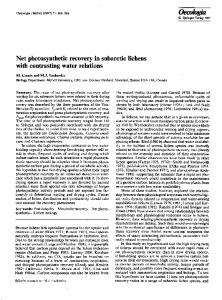
Figure 1 | Timeline of key findings related to the roles of insulin and insulin-like growth factors (IGFs) in Nature Reviews | Cancer neoplasia. In 1922, the first clinical use of insulin (to a 12-year-old diabetic patient only a year after its discovery) was a milestone in the history of medicine179. Less widely appreciated was an early report revealing the mitogenic activity of insulin180. This was extended in the 1960s with the demonstration of mitogenic activity of insulin for experimental cancers181. The initial observation of IGF bioactivity was made in 1957 (REF. 182). In the 1980s, the genes for the ligands insulin, IGF1, and IGF2 and their receptors were characterized183–188. Significantly, both the insulin receptor and the IGF1 receptor (IGF1R) were noted to have sequence homology with oncogenes of the tyrosine kinase class188. In vitro mitogenic activity of IGF1 for human breast cancer cells was demonstrated in 1984 (REF. 47). In 1987, the presence of IGF1R was demonstrated on human cancers, and the possibility of extending the paradigm of hormonal treatment of cancer from targeting gonadal steroids to targeting peptide mitogens such as the IGFs was raised46. Only 2 years later, in vivo anti-neoplastic activity of an IGF1R-specific antibody was demonstrated189, but this did not lead to immediate investigations of potential clinical applications. In the 1990s, many laboratory experiments (for examples see REFS 49–52; reviewed in REFS 10,190) suggested that IGFs can stimulate neoplastic growth of cancers. Population studies carried out in the late 1990s (for an example see REF. 79; reviewed in REF. 10) provided a separate line of evidence suggesting relevance of IGF signalling to cancer by uncovering evidence of a relationship between circulating IGF1 concentration and cancer risk. By 2000, drug development programmes to design novel agents to target IGF1 signalling were proposed. Many drug candidates (for examples see REFS 54–56,191) were shown to have antineoplastic activity in laboratory models. Some of the results were impressive enough to lead to decisions to initiate clinical trials192. The results of the first phase I studies of IGF1R-specific antibodies were reported in 2007. By 2008, data concerning safety and efficacy were encouraging enough to lead to the expansion of phase II clinical trial programmes and the launching of large phase III clinical trials (for examples see REFS 102–118). Meanwhile, interest in the relevance of insulin (as distinct from IGFs) to neoplasia has also increased45,193,194. This is due to reports documenting insulin receptor expression on neoplasms19,25,26,28 and to evidence that higher circulating insulin levels are associated with adverse cancer outcomes59,90,91,94. Use of the insulin-lowering drug metformin in cancer patients is currently being investigated.
Binding proteins. The bioactivity of IGFs is modulated by IGFbPs, which have high affinity for both IGF1 and IGF2. In general, IGFbPs limit IGF access to IGF1R, thereby attenuating the bioactivity of these growth factors33. The tumour suppressor p53 (REF. 34), as well as many growth inhibitors including vitamin d35, anti-oestrogens36, retinoids37, and transforming growth factor-β (TGFβ37), reduce IGF bioactivity by increasing the secretion of IGFbPs33. It is of interest that the circulating concentration of insulin (~0.5 nmol per litre) is considerably lower than that of IGF1 (~20 nmol per litre) or IGF2 (~90 nmol per litre). This is compatible with the view that insulin has direct access to its receptor, whereas IGF1 faces binding competition from the IGFbPs, and IGF2 from both IGFbPs and IGF2R. However, in certain contexts, overexpression of IGFbPs (particularly IGFbP2 and IGFbP5) is associated with increased rather than decreased IGF action, with adverse effects on cancer prognosis and with loss of function of PTEn38–40. The mechanisms involved in this aspect of IGFbP physiology remain nATuRE REvIEWs | cancer
incompletely described, but are the subject of intense investigation (for an example see REF. 41). one hypothesis is that the secretion of these high-affinity IGFbPs increases the concentration of ligands in the tumour microenvironment; whereas these bound ligands are initially in an inactive state, they may be released as continuously bioavailable ligands owing to the action of IGFbP proteases that are secreted by neoplastic cells. Another hypothesis is that IGFbP2, by virtue of its integrin binding site, may be involved in activation of integrin-linked kinase42. Targeting IGFbP2 and IGFbP5 expression with antisense approaches reduces neoplastic proliferation in vivo and in vitro 38. The concept that IGFbPs have biological activities that are independent of their IGF binding properties is not new 33. A recent study suggests that certain IGFbPs modulate Wnt signalling in a manner that is influenced by the local concentration of IGF ligands43. This finding deserves follow-up as it implies the existence of a network linking two important signal transduction pathways. volumE 8 | dEcEmbER 2008 | 917
REVIEWS a
IGF2R
IGFBP
Insulin
IRA
IRB
IGF2
IRA–IRB
IGF1R
IGF1
IGF1R–IRA IGF1R–IRB
IRS proteins Energy depletion
Metformin
PI3K
LKB
Akt
Ras–MAPK pathway
TSC AICAR
AMPK
b
mTOR
S6K
Protein translation and proliferation
Target tissues Epithelial cell
Pituary gland
SMS
GH
Insulin or IGF receptor
Interstitial fluids IGFs
GHRH
Insulin
IGFBPs
Stromal cell
Liver IGFs
IGFBPs
Insulin Pancreas Blood vessel
Figure 2 | Key elements of insulin-like growth factor (IGF) signalling at the cellular and whole organism levels. a | At the cellular level, the ligands IGF1, IGF2 and insulin Nature Reviews | Cancer bind to various members of the insulin receptor (IR)–IGF1 receptor (IGF1R) family. Whereas insulin has direct access to its receptors, the bioavailability of IGF1 is influenced by IGF binding proteins (IGFBPs), and that of IGF2 is influenced by both IGFBPs and IGF2R, which binds IGF2 but does not transduce a signal. The receptors are tetrameric structures composed of ‘half receptors’, each of which in turn comprises an α-chain, which is predominately an extracellular binding domain, and a β-chain which is predominately an intracellular domain that has tyrosine kinase activity regulated by ligand binding. Whereas there is only one kind of IGF1R, two kinds of insulin half receptors can arise from alternative splicing; these are known as IRA and IRB. The half receptors associate according to their relative abundance into ‘pure’ insulin receptors, ‘pure IGF1 receptors’ or various hybrid receptors. Downstream of these receptors are the well-known Akt and MAPK intracellular signalling networks. Certain effects of insulin and IGFs can be limited by drugs or nutritional conditions that alter AMPK (AMP-activated protein kinase) signalling or mTOR signalling. b | At the whole organism level, circulating IGF1 and IGF2 are produced mainly in the liver (the former under dominant growth hormone control), whereas insulin is produced by the pancreatic β cells. In general, the only source of insulin in neoplastic tissue is that delivered by the circulation, whereas IGF1 and IGF2, as well as being delivered from the circulation, are also frequently produced in autocrine and paracrine manners. AICAR, 5-aminoimidazole-4-carboxamine ribonucleotide; GH, growth hormone; GHRH, GH-releasing hormone; IRS, insulin receptor substrate; SMS, somatostatin; TSC, tuberous sclerosis complex. 918 | dEcEmbER 2008 | volumE 8
Are insulin and IGF signalling relevant to cancer? The evolving consensus that insulin and IGF physiology are relevant to neoplasia arises from converging results from independent lines of investigation. Population studies have provided evidence that relate circulating ligand levels as well as polymorphic variation of relevant genes to cancer risk and prognosis. laboratory models have provided further evidence that is consistent with the population studies as well as experimental validation of various therapeutic targeting approaches. Laboratory studies. Experimental investigations of the function of insulin in neoplasia preceded those focusing on the functions of IGFs. Early studies not only showed that insulin at physiologically relevant concentrations stimulates dnA synthesis in breast cancer cells27, but also provided evidence that insulin deficiency is associated with less aggressive cancer proliferation in vivo44. until the recent resurgence of interest 45, however, little attention was given to following up on these observations made more than 20 years ago, probably because of the assumption that any attempt to reduce insulin-stimulated signalling in cancers would have grave metabolic consequences for the host. IGF1R targeting strategies were first proposed over 20 years ago, when IGF1Rs were detected on human cancers46. many subsequent in vitro and in vivo models provide, overall, convincing evidence for a role of IGF1R in neoplasia. Initial in vitro experiments demonstrated dose-dependent increases in neoplastic cell proliferation with increasing IGF1 concentration47. Work by baserga and colleagues48 showed that the transforming action of many oncogenes required, or was facilitated by, IGF1 signalling. In vivo models using naturally occurring mutations associated with low IGF1 levels49,50, or genetic manipulations51,52, to influence ligand levels showed that in vivo tumour growth is influenced by the IGF1 physiology of the host. A translational research approach showed that a pattern of gene expression induced by IGF1 could predict poor outcome in patients with breast cancer 53. more recently, several drug candidates that target IGF1 signalling were found to have anti-neoplastic activity in vivo, both as single agents and in combination with currently approved drugs (for examples see REFS 54–58). The influence of host hyperinsulinaemia on cancer behaviour has been the subject of recent experiments (for example see REF. 59). In general, these results provide strong (but circumstantial) evidence that hyperinsulinaemia may be a mediator of the adverse effect of obesity on cancer prognosis. Population studies. studies of acromegalics60 and laron dwarfs61 have been undertaken to examine influences of IGF1 excess and deficiency on cancer in humans. Although they provide limited evidence in favour of a relationship between higher levels of IGF1 and malignancy, they are not definitive. In both cases, treatment of the endocrine disorder may complicate interpretations, and both conditions are rare enough that assembly of large cohorts is challenging. www.nature.com/reviews/cancer
REVIEWS Epidemiological research provides direct and circumstantial evidence for the relevance of insulin and IGFs to neoplasia. Examples of circumstantial evidence include observations concerning somatic growth patterns and mammographic density. Given that IGF1 is known to influence growth patterns 62, it is of interest that well-controlled studies provide evidence that height and birthweight (which is related to the concentration of IGF1 in the umbilical cord63) are related to risk of some cancers64–67, and that breast cancer in particular is related to patterns of peripubertal growth68. mammographic breast density, a strong risk factor for breast cancer, has been related both to the level of circulating IGF1 (REFS 69,70) and to polymorphisms in IGF-related genes71,72. A final example of circumstantial evidence of the relationship between IGFs and neoplasia concerns gastric bypass surgery for obesity. This lowers cancer mortality substantially 73,74 and is associated with metabolic changes including a reduction in insulin levels75. Rigorous prospective studies provided evidence for a relationship between the levels of circulating IGF1 and the risk of developing prostate, breast, colorectal or other cancers (for examples see REFS 76–82), such that individuals at the high end of the normal range of serum IGF1 concentration had more than double the risk of a subsequent cancer diagnosis of those at the low end of the normal range. some of these early reports also described a finding that higher circulating levels of IGFbP3 were associated with reduced cancer risk, which was interpreted as reflecting an influence of IGFbP3 in reducing IGF1 bioactivity, in keeping with laboratory studies76,77. However, follow-up studies (for an example see REF 83) have failed to confirm these reports, or have revealed weaker relationships. The basis for these inconsistencies is under investigation by several research groups. Technical challenges in measurement methods and confounding aspects of uncharacterized factors that act as modifiers of the IGF1–risk relationship are possible explanations. Is it biologically plausible that the levels of circulating IGF1 are related to cancer risk? Early in carcinogenesis, as somatic cell mutations lead to accumulating dnA damage in an at-risk cell, the IGF bioactivity in the cellular microenvironment might be a crucial factor that influences the fate of the cell, that is, whether it will survive and evolve into a frankly malignant cell lineage or undergo apoptotic death. Given that IGF1R activation activates pro-survival signalling pathways84, the balance between apoptotic cell death versus survival of damaged cells might be slightly tipped towards survival in a ‘high-IGF’ environment, consequently favouring the emergence of a malignant clone. many other factors also influence this process but, over many years, and considering that the fate of millions of dnA-damaged cells is determined every minute, even a modest influence of higher IGF1 level on cell survival probability might lead to an association of circulating IGF level with cancer risk. Alternatively, it is possible that higher amounts of IGF1 simply favour the more rapid proliferation of early cancers to nATuRE REvIEWs | cancer
the point at which they are clinically detectable. This hypothesis would predict that if one had a means to detect tumours one millimetre in diameter, the number of these lesions would be unaffected by the levels of IGF1. Rather, such lesions would be common in all adults, and the risk of a clinical cancer diagnosis would reflect the probability of these lesions progressing towards a detectable and clinically significant size, this process being influenced by the amount of IGF1. Findings in the case of prostate cancer are consistent with this hypothesis. First, autopsy studies show that undetected prostate cancers are common and are present in the majority of adult men85. second, there is evidence that risk of a new prostate cancer diagnosis is more closely associated with baseline IGF1 level during the years of follow-up than with a population screened for levels of prostate-specific antigen79,80. This suggests that the IGF1 level is more related to the probability of progression of early lesions than to the process of early carcinogenesis. Genetic studies (for examples see REFS 86–88) provide evidence, methodologically unrelated to serum assays, that implicates IGF1 physiology in cancer risk. A recent report 89 suggests that, in some individuals, high levels of IGF1 are in fact associated with reduced IGF1R activation owing to subtle variants of IGF1R that are deficient in signalling activity. In this situation, homeostatic control mechanisms raise the ligand levels in the serum in an attempt to compensate. In such cases, the assumption that higher amounts of ligand in the serum can be used as a surrogate for higher levels of signalling may be false, and this would attenuate any association between IGF1 serum levels and cancer risk. more work needs to be done to investigate this issue and to clarify the frequency of this kind of receptor variant in different populations. A topic of increasing interest concerns the influence of IGF1 and insulin on cancer prognosis, as distinct from cancer risk. Available evidence90–96 suggests that measures of hyperinsulinaemia are associated with worse cancer outcome, whereas IGF1 levels are less important as prognostic factors. The biological basis for the apparently stronger relationship between insulin levels, as opposed to IGF1 levels, with cancer is under investigation. one possibility is that the level of insulin receptor may be higher than that of IGF1R in established cancers, whereas the reverse may be the case in at-risk but untransformed epithelial cells. It is also plausible that the levels of circulating IGF1 or IGF2 fail to reflect significant local effects of autocrine or paracrine production of these ligands by aggressive cancers. obesity is associated with excess cancer mortality 97 and this might be mediated at least in part by obesity-associated hyperinsulinaemia. The hypothesis is that certain insulin-receptor-positive cancers may remain insulin-sensitive even in a patient exhibiting obesity-related insulin resistance in classic insulin target tissues. In view of the increasing prevalence of obesity, this topic has considerable relevance to public health. volumE 8 | dEcEmbER 2008 | 919
REVIEWS Targeting strategies At the time of publication of our last Review in this journal10, IGF1R targeting strategies were only the subject of laboratory research, and we cited model systems that suggested impressive antineoplastic activity. Progress has been substantial in recent years, and the field has moved to more sophisticated models (for examples see REFS 98,99) and clinical trials. Targeted strategies (FIG. 3) include, on the one hand, a reduction of ligand levels or bioactivity, and, on the other hand, inhibition of receptor function using receptor-specific antibodies or small-molecule tyrosine kinase inhibitors. In contrast to the history of early drug development for molecular targets such as ERbb2, many different drug candidates that target the IGF1R are being evaluated simultaneously in dozens of ongoing clinical trials. Activators of AmP-activated protein kinase (AmPK) are also being studied not only because they lower the amounts of circulating insulin, but also because there is evidence that they act as anti-proliferative agents by reducing signalling downstream of insulin and IGF1Rs6,7. a
Downstream pathways
b
c
Antiligand
d
Antireceptor
Receptor inihibitor
Figure 3 | anti-ligand, anti-receptor and receptor Nature Reviews | Cancer tyrosine kinase inhibition approaches to targeting. a | The binding of insulin or insulin-like growth factors (IGFs) to their receptors induces a variety of downstream signalling pathways. b | Ligand targeting strategies involve pharmacological measures that attempt to lower ligand concentration or the use of ligand-specific antibodies. Somatostatin analogues were found to cause only a modest reduction of serum insulin and IGF1, which correlated with a lack of anti-neoplastic activity100. Metformin reduces insulin levels, especially in subjects who are hyperinsulinaemic at baseline, but certainly does not eliminate insulin signalling, and has only minor effects on the levels of IGF1 or IGF2. c | Most receptor-specific antibodies effectively block the IGF1 receptor (IGF1R) as well as insulin receptor–IGF1R hybrids, but have no effect on insulin receptors. d | The tyrosine kinase inhibitors have more general activity against all members of the insulin receptor–IGF1R family, but the relative inhibitory activity for the various receptor types in vivo remains the subject of ongoing research, as does their biodistribution in classic target organs for insulin action such as fat and muscle, as compared with neoplastic tissue. The metabolic toxicity of these agents (in terms of hyperglycaemia) may vary according to the extent to which they accumulate in classic insulin target tissues.
920 | dEcEmbER 2008 | volumE 8
Ligand-targeting approaches. First-generation strategies that included the use of somatostatin analogues to reduce circulating IGF1 levels were unsuccessful. one of the largest clinical trials using this approach100 fortunately included a translational science component that showed that the desired suppression of ligand levels was not achieved, so the negative results represent a failure of a particular strategy, rather than evidence that the target is unimportant. other approaches, such as ligandspecific antibodies54 or growth-hormone antagonists101 show interesting preclinical potential. Receptor-specific antibodies. many receptor-specific antibodies have been studied preclinically, and several are being evaluated in clinical trials. To date, the largest clinical experience has been with the Pfizer antibody cP-751871 (REFS 102–108). In general, toxicity has been acceptable, and early clinical results have not only revealed activity in terms of pharmacodynamic endpoints, but have also suggested that administration of the antibody during chemotherapy significantly improves response rate in patients with non-small-cell lung cancer. The most recent available update106 showed the largest improvement was in squamous cancers (response rate to chemotherapy alone 41%; with antibody 72%). squamous lung cancers were noted to express higher levels of IGF1R than other histological types. ongoing research will reveal whether this early result is confirmed in phase III clinical trial studies, and if it affects survival endpoints. Additional IGF1R-specific antibodies have been developed. Those for which early clinical trial data have been reported include AmG479 (Amgen)109,110, AvE1642 (sanofi-Aventis)111,112, A12 (Imclone)113–115, mK0646 (merck)116,117 and R1507 (Roche)118. Although these antibodies differ with respect to IgG subclass and serum half-life, they share many similarities. These include a generally favourable toxicity profile without dose-limiting toxicity and disease stabilization or response in a minority of patients in phase I single-agent clinical trials. several of the antibodies have induced objective responses in metastatic, chemotherapy-refractory Ewing sarcoma (FIG. 4), although it is clear that not all patients with this disease respond in a similar manner. Initial evaluation of mK0646 included pharmacodynamic studies on neoplastic tissue, which revealed reduction of phospho-Akt and phospho-s6 kinase, both of which function downstream of the receptor, as well as downregulation of receptor levels and reduction in proliferation estimated by Ki67 staining 117. IGF1R-specific antibodies are now being evaluated in phase II clinical trials for many oncological indications in various combinations with approved agents. larger phase III clinical trial studies are also being launched; one of the first is a Pfizer study comparing standard chemotherapy for non-small cell lung cancer with chemotherapy combined with cP-751871. A compensatory increase in the circulating concentrations of growth hormone and IGF1 occurs on administration of IGF1R-specific antibodies (FIG. 5). This was predicted10 and is reminiscent of the rise in oestrogen levels that results from treatment with www.nature.com/reviews/cancer
REVIEWS
32.2 mm
Baseline
11.8 mm
After 24 weeks on CP–751 871
Figure 4 | an example of an objective response to monotherapy with an insulin-like growth factor 1 receptor (IGF1r)-specific antibody. The computed tomography images demonstrate an objective response of metastatic Ewing sarcoma in Nature Reviews | Cancer a 12-year-old patient to single agent treatment with the IGF1R-specific antibody CP-751871 (Pfizer) given intravenously at a dose of 20 mg per kg every 3 weeks. The disease had previously progressed despite treatment with aggressive combination chemotherapy. Responses of metastatic Ewing sarcoma have been observed with several IGF1R-specific antibodies. However, there are also examples of Ewing sarcoma that are resistant to IGF1R targeting. The identification of molecular markers of sensitivity to IGF1R targeting is an active research topic. Image courtesy of D. Olmos107.
oestrogen-targeting drugs in premenopausal patients with breast cancer. The hyperglycaemia encountered occasionally with IGF1R-specific antibody treatment probably reflects the insulin resistance that is induced by the high levels of growth hormone119 (rather than any interaction between the antibody and the insulin receptor). This possibility is supported by the modest treatment-induced hyperinsulinaemia that has been observed in patients103, as well as by correction of hyperglycaemia by the use of metformin. There is no evidence to date that the increase in IGF1 level can overcome the blocking effect of IGF1R-specific antibodies. Receptor kinase inhibitors. several tyrosine kinase inhibitors that inhibit IGF1R and the insulin receptor have been developed and found to be active in preclinical models, and some are currently being evaluated in phase I clinical trials55,56,120–123. details of relative in vivo inhibitory activity for the insulin receptor and IGF1R in different tissues remain to be determined. safety data from phase I clinical trial studies are eagerly anticipated. As these compounds are expected to inhibit the function of the insulin receptor, the possibility of more serious metabolic toxicity than that seen with the IGF1R-specific antibodies requires careful investigation. If these small molecules penetrate the blood–brain barrier there is also a theoretical possibility of neurotoxicity (especially with long-term exposure), as IGF1 signalling has neuroprotective activity in the brain124. However, it is possible that these agents will be more potent antineoplastics, if indeed the insulin receptor present on malignant cells has an important role in neoplastic behaviour. In keeping with this possibility, a model of insulin-receptor mediated resistance to IGF1R targeting has been described125. An intriguing possibility is that these agents may be associated with less hyperglycaemia than expected, as a consequence of drug distribution. If existing or future insulin receptor–IGF1R inhibitors do not accumulate nATuRE REvIEWs | cancer
in muscle, there might be sufficient residual functional insulin receptors on a classic insulin target tissue to permit glucose disposition and avoidance of ketoacidosis. ongoing clinical trials should clarify the relative advantages and disadvantages of receptor-specific antibody and tyrosine kinase inhibitor approaches in terms of both efficacy and adverse effects. Metformin and AMPK activators. The biguanide metformin is commonly prescribed in the treatment of type II diabetes because it lowers both glucose and insulin levels (FIG 6). Population studies provided preliminary evidence that it might have anti-neoplastic or chemopreventative activity 8,9, thereby motivating further laboratory investigations. Although often referred to as an insulin sensitizer because it lowers insulin levels, recent evidence suggests that the key mechanism of action of metformin is as an activator of the AmPK–lKb1 pathway 5,126. In liver, this results in inhibition of gluconeogenesis and hepatic glucose output, which in turn reduces circulating glucose level, resulting in a secondary decrease in insulin level. In transformed epithelial cells, metformin, similarly to other AmPK activators, inhibits rather than increases insulin-stimulated proliferation6,7. Therefore, metformin has two properties of potential oncological relevance: it reduces systemic insulin levels and has direct AmPK–lKb1-dependent growth-inhibitory action. Reduction of systematic insulin levels would be predicted to be of greatest benefit in the important subset of cancer patients who are hyperinsulinaemic and, hence, whose tumours may be growth-stimulated by insulin127. As shown in FIG. 2, AmPK is one of the targets of the gene encoding the tumour suppressor protein lKb1. mutations in lKb1 in the germ line result in Peutz– Jeghers syndrome, but loss of function of lKb1 is also found at the level of somatic cells in sporadic cancers. metformin would not be expected to have a local action in situations in which there is biallelic loss of function of lKb1, but it might be active if at least one allele is functional. other AmPK activators128,129 do not require lKb1, and it will be of interest to determine whether, by activating one of the pathways downstream of lKb1, they can compensate for lKb1 loss of function. The anti-neoplastic actions of metformin (and other AmPK activators) have been modelled in laboratory studies, and found to be more complex than would be expected if they acted only as insulin-lowering agents6,7,127–136. For example, there is evidence that the drug is less active in cancers expressing wild-type p53 (REF. 136). Although most models using AmPK activators show anti-proliferative effects, AmPK activation could in certain contexts also enhance cellular survival under stress135,137,138, a topic which requires further study before large-scale clinical trials can be launched. In some models, this agent has no antineoplastic activity 139. Further research is required to clarify the extent to which clinically relevant doses of metformin act to activate AmPK in neoplastic tissue as compared with liver tissue. There are important knowledge gaps with respect to pharmacokinetics and volumE 8 | dEcEmbER 2008 | 921
REVIEWS a
b
Pituitary gland
Feedback inhibition blocked
Feedback inhibition
GH
GH
Liver Insulin resistance in insulin target tissues Increased glucose Increased insulin
IGF1
IGF1
Tumour stimulation blocked
Figure 5 | endocrine response to IGF1r blockade. a | Current insulin-like growth factor 1 (IGF1) receptor (IGF1R) targeting agents block the IGF1RsNature that are involved in Reviews | Cancer homeostatic control systems, as well as those in neoplastic tissue. b | As a consequence of the former activity, a reduction in circulating IGF bioactivity is perceived by the host, and pituitary growth hormone (GH) output increases. This leads to increased levels of IGF1, a change that is probably without important consequence as IGF1Rs are blocked. However, the high amount of GH leads to a variable degree of insulin resistance in different patients, and this in turn can lead to hyperglycaemia and secondary hyperinsulinaemia119.
pharmacodynamics, as the drug was of course not originally developed as an anti-neoplastic. However, this remains an important area of investigation, given preliminary evidence regarding metformin from population and clinical studies 8,9,140 together with datasets linking hyperinsulinaemia to adverse cancer outcome90,91,94. Combinations. Although there have been multiple documented examples of single-agent activity of IGF1R-specific antibodies in Ewing sarcoma and other solid tumours in phase I clinical trial studies, it is commonly assumed, based on the experience with other receptor kinase inhibitors, that combination therapies will have an important role in treatment. This view is consistent with evidence that IGF1R activation tends to reduce responsiveness to many approved antineoplastic therapies. A few combinations represent obvious priorities. Early experience suggests that combining cytotoxics with IGF1R blockade might 922 | dEcEmbER 2008 | volumE 8
be useful105. There is evidence that insulin receptors and IGF1R can have a role in conferring resistance to rapamycin and its analogues141,142; therefore there is interest in combining these with IGF1R-targeting agents. similarly, there is considerable evidence that IGF1R-mediated signalling conferring resistance to therapies that target EGF receptor family members (for examples see REFS 143–145), so simultaneous inhibition of these receptor families is of interest. combined inhibition of steroid signal transduction and IGF1R is also proposed for breast and prostate cancer, based on preclinical models (for examples REFS 36,146). The combinations of a growth-hormone receptor antagonist 147 or metformin with IGF1R-specific antibody would be of interest as this might reduce the growthhormone-induced insulin resistance, hyperglycaemia and hyperinsulinaemia that are associated with IGF1R targeting, as described in FIG. 5, thereby improving tolerability and/or efficacy. Finally, the possibility that IGF1R inhibition might enhance radiotherapy outcomes is being examined148. Challenges. Although initial evidence of possible clinical efficacy has justified rapid expansion of early clinical-trial programmes, there are significant challenges in clinical drug development. Preclinical research provides few clues as to what potential clinical indications should be prioritized. There is broad therapeutic potential for many cancers across different organ sites. Although this increases interest in the target, it complicates phase II clinical trial evaluation by requiring a wide scope of studies of many neoplastic diseases. There is at this time no validated molecular marker for sensitivity or resistance that would allow restriction of clinical trials to those patients who are most likely to benefit, although this is an active area of research. Efforts to identify predictors of response are being embedded in ongoing clinical trials. some approaches involve undirected surveys of gene expression variation in relation to response, whereas others are hypothesis driven. An example of a hypothesisdriven approach is the notion that intratumoural overexpression of IGF2 might indicate the presence of an autocrine loop, implying addiction to IGF1R activation and a higher probability of response to agents that effectively target this receptor (REF. 10). notwithstanding efforts to develop novel molecular markers of sensitivity, early phase II clinical-trial results evaluating the Pfizer IGF1R-specific antibody cP-751871 in lung cancer have yielded initial data suggesting that the response rate might vary simply according to the histopathology, with higher activity in squamous cancers than in other lung cancer histologies105. more research is required to clarify the role of molecular pathology downstream of IGF1R in resistance to therapies. It is plausible, for example, that PTEn loss of function could result in constitutive downstream pathway activation, rendering IGF1R targeting futile. In this case, PTEn loss of function would be a resistance marker. However, there is some evidence that PTEn loss of function results in hypersensitivity to www.nature.com/reviews/cancer
REVIEWS Liver
Metformin
LKB1–AMPK
Research frontiers many topics concerning the roles of insulin and IGFs in neoplasia have become well-established areas of research involving large teams and productive academic– private-sector collaborations. below and in BOX 1 are a few examples of research topics at earlier stages of development.
Circulation
+
↓ IGF bioactivity
Gluconeogenesis ↑ IGFBP1
↓ Glucose ↓ Insulin
F ↓ IG IGF1R
↓ IGF HR
↓ In
suli
n
IR
IRS1 ↓
↓ Glucose Epithelial cell
Akt ↓
LKB1–AMPK
+
mTOR ↓ S6K ↓
Metformin
Figure 6 | Metformin actions that are relevant to neoplasia. Metformin is well known to be useful in the treatment of type II diabetes. By activating AMP-activated Nature Reviews | Cancer protein kinase (AMPK) in the liver it suppresses gluconeogenesis, leading to decreased hepatic glucose output and therefore to reduced blood glucose, with a secondary decrease in insulin levels. This effect is mainly seen in subjects with high baseline glucose levels, and is less marked in subjects with normal baseline glucose levels. Separately, metformin (and other AMPK activators) can increase AMPK activity in neoplastic cells, leading to downstream effects that include inhibition of mTOR signalling, protein synthesis and proliferation. However, the extent to which this latter mechanism operates in vivo is a matter of ongoing research, and pharmacokinetic and pharmacodynamic studies are in progress using in vivo models and clinically. Metformin requires functional LKB1 to be active, but other AMPK activators might bypass this requirement by acting directly with AMPK. HR, hybrid receptor; IGF, insulin-like growth factor; IGFBP1, IGF binding protein 1; IGF1R, IGF1 receptor; IR, insulin receptor; IRS1, insulin receptor substrate 1; S6K, S6 kinase.
upstream stimulation rather than to constitutive pathway activation, and that it is not necessarily associated with resistance to treatment 149. drug dose and schedule are also challenging. For example, for the IGF1R-specific antibodies, choosing the highest tolerated dose in phase I clinical trials for efficacy studies is not an effective strategy because these drugs are so well-tolerated that one could easily escalate to impractical dose ranges. one approach is to aim for a serum concentration in humans that was associated in animal models with activity. Another is to rely on pharmacodynamic endpoints, such as the degree of compensatory increase of growth hormone secretion or IGF1 level (FIG. 5), the degree of receptor downregulation in leukocytes102 or the degree of pathway inhibition in neoplastic tissue. However, none of these methods can be regarded as definitive. In terms of scheduling, it is unclear at this time whether IGF1R targeting will find an application in long-term therapy analogous to the steroid hormone targeting agents that are currently being used in breast or prostate cancer, or whether it will typically be given over a limited period of time in conjunction with chemotherapy. In this context, scheduling might be important to enhance synergism or avoid antagonism when IGF1R targeting is combined with other treatments. nATuRE REvIEWs | cancer
Diabetes, insulin resistance and neoplasia. studies of cancer endpoints in relation to a clinical diagnosis of diabetes have yielded inconsistent results, although experimental studies have provided limited evidence that insulin deficiency is associated with less aggressive cancer behaviour 44 and diet-induced hyperinsulinaemia is associated with accelerated growth of experimental neoplasms59. The inconsistencies are probably related to failure to accurately measure the relevant variable. diabetic patients are an extremely heterogeneous group in terms of their degree of glycaemic control, medication, diet and insulin levels. moving beyond studies related to a clinical diagnosis of diabetes is a first step. Progress in this area requires the measurement of quantitative metabolic variables, such as plasma levels of glucose, c-peptide (a fragment of proinsulin released during insulin biosynthesis), insulin, leptin and others, to determine those that relate to clinical endpoints involving cancer risk or prognosis. Results of such association studies could then lead to experimental work to establish which metabolic measures associated with cancer outcomes are actually mediators, and ultimately to studies of lifestyle or pharmacological interventions to target the mediators. Recent results indicate the potential of this approach, especially with respect to colon95,96, prostate25,59,90,150 and breast cancers26,45,91,94. similar work is in progress for other tumour types, particularly gastrointestinal, renal and endometrial tumours. Although not definitive, the cited data are consistent with the possibility that increased insulin levels seen in association with type II diabetes or obesity might at least in part lead to aggressive tumour behaviour. Evidence for increased lung cancer among diabetics enrolled in a clinical trial of inhaled insulin151 is also consistent with this hypothesis. certain genetic loci that were recently associated with diabetes risk (for an example see REF. 152) influence insulin secretion; these loci also deserve study with respect to neoplasia. However, control of variations in nutrient intake would be necessary as these could potentially be important confounders. There is early evidence that other loci linked to type II diabetes are also related to neoplasia153. A particular area of interest concerns the insulin resistance and hyperinsulinaemia that arises in the context of castration therapy for men with prostate cancer. This has been studied with respect to its adverse effects on non-prostate cancer morbidity and mortality in these men154. It is now timely to recognize that progression to castration-resistant prostate cancer occurs in men with a degree of hyperinsulinaemia. In view of the evidence for insulin responsiveness of prostate cancer in mouse models59 and insulin receptor expression by human prostate cancer 25, it is possible that hyperinsulinaemia facilitates progression volumE 8 | dEcEmbER 2008 | 923
REVIEWS Box 1 | Metabolic imaging Positron-emission tomography (PET) scanning with labelled glucose is routinely used for imaging of tumours, but often little attention is given by clinicians to the physiological basis for the differential glucose uptake between normal and neoplastic tissue that underlies the method. The possibility that tumour imaging might be enhanced by a pre-scan bolus of insulin that would increase glucose uptake by neoplasms has not been rigorously examined; one of the few studies that addressed this issue176 observed no such effect, but rather detected only insulin-stimulated increase in glucose uptake into muscle. These results are based on a small number of subjects and lack formal time course or dose–response measurements. They do not exclude the possibility that carefully designed studies might uncover a subset of cancers in which glucose uptake is stimulated by insulin, insulin-like growth factors (IGFs) or other hormones. Although current data imply that the high rate of glucose uptake by cancers is constitutive rather than insulin regulated, there are important opportunities to correlate in vivo uptake of glucose or other energy sources with molecular pathology of the neoplasm. One recent study177 finds evidence for a decrease in glucose uptake following treatment with an IGF1 receptor (IGF1R)-specific antibody. A new possibility under development is the imaging of IGF1R-positive cancers by the use of radiolabelled IGF1, a method that might have application in selecting patients for IGF1R-targeted therapies, provided that early evidence for a relation between response and receptor level106 is confirmed and that technical challenges in the optimization of imaging can be addressed. An impressive initial result documented IGF1R expression in mouse models of cancer that developed resistance to trastuzumab in vivo178, consistent with prior in vitro models143.
to a castration-resistant state. In this context, it will be of interest to investigate the possibility that interpatient variability in the degree of castration-induced hyperinsulinaemia is related to time to progression to castration-resistant prostate cancer. Furthermore, if androgen sufficiency is associated with insulin sensitivity and castration with insulin resistance, recent evidence that castration-resistant prostate cancer produces androgens locally 155 raises the possibility that in castrated, insulin-resistant, hyperinsulinaemic prostate cancer patients, the tumour may retain a degree of relative insulin sensitivity, which could contribute to neoplastic behaviour. This represents a special case of the more general concept that cancers in insulin-resistant, hyperinsulinaemic patients may retain a degree of insulin sensitivity. This deserves further study because of the increasing prevalence of hyperinsulinaemia, and because of the implications for targeted therapies. Energy balance and cancer: a role for insulin? Energy metabolism of cancer has been an intriguing topic since Warburg’s original observations156. many groups (for examples see REFS 14,157) are using modern methods to investigate issues in cellular energetics, including the preferential use of glycolysis as distinct from oxidative phosphorylation to generate ATP. However, other investigators are dealing with energy metabolism at the whole organism level, exploring how the nutritional status and energy balance of the host influences tumour biology 127,158,159. It is well-known that caloric restriction has important antineoplastic actions in rodent models160,161, but the physiological basis of this finding has not been clearly established. Furthermore, it is unclear how energy balance at the whole organism level influences cellular energy metabolism within neoplastic tissue. 924 | dEcEmbER 2008 | volumE 8
surviving episodes of starvation has represented a fundamental challenge throughout evolution. mToR, AmPK and insulin signalling represent three interrelated components of a regulatory mechanism that controls cellular behaviour according to nutrient availability. Insulin receptor activation is associated with the presence of nutrients, and favours the uptake of fuels and energy consuming processes such as protein translation and proliferation. In specialized tissues, such as the liver and fat, insulin signalling encourages energy storage in the form of glycogen or lipids. by contrast, inhibition of mToR or activation of AmPK both act to constrain energy consumption at the cellular level during times of nutrient deprivation, inhibiting protein synthesis and proliferation162–165. Rapamycin analogues are in use as antineoplastics: their antiproliferative action has some similarities to the physiological antiproliferative effect of nutrient deprivation. similarly, AIcAR (5-aminoimidazole-4-carboxamine ribonucleotide) and metformin simulate aspects of nutritional deprivation and have antineoplastic activity in some (but not all) models7,127–136,166. In this context, it is logical to question whether reduction of insulin signalling, which occurs physiologically at times of caloric restriction, might also have an antiproliferative effect and contribute to the anti-neoplastic consequence of caloric restriction observed in rodent models160. It is of interest that, whereas AmPK and mToR signalling systems regulate cellular behaviour in response to nutrient availability, in higher organisms these same systems are used by specialized cells of the central nervous system to regulate appetite and food intake167. Furthermore, it is relevant that, although homeostatic control systems maintain concentrations of blood glucose and other nutrients during caloric restriction (by using energy stores), AmPK activation and reduction of mToR signalling are detectable in various tissues159. Therefore it appears that hormonal signals rather than simple nutrient depletion are key elements in AmPK and mToR regulation in vivo. Thus, the energy balance of the organism can influence energy metabolism at the cellular level through changes in levels of insulin, glucagon and other hormones, including the fasting-induced hormone fibroblast growth factor 21 (REF. 168). Although it is not surprising that large variations in energy intake can influence the levels of insulin and IGF1, relatively subtle variations can also have significant effects169. Exercise. Whereas there is little convincing evidence that exercise has major effects on outcome among patients with advanced cancer, there is epidemiological170 and experimental171 evidence that it has a favourable impact on the risk of cancer and/or the natural history of early cancers. A current challenge is to identify the physiological basis for this effect. often, reference is made to the general hypothesis that the mechanisms underlying the benefit of exercise involve favourable effects on insulin and IGF levels. Although it remains probable that many lifestyle factors that influence cancer risk or prognosis act at least in part through mechanisms involving insulin or IGF physiology, the mechanistic details require further study. Exercise has www.nature.com/reviews/cancer
REVIEWS many metabolic consequences; exercise-induced muscle hypertrophy is actually associated with an increase in local IGF1 concentrations, but a relatively minor effect on the amount of circulating IGF1. levels of circulating insulin and IGF1 might be more closely related to energy balance than to the absolute amount of exercise undertaken, so future studies in this area must examine energy intake and exercise jointly. It is plausible that the benefit of exercise is greatest when it is not balanced by an increase in energy intake, and when associated with decreases in levels of IGF1 and especially insulin. A recent study 171 has suggested that exercise increases AmPK activation in cancers, presumably through hormonal mediators. This action would be expected to limit signalling downstream of the insulin receptor and IGF1R, with anti-proliferative consequences. This is of particular interest in the context of the observation that administration of AmPK agonists has effects on muscle physiology similar to those provided by exercise172.
Conclusion In the past decade, the study of insulin and IGFs in neoplasia has grown from a relatively obscure area to a major research topic. Progress has been rapid: in the past year, there have been more than 20 publications per month in the field. Although many gaps in our knowledge concerning the fundamental roles of these peptides in neoplasia remain to be addressed, efforts to
Steiner, D. F., Chan, S. J., Welsh, J. M. & Kwok, S. C. Structure and evolution of the insulin gene. Annu. Rev. Genet. 19, 463–484 (1985). 2. De Meyts, P. Insulin and its receptor: structure, function and evolution. Bioessays 26, 1351–1362 (2004). 3. Dong, M. Q. et al. Quantitative mass spectrometry identifies insulin signaling targets in C. elegans. Science 317, 660–663 (2007). 4. Toyoshima, Y. et al. The role of insulin receptor signaling in zebrafish embryogenesis. Endocrinol. 7 Aug 2008 (doi:10.1210/en.2008-0329). 5. Shaw, R. J. et al. The kinase LKB1 mediates glucose homeostasis in liver and therapeutic effects of metformin. Science 310, 1642–1646 (2005). Genetic evidence from a knockout model that advances understanding of the mechanism of action of metformin. 6. Dowling, R. J., Zakikhani, M., Fantus, I. G., Pollak, M. & Sonenberg, N. Metformin inhibits mammalian target of rapamycin-dependent translation initiation in breast cancer cells. Cancer Res. 67, 10804–10812 (2007). 7. Zakikhani, M., Dowling, R., Fantus, I. G., Sonenberg, N. & Pollak, M. Metformin is an AMP kinasedependent growth inhibitor for breast cancer cells. Cancer Res. 66, 10269–10273 (2006). This report provides evidence that metformin does not act as an ‘insulin sensitizer’ for neoplastic cells in vitro; rather it reduces mitogenic activity of insulin by an AMPK-dependent mechanism. 8. Bowker, S. L., Majumdar, S. R., Veugelers, P. & Johnson, J. A. Increased cancer-related mortality for patients with type 2 diabetes who use sulfonylureas or insulin. Diabetes Care 29, 254–258 (2006). 9. Evans, J. M., Donnelly, L. A., Emslie-Smith, A. M., Alessi, D. R. & Morris, A. D. Metformin and reduced risk of cancer in diabetic patients. BMJ 330, 1304–1305 (2005). This population study provides hypothesis-generating evidence for an unexpected reduction of cancer risk associated with use of metformin among diabetic subjects. 10. Pollak, M. N., Schernhammer, E. S. & Hankinson, S. E. Insulin-like growth factors and neoplasia. Nature Rev. Cancer 4, 505–518 (2004). 1.
nATuRE REvIEWs | cancer
11.
12. 13. 14.
15. 16. 17.
18.
19. 20.
21.
22.
translate currently available information towards clinical application have been impressive. more than a dozen new drugs that target IGF signalling have recently entered clinical trials, and some of the early results have been encouraging enough to justify the expansion of clinical investigation programmes. We anticipate further progress by epidemiologists, basic scientists and clinicians in the field over the coming years, and speculate that the medical relevance of insulin and IGFs will extend beyond their classic actions in the regulation of somatic growth and carbohydrate metabolism. Investigation of the roles of insulin and IGFs in neoplasia might have relevance to the challenge of global cancer control. Whereas the high prevalence of neoplastic disease in affluent countries is well recognized, the World Health organization estimates that two-thirds of cancer cases are found in those developing nations in which obesity is becoming more common than malnutrition, and in which cancer mortality now exceeds that of AIds, malaria and tuberculosis combined173. It is generally recognized that changing dietary and lifestyle practices are leading to widespread increases in the prevalence of obesity and hyperinsulinism, and there is indirect evidence for a secular trend towards increasing levels of IGF1174,175. Therefore, the metabolic profiles related to insulin and IGF physiology that have been associated with increased cancer risk and more aggressive cancer behaviour are becoming more common globally.
Chitnis, M. M., Yuen, J. S. P., Protheroe, A. S., Pollak, M. & Macaulay, V. M. The type 1 insulin-like growth factor receptor pathway. Clin. Cancer Res. 14, 6364–6370 (2008). Sachdev, D. & Yee, D. Disrupting insulin-like growth factor signaling as a potential cancer therapy. Mol. Cancer Ther. 6, 1–12 (2007). Kroemer, G. & Pouyssegur, J. Tumor cell metabolism: cancer’s Achilles’ heel. Cancer Cell 13, 472–482 (2008). DeBerardinis, R. J., Lum, J. J., Hatzivassiliou, G. & Thompson, C. B. The biology of cancer: metabolic reprogramming fuels cell growth and proliferation. Cell. Metab. 7, 11–20 (2008). Shaw, R. J. Glucose metabolism and cancer. Curr. Opin. Cell Biol. 18, 598–608 (2006). Brogiolo, W. et al. An evolutionarily conserved function of the Drosophila insulin receptor and insulin-like peptides in growth control. Curr. Biol. 11, 213–221 (2001). Teleman, A. A., Hietakangas, V., Sayadian, A. C. & Cohen, S. M. Nutritional control of protein biosynthetic capacity by insulin via Myc in Drosophila. Cell. Metab. 7, 21–32 (2008). Benyoucef, S., Surinya, K. H., Hadaschik, D. & Siddle, K. Characterization of insulin/IGF hybrid receptors: contributions of the insulin receptor L2 and Fn1 domains and the alternatively spliced exon 11 sequence to ligand binding and receptor activation. Biochem. J. 403, 603–613 (2007). Belfiore, A. The role of insulin receptor isoforms and hybrid insulin/IGF-I receptors in human cancer. Curr. Pharm. Des. 13, 671–686 (2007). Eccles, S. A. et al. NVP-AUY922: a novel heat shock protein 90 inhibitor active against xenograft tumor growth, angiogenesis, and metastasis. Cancer Res. 68, 2850–2860 (2008). Lang, S. A. et al. Targeting heat shock protein 90 in pancreatic cancer impairs insulin-like growth factor-I receptor signaling, disrupts an interleukin-6/signaltransducer and activator of transcription 3/hypoxiainducible factor-1α autocrine loop, and reduces orthotopic tumor growth. Clin. Cancer Res. 13, 6459–6468 (2007). Martins, A. S. et al. A pivotal role for heat shock protein 90 in Ewing sarcoma resistance to anti-insulin-like
growth factor 1 receptor treatment: in vitro and in vivo study. Cancer Res. 68, 6260–6270 (2008). 23. De Souza, A. T. et al. M6P/IGF2R gene is mutated in human hepatocellular carcinomas with loss of heterozygosity. Nature Genet. 11, 447–449 (1995). 24. Hellawell, G. O. et al. Expression of the type 1 insulinlike growth factor receptor is up-regulated in primary prostate cancer and commonly persists in metastatic disease. Cancer Res. 62, 2942–2950 (2002). 25. Cox, M. et al. Insulin receptor expression by human prostate cancers. Prostate 10 Sep 2008 (doi:10.1002/pros.20852). 26. Law, J. H. et al. Phosphorylated insulin-like growth factor-1/insulin receptor is present in all breast cancer subtypes and is related to poor survival. Cancer Res. (in the press). 27. Osborne, C. K., Bolan, G., Monaco, M. E. & Lippman, M. E. Hormone responsive human breast cancer in long-term tissue culture: effect of insulin. Proc. Natl Acad. Sci. USA 73, 4536–4540 (1976). 28. Frasca, F. et al. The role of insulin receptors and IGF-I receptors in cancer and other diseases. Arch. Physiol. Biochem. 114, 23–37 (2008). 29. Arcaro, A. et al. Novel role for insulin as an autocrine growth factor for malignant brain tumour cells. Biochem. J. 406, 57–66 (2007). 30. Ohsugi, M. et al. Reduced expression of the insulin receptor in mouse insulinoma (MIN6) cells reveals multiple roles of insulin signaling in gene expression, proliferation, insulin content, and secretion. J. Biol. Chem. 280, 4992–5003 (2005). 31. Kaneda, A. et al. Enhanced sensitivity to IGF-II signaling links loss of imprinting of IGF2 to increased cell proliferation and tumor risk. Proc. Natl Acad. Sci. USA 104, 20926–20931 (2007). 32. Zhang, L. et al. Gene expression profiles in normal and cancer cells. Science 276, 1268–1272 (1997). A demonstration that overexpression of IGF1R is a common event in colorectal cancer. 33. Firth, S. M. & Baxter, R. C. Cellular actions of the insulin-like growth factor binding proteins. Endocr. Rev. 23, 824–854 (2002). 34. Buckbinder, L. et al. Induction of the growth inhibitor IGF-binding protein 3 by p53. Nature 377, 646–649 (1995).
volumE 8 | dEcEmbER 2008 | 925
REVIEWS 35. Rozen, F., Yang, X., Huynh, H. T. & Pollak, M. Antiproliferative action of vitamin-D-related compounds and insulin-like growth factor binding protein 5 accumulation. J. Natl Cancer Inst. 89, 652–656 (1997). 36. Huynh, H. T., Yang, X. F. & Pollak, M. Estradiol and antiestrogens regulate a growth inhibitory insulin-like growth factor binding protein 3 autocrine loop in human breast cancer cells. J. Biol. Chem. 271, 1016–1021 (1996). 37. Gucev, Z. S., Oh, Y., Kelley, K. M. & Rosenfeld, R. G. Insulin-like growth factor binding protein 3 mediates retinoic acid and transforming growth factor β2-induced growth inhibition in human breast cancer cells. Cancer Res. 56, 1545–1550 (1996). 38. So, A. et al. Insulin-like growth factor binding protein-2 is a novel therapeutic target associated with breast cancer. Clin. Cancer Res. 14, 6944–6954 (2008). 39. Mehrian-Shai, R. et al. Insulin growth factor-binding protein 2 is a candidate biomarker for PTEN status and PI3K/Akt pathway activation in glioblastoma and prostate cancer. Proc. Natl Acad. Sci. USA 104, 5563–5568 (2007). 40. Levitt, R. J., Georgescu, M. M. & Pollak, M. PTENinduction in U251 glioma cells decreases the expression of insulin-like growth factor binding protein-2. Biochem. Biophys. Res. Commun. 336, 1056–1061 (2005). 41. Perks, C. M., Vernon, E. G., Rosendahl, A. H., Tonge, D. & Holly, J. M. IGF-II and IGFBP-2 differentially regulate PTEN in human breast cancer cells. Oncogene 26, 5966–5972 (2007). 42. Holmes, K. M. et al. Integrin-linked kinase contributes to glioma progression in a glial-specific tumour mouse model. Proc. 99th Annu. Meet. Am. Assoc. Cancer Res., Abstr. 2909 (AACR, Philadelphia, 2008). 43. Zhu, W. et al. IGFBP-4 is an inhibitor of canonical Wnt signalling required for cardiogenesis. Nature 454, 345–349 (2008). 44. Heuson, J. C. & Legros, N. Influence of insulin deprivation on growth of the 7,12-dimethylbenz(a) anthracene-induced mammary carcinoma in rats subjected to alloxan diabetes and food restriction. Cancer Res. 32, 226–232 (1972). 45. Hede, K. Doctors seek to prevent breast cancer recurrence by lowering insulin levels. J. Natl Cancer Inst. 100, 530–532 (2008). 46. Pollak, M., Perdue, J. F., Margolese, R. G., Baer, K. & Richard, M. Presence of somatomedin receptors on primary human breast and colon carcinomas. Cancer Lett. 38, 223–230 (1987). This paper provides the initial description of IGF1 binding sites on human tumour specimens, and proposed the possibility of therapies that target IGF1 receptors. 47. Myal, Y., Shiu, R. P., Bhaumick, B. & Bala, M. Receptor binding and growth-promoting activity of insulin-like growth factors in human breast cancer cells (T-47D) in culture. Cancer Res. 44, 5486–5490 (1984). 48. Sell, C. et al. Simian virus 40 large tumor antigen is unable to transform mouse embryonic fibroblasts lacking type 1 insulin-like growth factor receptor. Proc. Natl Acad. Sci. USA 90, 11217–11221 (1993). 49. Majeed, N. et al. A germ line mutation that delays prostate cancer progression and prolongs survival in a murine prostate cancer model. Oncogene 24, 4736–4740 (2005). 50. Yang, X. F., Beamer, W., Huynh, H. T. & Pollak, M. Reduced growth of human breast cancer xenografts in hosts homozygous for the ‘lit’ mutation. Cancer Res. 56, 1509–1511 (1996). This is an early example of laboratory evidence for an influence of the host growth hormone–IGF1 axis on tumour growth in vivo. 51. Wu, Y. et al. Reduced circulating insulin-like growth factor I levels delay the onset of chemically and genetically induced mammary tumors. Cancer Res. 63, 4384–4388 (2003). 52. Pollak, M., Blouin, M. J., Zhang, J. C. & Kopchick, J. J. Reduced mammary gland carcinogenesis in transgenic mice expressing a growth hormone anatgonist. Br. J. Cancer 85, 428–430 (2001). 53. Creighton, C. J. et al. Insulin-like growth factor-I activates gene transcription programs strongly associated with poor breast cancer prognosis. J. Clin. Oncol. 26, 4078–4085 (2008). 54. Goya, M. et al. Growth inhibition of human prostate cancer cells in human adult bone implanted into nonobese diabetic/severe combined immunodeficient mice by a ligand-specific antibody to human insulin-like growth factors. Cancer Res. 64, 6252–6258 (2004).
926 | dEcEmbER 2008 | volumE 8
55. Haluska, P. et al. In vitro and in vivo antitumor effects of the dual insulin-like growth factor-I/insulin receptor inhibitor, BMS-554417. Cancer Res. 66, 362–371 (2006). 56. Ji, Q. S. et al. A novel, potent, and selective insulin-like growth factor-I receptor kinase inhibitor blocks insulinlike growth factor-I receptor signaling in vitro and inhibits insulin-like growth factor-I receptor dependent tumor growth in vivo. Mol. Cancer Ther. 6, 2158–2167 (2007). 57. Rowinsky, E. K. et al. IMC-A12, a human IgG1 monoclonal antibody to the insulin-like growth factor I receptor. Clin. Cancer Res. 13, 5549s–5555s (2007). 58. Cohen, B. D. et al. Combination therapy enhances the inhibition of tumor growth with the fully human antitype 1 insulin-like growth factor receptor monoclonal antibody CP-751, 871. Clin.Cancer Res. 11, 2063–2073 (2005). 59. Venkateswaran, V. et al. Association of diet-induced hyperinsulinemia with accelerated growth of prostate cancer (LNCaP) xenografts. J. Natl Cancer Inst. 99, 1793–1800 (2007). This paper provides experimental evidence consistent with a stimulatory effect of a high-sucrose diet on prostate cancer proliferation, mediated by diet-induced hyperinsulinaemia. 60. Jenkins, P. J. Cancers associated with acromegaly. Neuroendocrinology 83, 218–223 (2006). 61. Shevah, O. & Laron, Z. Patients with congenital deficiency of IGF-I seem protected from the development of malignancies: a preliminary report. Growth Horm. IGF Res. 17, 54–57 (2007). 62. Schernhammer, E. S. et al. Body shape throughout life and correlations with IGFs and GH. Endocr. Relat. Cancer 14, 721–732 (2007). 63. Vatten, L. J., Nilsen, S. T., Odegard, R. A., Romundstad, P. R. & Austgulen, R. Insulin-like growth factor-I and leptin in umbilical cord plasma and infant birth size at term. Pediatrics 109, 1131–1135 (2002). 64. McCormack, V. A. et al. Fetal growth and subsequent risk of breast cancer: results from long term follow up of Swedish cohort. BMJ 326, 248 (2003). 65. Sandhu, M. S., Luben, R., Day, N. E. & Khaw, K. T. Self-reported birth weight and subsequent risk of colorectal cancer. Cancer Epidemiol. Biomarkers Prev. 11, 935–938 (2002). 66. Tibblin, G., Eriksson, M., Cnattingius, S. & Ekbom, A. High birthweight as a predictor of prostate cancer risk. Epidemiology 6, 423–424 (1995). 67. Gunnell, D. et al. Height, leg length, and cancer risk: a systematic review. Epidemiol. Rev. 23, 313–342 (2001). 68. Ahlgren, M., Melbye, M., Wohlfahrt, J. & Sorensen, T. I. Growth patterns and the risk of breast cancer in women. N. Engl. J. Med. 351, 1619–1626 (2004). 69. Diorio, C. et al. Insulin-like growth factor-I, IGF-binding protein-3, and mammographic breast density. Cancer Epidemiol. Biomarkers Prev. 14, 1065–1073 (2005). 70. Byrne, C. et al. Plasma insulin-like growth factor-I, insulin-like growth factor-binding protein-3 and mammographic density. Cancer Res. 60, 3744–3748 (2000). 71. Diorio, C., Brisson, J., Berube, S. & Pollak, M. Genetic polymorphisms involved in insulin-like growth factor (IGF) pathway in relation to mammographic breast density and IGF levels. Cancer Epidemiol. Biomarkers Prev. 17, 880–888 (2008). 72. Tamimi, R. M. et al. Common genetic variation in IGF1, IGFBP-1, and IGFBP-3 in relation to mammographic density: a cross-sectional study. Breast Cancer Res. 9, R18, (2007). 73. Adams, T. D. et al. Long-term mortality after gastric bypass surgery. N. Engl. J. Med. 357, 753–761 (2007). 74. Sjostrom, L. et al. Effects of bariatric surgery on mortality in Swedish obese subjects. N. Engl. J. Med. 357, 741–752 (2007). 75. de la Torre, N. G. et al. Effects of weight loss after bariatric surgery for morbid obesity on vascular endothelial growth factor-A, adipocytokines, and insulin. J. Clin. Endocrinol. Metab. 19 Aug 2008 (doi:10.1210/jc.2007-1370). 76. Ma, J. et al. Prospective study of colorectal cancer risk in men and plasma levels of insulin-like growth factor (IGF)-I, and IGF-binding protein-3. J. Natl Cancer Inst. 91, 620–625 (1999). 77. Giovannucci, E. et al. A prospective study of plasma insulin-like growth factor-1 and binding protein-3 and risk of colorectal neoplasia in women. Cancer Epidemiol. Biomarkers Prev. 9, 345–349 (2000).
78. Palmqvist, R. et al. Plasma insulin-like growth factor-1, insulin-like growth factor binding protein-3, and risk of colorectal cancer: a prospective study in northern Sweden. Gut 50, 642–646 (2002). 79. Chan, J. M. et al. Plasma insulin-like growth factor-I and prostate cancer risk: a prospective study. Science 279, 563–566 (1998). This was the first prospective population study to investigate the relationship between circulating IGF1 levels and cancer risk; the finding led to a renewed interest in IGF1 physiology in the context of neoplasia and stimulated both epidemiological and laboratory research. 80. Chan, J. M. et. al. Insulin-like growth factor-I (IGF-I) and IGF binding protein-3 as predictors of advancedstage prostate cancer. J. Natl Cancer Inst. 94, 1099–1106 (2002). 81. Harman, S. M., Metter, E. J., Blackman, M. R., Landis, P. K. & Carter, H. B. Serum levels of insulin-like growth factor I (IGF-I), IGF-II, IGF-binding protein-3, and prostate-specific antigen as predictors of clinical prostate cancer. J. Clin. Endocrinol. Metab. 85, 4258–4265 (2000). 82. Stattin, P. et al. Plasma insulin-like growth factor-I, insulin-like growth factor-binding proteins, and prostate cancer risk: a prospective study. J. Natl Cancer Inst. 92, 1910–1917 (2000). 83. Schernhammer, E. S., Holly, J. M., Hunter, D. J., Pollak, M. N. & Hankinson, S. E. Insulin-like growth factor-I, its binding proteins (IGFBP-1 and IGFBP-3), and growth hormone and breast cancer risk in The Nurses Health Study II. Endocr. Relat. Cancer 13, 583–592 (2006). 84. Kurmasheva, R. T. & Houghton, P. J. IGF-I mediated survival pathways in normal and malignant cells. Biochim. Biophys. Acta 1766, 1–22 (2006). 85. Miller, A. B. Commentary: implications of the frequent occurrence of occult carcinoma of the prostate. Int. J. Epidemiol. 36, 282–284 (2007). 86. Cheng, I. et al. Common genetic variation in IGF1 and prostate cancer risk in the Multiethnic Cohort. J. Natl Cancer Inst. 98, 123–134 (2006). 87. Johansson, M. et al. Implications for prostate cancer of insulin-like growth factor-I (IGF-I) genetic variation and circulating IGF-I levels. J. Clin. Endocrinol. Metab. 92, 4820–4826 (2007). 88. Reeves, S. G. et al. IGF1 is a modifier of disease risk in hereditary non-polyposis colorectal cancer. Int. J. Cancer 123, 1339–1343 (2008). 89. Suh, Y. et al. Functionally significant insulin-like growth factor I receptor mutations in centenarians. Proc. Natl Acad. Sci. USA 105, 3438–3442 (2008). 90. Ma, J. et al. Prediagnostic body-mass index, plasma C-peptide concentration, and prostate cancer-specific mortality in men with prostate cancer: a long-term survival analysis. Lancet Oncol. 9, 1039–1047 (2008). This paper provides direct evidence for a relationship between plasma c-peptide levels, which reflect insulin secretion, and prostate cancer-specific death in men with this disease. 91. Goodwin, P. J. et al. Fasting insulin and outcome in early-stage breast cancer: results of a prospective cohort study. J. Clin. Oncol. 20, 42–51 (2002). This report provides early evidence for a relationship between high-fasting insulin levels and risk of relapse in breast cancer, a finding since confirmed by reference 94 and additional studies. 92. Freedland, S. J., Giovannucci, E. & Platz, E. A. Are findings from studies of obesity and prostate cancer really in conflict? Cancer Causes Control 17, 5–9 (2006). 93. Giovannucci, E. Metabolic syndrome, hyperinsulinemia, and colon cancer: a review. Am. J. Clin. Nutr. 86, s836–s842 (2007). 94. Pollak, M. et al. Insulin resistance, estimated by serum c-peptide level, is associated with reduced event-free survival for postmenopausal women in the NCIC MA14 adjuvant breast cancer trial. J. Clin. Oncol. 24 (June 20 Suppl.), 524 (2006). 95. Wolpin, B. M. et al. Insulin, the insulin-like growth factor axis, and mortality in patients with nonmetastatic colorectal cancer. J. Clin. Oncol. (in the press). 96. Fuchs, C. S. et al. Plasma insulin-like growth factors, insulin-like binding protein-3 and outcome in metastatic colorectal cancer: results from intergroup trial N9741. Clin. Cancer Res. (in the press). 97. Calle, E. E., Rodriguez, C., Walker-Thurmond, K. & Thun, M. J. Overweight, obesity, and mortality from cancer in a prospectively studied cohort of U. S. adults. N. Engl. J. Med. 348, 1625–1638 (2003).
www.nature.com/reviews/cancer
REVIEWS 98. Lopez, T. & Hanahan, D. Elevated levels of IGF-1 receptor convey invasive and metastatic capability in a mouse model of pancreatic islet tumorigenesis. Cancer Cell 1, 339–353 (2002). 99. Plymate, S. R. et al. An antibody targeting the type I insulin-like growth factor receptor enhances the castration-induced response in androgen-dependent prostate cancer. Clin. Cancer Res. 13, 6429–6439 (2007). 100. Pollak, M. N. et al. NCIC-CTG MA14 trial: Tamoxifen (tam) vs. tam + octreotide (oct) for adjuvant treatment of stage I or II postmenopausal breast cancer. J. Clin. Oncol. 26 (May 20 Suppl.), 532 (2008). 101. Divisova, J. et al. The growth hormone receptor antagonist pegvisomant blocks both mammary gland development and MCF-7 breast cancer xenograft growth. Breast Cancer Res. Treat. 98, 315–327 (2006). 102. Lacy, M. Q. et al. Phase I, Pharmacokinetic and pharmacodynamic study of the anti-insulin like growth factor type 1 receptor monoclonal antibody CP-751,871 in patients with multiple myeloma. J. Clin. Oncol. 26, 3196–3203 (2008). 103. Haluska, P. et al. Phase I dose escalation study of the anti insulin-like growth factor-I receptor monoclonal antibody CP-751,871 in patients with refractory solid tumors. Clin. Cancer Res. 13, 5834–5840 (2007). 104. De Bono, J. S. et al. Potential applications for circulating tumor cells expressing the insulin-like growth factor-I receptor. Clin. Cancer Res. 13, 3611–3616 (2007). 105. Karp, D. D. et al. High activity of the anti-IGF-IR antibody CP-751,871 in combination with paclitaxel and carboplatin in squamous NSCLC. J. Clin. Oncol. 26 (May 20 Suppl.), 8015 (2008). This abstract updated the latest results of a phase II clinical trial study of a combination of an IGF1R-specific antibody with chemotherapy in the treatment of squamous lung cancer; the data contributed to the justification of large phase III clinical trial studies currently underway. 106. Gualberto, A. et al. Characterization of NSCLC patients responding to anti-IGF-IR therapy. J. Clin. Oncol. 26 (May 20 Suppl.), 8000 (2008). 107. Olmos, D. et al. Safety, pharmacokinetics and preliminary activity of the anti-IGF-IR antibody CP-751, 871 in patients with sarcoma. J. Clin. Oncol. 26 (May 20 Suppl.), 10501 (2008). 108. Yin, D., et al. Population pharmacokinetics of CP-751,871, a monoclonal antibody against IGF-I receptor, in patients with multiple myeloma or solid tumors. J. Clin. Oncol. 26 (May 20 Suppl.), 2524 (2008). 109. Tolcher, A. W. et al. A phase I pharmacokinetic and pharmacodynamic study of AMG 479, a fully human monoclonal antibody against insulin-like growth factor type 1 receptor (IGF-1R), in advanced solid tumors. J. Clin. Oncol. 25 (Jun 20 Suppl.), 3002 (2007). 110. Sarantopoulos, J. et al. A phase IB study of AMG 479, a type 1 insulin-like growth factor receptor (IGF1R) antibody, in combination with panitumumab (P) or gemcitabine (G). J. Clin. Oncol. 26 (May 20 Suppl.), 3583 (2008). 111. Moreau, P. et al. Phase I study of AVE1642 anti IGF-1R monoclonal antibody in patients with advanced multiple myeloma. Proceedings of ASH, abstract 1166 (2007). 112. Tolcher, A. W. et al. A phase I study of AVE1642, a humanized monoclonal antibody IGF-1R antagonist, in patients with advanced solid tumor. J. Clin. Oncol. 26 (May 20 Suppl.), 3582 (2008). 113. Higano, C. S. et al. A phase I, first in man study of weekly IMC-A12, a fully human insulin like growth factor-I receptor IgG1 monoclonal antibody, in patients with advanced solid tumors. J. Clin. Oncol. 25 (Jun 20 Suppl.), 3505 (2007). 114. Rothenberg, M. L. et al. Phase I dose-escalation study of the anti-IGF-IR recombinant human IgG1 monoclonal antibody (Mab) IMC-A12, administered every other week to patients with advanced solid tumors. Proc. AACR NCI EORTC Int. Conf. Mol. Targets Cancer Therapeut., abstract C84 (2007). 115. Higano, C. et al. A phase I study of the recombinant human IgG1 anti-IGF-IR monoclonal antibody (Mab) IMC-A12, administered on a weekly basis to patients with advanced solid tumors: interim analysis. Proc. AACR NCI EORTC Int. Conf. Mol. Targets Cancer Therapeut., abstract B19 (2007). 116. Hidalgo, M. et al. A phase I sudy of MK-0646, a humanized monoclonal antibody against the insulin-like growth factor receptor type 1 in advanced solid tumor
nATuRE REvIEWs | cancer
patients in a q2 wk schedule. J. Clin. Oncol. 26 (May 20 Suppl.), 3520 (2008). 117. Atzori, F. et al. A phase I, pharmacokinetic and pharmacodynamic study of weekly MK-0646, an insulin-like growth factor-1 receptor monoclonal antibody in patients with advanced solid tumors. J. Clin. Oncol. 26 (May 20 Suppl.), 3519 (2008). 118. Rodon, J. et al. A phase I study of q3W R1507, a human monoclonal antibody IGF-1R antagonist in patients with advanced cancer. J. Clin. Oncol. 26 (May 20 Suppl.), 3590 (2007). 119. del Rincon, J. P. et al. Growth hormone regulation of p85α expression and phosphoinositide 3-kinase activity in adipose tissue: mechanism for growth hormone-mediated insulin resistance. Diabetes 56, 1638–1646 (2007). 120. Zimmermann, K., et al . Balancing oral exposure with Cyp3A4 inhibition in benzimidazole-based IGF-IR inhibitors. Bioorg. Med. Chem. Lett. 18, 4075–4080 (2008). 121. Mulvihill, M. J. et al. Novel 2-phenylquinolin-7-yl-derived imidazo[1,5-a]pyrazines as potent insulin-like growth factor-I receptor (IGF-IR) inhibitors. Bioorg. Med. Chem. 16, 1359–1375 (2008). 122. Hofmann, F. & Garcia-Echeverria, C. Blocking the insulin-like growth factor-I receptor as a strategy for targeting cancer. Drug Discov. Today 10, 1041–1047 (2005). 123. Vasilcanu, R. et al. Picropodophyllin induces downregulation of the insulin-like growth factor 1 receptor: potential mechanistic involvement of Mdm2 and β-arrestin1. Oncogene 27, 1629–1638 (2008). 124. D’Ercole, A. J. & Ye, P. Expanding the mind: IGF-I and brain development. Endocrinol. 7 Aug 2008 (doi:10.1210/en.2008-0820). 125. Zhang, H., Pelzer, A. M., Kiang, D. T. & Yee, D. Down-regulation of type I insulin-like growth factor receptor increases sensitivity of breast cancer cells to insulin. Cancer Res. 67, 391–397 (2007). 126. Hardie, D. G. Neither LKB1 nor AMPK are the direct targets of metformin. Gastroenterology 131, 973 (2006). 127. Algire, C., Zakikhani, M., Blouin, M.-J., Shuai, J. H. & Pollak, M. Metformin attenuates the stimulatory effect of a high energy diet on in vivo H59 carcinoma growth. Endocr. Relat. Cancer 15, 833–839 (2008). The in vivo model described in this paper suggests that antineoplastic activity of metformin might be confined to metabolically-defined subsets of individuals. 128. Rattan, R., Giri, S., Singh, A. K. & Singh, I. 5-Aminoimidazole-4-carboxamide-1-β-dribofuranoside inhibits cancer cell proliferation in vitro and in vivo via AMP-activated protein kinase. J. Biol. Chem. 280, 39582–39593 (2005). 129. Cool, B. et al. Identification and characterization of a small molecule AMPK activator that treats key components of type 2 diabetes and the metabolic syndrome. Cell. Metab. 3, 403–416 (2006). 130. Blagosklonny, M. V. & Campisi, J. Cancer and aging: more puzzles, more promises? Cell Cycle 7, 2615–2618 (2008). 131. Gotlieb, W. H. et al. In vitro metformin anti-neoplastic activity in epithelial ovarian cancer. Gynecol. Oncol. 110, 246–250 (2008). 132. Huang, X. et al. Important role of the LKB1–AMPK pathway in suppressing tumourigenesis in PTEN deficient mice. Biochem. J. 412, 211–221 (2008). 133. Ben Sahra, I. et al. The antidiabetic drug metformin exerts an antitumoral effect in vitro and in vivo through a decrease of cyclin D1 level. Oncogene 27, 3576–3586 (2008). 134. Anisimov, V. N. et al. Effect of metformin on life span and on the development of spontaneous mammary tumors in HER-2/neu transgenic mice. Exp. Gerontol. 40, 685–693 (2005). 135. Phoenix, K. N., Vumbaca, F. & Claffey, K. P. Therapeutic metformin/AMPK activation promotes the angiogenic phenotype in the ERα negative MDA-MB-435 breast cancer model. Breast Cancer Res.Treat. 7 Feb 2008 (doi:10.1007/s10549-008-9916-5). 136. Buzzai, M. et al. Systemic treatment with the antidiabetic drug metformin selectively impairs p53deficient tumor cell growth. Cancer Res. 67, 6745–6752 (2007). 137. Liang, J. et al. The energy sensing LKB1–AMPK pathway regulates p27kip1 phosphorylation mediating the decision to enter autophagy or apoptosis. Nature Cell Biol. 9, 218–224 (2007).
138. Borger, D. R., Gavrilescu, L. C., Bucur, M. C., Ivan, M. & Decaprio, J. A. AMP-activated protein kinase is essential for survival in chronic hypoxia. Biochem. Biophys. Res. Commun. 370, 230–234 (2008). 139. Anisimov, V. N. et al. Metformin slows down aging and extends life span of female SHR mice. Cell Cycle 7, 2769–2773 (2008). 140. Jiralerspong, S. et al. The effects of metformin on pathologic complete response rates in diabetic breast cancer patients receiving neoadjuvant systemic therapy. J. Clin. Oncol. 26 (May 20 Suppl.), 528 (2008). 141. Wan, X., Harkavy, B., Shen, N., Grohar, P. & Helman, L. J. Rapamycin induces feedback activation of Akt signaling through an IGF-1R-dependent mechanism. Oncogene 26, 1932–1940 (2007). 142. O’Reilly, K. E. et al. mTOR inhibition induces upstream receptor tyrosine kinase signaling and activates Akt. Cancer Res. 66, 1500–1508 (2006). This reference shows that rapamycin activates the Akt pathway by a mechanism not originally anticipated that might be dependent on IGF1 signalling, providing a rationale for combination therapies involving rapamycin analogues and IGF1 and/or insulin receptor-targeting agents. 143. Lu, Y., Zi, X., Zhao, Y., Mascarenhas, D. & Pollak, M. Insulin-like growth factor-I receptor signaling and resistance to trastuzumab (Herceptin). J. Natl Cancer Inst. 93, 1852–1857 (2001). This paper provided early evidence for IGF1R-mediated resistance to other targeted therapies. It led to other reports that extended the findings, and contributed to the rationale for clinical trials now under way that co-target the IGF1R with other receptors, including members of the EGF receptor family. 144. Guix, M. et al. Acquired resistance to EGFR tyrosine kinase inhibitors in cancer cells is mediated by loss of IGF-binding proteins. J. Clin. Invest. 118, 2609–2619 (2008). 145. Buck, E. et al. Feedback mechanisms promote cooperativity for small molecule inhibitors of epidermal and insulin-like growth factor receptors. Cancer Res. 68, 8322–8332 (2008). 146. Lisztwan, J., Pornon, A., Chen, B., Chen, S. & Evans, D. B. The aromatase inhibitor letrozole and inhibitors of insulin-like growth factor I receptor synergistically induce apoptosis in in vitro models of estrogen-dependent breast cancer. Breast Cancer Res. 10, R56 (2008). 147. Kopchick, J. J., Parkinson, C., Stevens, E. C. & Trainer, P. J. Growth hormone receptor antagonists: discovery, development, and use in patients with acromegaly. Endocr. Rev. 23, 623–646 (2002). 148. Cosaceanu, D. et al. Ionizing radiation activates IGF-1R triggering a cytoprotective signaling by interfering with Ku-DNA binding and by modulating Ku86 expression via a p38 kinase-dependent mechanism. Oncogene 26, 2423–2434 (2007). 149. Burtrum, D., Pytowski, B. & Ludwig, D. Abrogation of PTEN does not confer resistance to anti-IGF-IR targeted therapy in IGF responsive tumor cells. Proc. 98th Annu. Meet. Am. Assoc. Cancer Res., Abstr. 425 (AACR, Philadelphia, 2007). 150. Jee, S. H. et al. Fasting serum glucose level and cancer risk in Korean men and women. JAMA 293, 194–202 (2005). 151. Kling, J. Inhaled insulin’s last gasp? Nature Biotechnol. 26, 479–480 (2008). 152. Bouatia-Naji, N. et al. A polymorphism within the G6PC2 gene is associated with fasting plasma glucose levels. Science 320, 1085–1088 (2008). 153. Folsom, A. R. et al. Variation in TCF7L2 and increased risk of colon cancer: the Atherosclerosis Risk in Communities (ARIC) Study. Diabetes Care 31, 905–909 (2008). 154. Smith, M. R., Lee, H. & Nathan, D. M. Insulin sensitivity during combined androgen blockade for prostate cancer. J. Clin. Endocrinol. Metab. 91, 1305–1308 (2006). This paper provides solid clinical evidence for hyperinsulinaemia as a consequence of androgen deprivation. 155. Locke, J. A. et al. Androgen levels increase by intratumoral de novo steroidogenesis during progression of castration-resistant prostate cancer. Cancer Res. 68, 6407–6415 (2008). 156. Warburg, O. On respiratory impairment in cancer cells. Science 124, 269–270 (1956). 157. Christofk, H. R. et al. The M2 splice isoform of pyruvate kinase is important for cancer metabolism and tumour growth. Nature 452, 230–233 (2008).
volumE 8 | dEcEmbER 2008 | 927
REVIEWS 158. Moore, T. et al. Dietary energy balance modulates signaling through the Akt/mammalian target of rapamycin pathways in multiple epithelial tissues. Cancer Prev. Res. 1, 65–76, (2008). 159. Jiang, W., Zhu, Z. & Thompson, H. J. Dietary energy restriction modulates the activity of AMP-activated protein kinase, Akt, and mammalian target of rapamycin in mammary carcinomas, mammary gland, and liver. Cancer Res. 68, 5492–5499 (2008). This paper extends prior efforts to clarify the mechanisms underlying the protective effect of dietary energy restriction on neoplastic growth, documenting changes in gene expression within tumours as a function of host diet. 160. Kritchevsky, D. & Klurfeld, D. M. Influence of caloric intake on experimental carcinogenesis: a review. Adv. Exp. Med. Biol. 206, 55–68 (1986). 161. Patel, A. C., Nunez, N. P., Perkins, S. N., Barrett, J. C. & Hursting, S. D. Effects of energy balance on cancer in genetically altered mice. J. Nutr. 134, 3394S–3398S, (2004). 162. Carling, D. LKB1: a sweet side to Peutz-Jeghers syndrome? Trends Mol. Med. 12, 144–147 (2006). 163. Hardie, D. G. AMPK and Raptor: matching cell growth to energy supply. Mol. Cell 30, 263–265 (2008). 164. Gwinn, D. M. et al. AMPK phosphorylation of raptor mediates a metabolic checkpoint. Mol. Cell 30, 214–226 (2008). 165. Kahn, B. B., Alquier, T., Carling, D. & Hardie, D. G. AMP-activated protein kinase: ancient energy gauge provides clues to modern understanding of metabolism. Cell. Metab. 1, 15–25 (2005). 166. Anisimov, V. N. et al. Metformin decelerates aging and development of mammary tumors in HER-2/neu transgenic mice. Bull. Exp. Biol. Med. 139, 721–723 (2005). 167. Morton, G. J., Cummings, D. E., Baskin, D. G., Barsh, G. S. & Schwartz, M. W. Central nervous system control of food intake and body weight. Nature 443, 289–295 (2006). 168. Inagaki, T. et al. Inhibition of growth hormone signaling by the fasting-induced hormone FGF21. Cell. Metab. 8, 77–83 (2008). 169. Giovannucci, E. et al. C. Nutritional predictors of insulin-like growth factor I and their relationships to cancer in men. Cancer Epidemiol. Biomarkers Prev. 12, 84–89 (2003). 170. Bernstein, L. et al. Lifetime recreational exercise activity and breast cancer risk among black women and white women. J. Natl Cancer Inst. 97, 1671–1679 (2005). 171. Zhu, Z. et al. Effect of nonmotorized wheel running on mammary carcinogenesis: circulating biomarkers, cellular processes, and molecular mechanisms in rats. Cancer Epidemiol. Biomarkers Prev. 17, 1920–1929 (2008). 172. Narkar, V. A. et al. AMPK and PPARγ agonists are exercise mimetics. Cell 134, 405–415 (2008).
928 | dEcEmbER 2008 | volumE 8
173. Global action against cancer. (World Health Organization Press, Geneva, 2005). 174. Marques-Vidal, P., Madeleine, G., Romain, S., Gabriel, A. & Bovet, P. Secular trends in height and weight among children and adolescents of the Seychelles, 1956–2006 BMC. Public Health 8, 166 (2008). 175. Cardoso, H. F. Secular changes in body height and weight of Portuguese boys over one century. Am. J. Hum. Biol. 20, 270–277 (2008). 176. Turcotte, E., Leblanc, M., Carpentier, A. & Benard, F. Optimization of whole-body positron emission tomography imaging by using delayed 2-deoxy-2[F-18]fluoro-d-glucose injection following I. V. Insulin in diabetic patients. Mol. Imaging Biol. 8, 348–354 (2006). 177. Shang Y et al. Antixenograft tumor activity of a humanized anti-insulin-like growth factor-I receptor monoclonal antibody is associated with decreased AKT activation and glucose uptake. Mol. Cancer Ther. 7, 2599–2608 (2008). 178. Cornelissen, B., McLarty, K., Kersemans, V. & Reilly, R. M. The level of insulin growth factor-1 receptor expression is directly correlated with the tumor uptake of 111In-IGF-1(E3R) in vivo and the clonogenic survival of breast cancer cells exposed in vitro to trastuzumab (Herceptin). Nucl. Med. Biol. 35, 645–653 (2008). 179. Raju, T. N. A mysterious something: the discovery of insulin and the 1923 Nobel Prize for Frederick G. Banting (1891–1941) and John J. R. Macleod (1876–1935). Acta Paediatr. 95, 1155–1156 (2006). 180. Elias, J. J. Effect of insulin and cortisol on organ cultures of adult mouse mammary gland. Proc. Soc. Exp. Biol. Med. 101, 500–502 (1959). 181. Heuson, J. C., Coune, A. & Heimann, R. Cell proliferation induced by insulin in organ culture of rat mammary carcinoma. Exp. Cell Res. 45, 351–360 (1967). 182. Salmon, W. D. & Daughaday, W. H. A hormonally controlled serum factor which stimulated sulfate incorporation by cartilage in vitro. J. Lab. Clin. Med. 49, 825–836 (1957). 183. Jansen, M. et al. Sequence of cDNA encoding human insulin-like growth factor I precursor. Nature 306, 609–611 (1983). 184. Dull, T. J., Gray, A., Hayflick, J. S. & Ullrich, A. Insulinlike growth factor II precursor gene organization in relation to insulin gene family. Nature 310, 777–781 (1984). 185. Ullrich, A., Dull, T. J., Gray, A., Brosius, J. & Sures, I. Genetic variation in the human insulin gene. Science 209, 612–615 (1980). 186. Bell, G. I. et al. Sequence of the human insulin gene. Nature 284, 26–32 (1980). 187. Ullrich, A. et al. Insulin-like growth factor I receptor primary structure: comparison with insulin receptor
suggests structural determinants that define functional specificity. EMBO J. 5, 2503–2512 (1986). 188. Ullrich, A., et al . Human insulin receptor and its relationship to the tyrosine kinase family of oncogenes. Nature 313, 756–761 (1985). Gave initial evidence for homology between the insulin and IGF1 receptors and characterized oncogenes. This key finding led to study of relationships between signalling systems controlling metabolism and those controlling proliferation. 189. Arteaga, C. L. et al. Blockade of the type l somatomedin receptor inhibits growth of human breast cancer cells in athymic mice. J. Clin. Invest. 84, 1418–1423 (1989). This report provided the first evidence that an IGF1R-specific antibody could reduce tumour growth rate in an animal model. 190. Samani, A. A., Yakar, S., LeRoith, D. & Brodt, P. The role of the IGF system in cancer growth and metastasis: overview and recent insights. Endocr. Rev. 28, 20–47 (2007). 191. Pandini, G., et al Functional responses and in vivo anti-tumour activity of h7C10: a humanised monoclonal antibody with neutralising activity against the insulinlike growth factor-1 (IGF-1) receptor and insulin/IGF-1 hybrid receptors. Eur. J. Cancer 43, 1318–1327 (2007). 192. Osborne, R. Commercial interest waxes for IGF-1 blockers. Nature Biotechnol. 26, 719–720 (2008). 193. Calle, E. & Kaaks, R. Overweight, obesity and cancer: epidemiological evidence and proposed mechanisms. Nature Rev. Cancer 4, 579–591 (2004). 194. Hursting, S. D. et al. Energy balance and carcinogenesis: underlying pathways and targets for intervention. Curr. Cancer Drug Targets 7, 484–491 (2007).
Acknowledgements
I thank L. Lui for valuable assistance in manuscript preparation, and students and colleagues for useful discussions. I apologize to those whose work could not be cited due to space constraints. The author has received research support from the National Cancer Institute of Canada, the Canadian Breast Cancer Research Alliance, the Prostate Cancer Foundation (Santa Monica, California, USA), the Prostate Cancer Foundation of Canada, and the National Cancer Institute (Washington, DC, USA).
DATABASES Entrez Gene: http://www.ncbi.nlm.nih.gov/entrez/query. fcgi?db=gene AMPK | IGF1 | IGF2 | IGFBP2 | IGFBP3 | IGFBP5 | IGF1R | IGF2R | LKB1 | mTOR | PTEN
FURTHER INFORMATION Michael Pollak’s homepage: http://www.mcgill.ca/pollak-lab/ all lInKs are acTIve In The onlIne pdF
www.nature.com/reviews/cancer