Oct 16, 2014 - Falode, John Adeolu, Department of Biochemistry,. Federal University, Oye â Ekiti, Ekiti ..... John Wright, Bristol,. England. 1987; 473-495. 11.
JSM Biochemistry & Molecular Biology
Central Research Article
Rhodanese and Mercaptopyruvate Sulphure Transferase Enzymes in the Tissues of Chichilidae (Tilapia Zillii, Sarotherodon Galilaeus) and Hepsetidae (Hepsetus Odoe) in Igun River, Ilesa, Southwestern Nigeria
*Corresponding author Falode, John Adeolu, Department of Biochemistry, Federal University, Oye – Ekiti, Ekiti State, Nigeria; Tel: 2348060756208, E-mail: Submitted: 20 August 2014 Accepted: 14 October 2014 Published: 16 October 2014 ISSN: 2333-7109 Copyright © 2013 Falode et al. OPEN ACCESS
Keywords • Rhodanese • Mercaptopyruvate sulphurtransferase • Cyanide • Tilapia • Detoxification
John Adeolu Falode1*, Okonji, Raphael Emuebie2 and Komolafe O O3 1
Department of Biochemistry, Federal University, Nigeria Department of Biochemistry, Obafemi Awolowo University, Nigeria 3 Department of Zoology, Obafemi Awolowo University, Nigeria 2
Abstract The cyanide detoxifying enzymes (rhodanese and mercaptopyruvate sulphurtransferase [MST]) were estimated in different tissues of three fishes (Tilapia zillii, Hepstus odea and Sarothodelon galileaus) from Igun River in Ilesa, South-Western region of Nigeria. The enzyme activities were carried out by measuring the amount of thiocyanate produced by the two enzymes using specific substrates in each case. The results also showed that both enzymes have a statistically high specific activity in Hepstus odea in all the tissues, followed by Sarothodelon galileaus and Tilapia zillii. Moreover, the tissue with the highest protein concentration is the gut, then the gills and the flesh, although the differences in their protein concentrations were statistically insignificant. The gut also showed the highest MST specific activity statistically, afterwards the gills and the flesh. Conversely, the rhodanese specific activity was however revealed to be high in the flesh than other tissues, but not statistically significantly different from the other tissues. The study showed the activities of two cyanide detoxifying enzymes (rhodanese and mercaptopyruvate sulphurtransferase) in the different fishes indicating the existence of a strong cyanide detoxifying mechanisms
INTRODUCTION From the distant past, cyanide has been known as a highly toxic compound that is readily absorbed and cause death by preventing the use of oxygen by tissues [1,2]. This toxicant is widespread in the environment. Many naturally occurring substances as well as industrial products contain cyanide [1]. More than 2,000 species of plants are known to contain cyanogenic glycosides [3]. It has been reported that ingestion of cyanogenic glycosides in forage crops can result in the death of grazing animals [4]. Many studies report the death of birds from cyanide poisoning through several routes, including exposure to cyanide salts or ingestion of cyanogenic plants [5]. Numerous accidental spills of sodium
cyanide or potassium cyanide into rivers and streams have resulted in massive kills of fishes, amphibians, aquatic insects, and aquatic vegetation [6]. Adverse effects of cyanide on fish include delayed mortality, pathology, impaired swimming ability and relative performance, susceptibility to predation, disrupted respiration, osmoregulatory disturbances, and altered growth patterns [2]. Cyanide acts rapidly in aquatic environments, does not persist for extended periods, and is highly species selective; organisms usually recover quickly on removal to clean water. The critical sites for cyanide toxicity in freshwater organisms include the gills, egg capsules and other sites where gaseous exchange and osmoregulatory processes occur [7].
Cite this article: Falode JA, Emuebie OR, Komolafe OO (2014) Rhodanese and Mercaptopyruvate Sulphure Transferase Enzymes in the Tissues of Chichilidae (Tilapia Zillii, Sarotherodon Galilaeus) and Hepsetidae (Hepsetus Odoe) in Igun River, Ilesa, Southwestern Nigeria. JSM Biochem Mol Biol 2(1): 1010.
Falode et al. (2014) Email:
Central Two major enzymes (rhodanese: EC. 2.8.1.1., thiosulphate: cyanide sulphurtransferase and mercaptopyruvate sulphurtransferase: EC. 2.8.1.2) are involved in cyanide metabolism. Rhodanese is a ubiquitous enzyme that is known to be responsible for the biotransformation of Cyanide to thiocyanate using thiosulphate as the donor substrate [8,9]. The enzyme rhodanese is well characterized among the cyanide detoxifying enzymes. The liver has always been considered to be the major source of rhodanese and is believed to be the major site of cyanide detoxification [10]. Unlike rhodanese, MST has not been fully characterized. The enzyme is also widely distributed in prokaryotes and eukaryotes and it is located mainly in the cytosol compared to rhodanese which is a mitochondrial enzyme [11]; [8,11-12]. Recent works have shown that different genes codes for the two cyanide detoxifying enzymes with speculations that they have the same origin [12,13].
Reports have also shown that the pattern of distribution of these two enzymes might affect the physiological fate of cyanide [14]. Also, the activity of the enzymes in a particular tissue/organ may reflect the ability of that tissue/organ to detoxify cyanide [14].
We report here the pattern of distribution of rhodanese and 3-MST in different tissues from three types fishes captured from Igun River. The results might indicate the parts of the fish (tissues) that are extensively exposed to, and involved in cyanide metabolism.
MATERIALS AND METHODS
Study area and collection of fish samples Cast-net was used to collect fish samples in Igun River. The fish samples were stored in an ice-chest covered with ice before transporting to the laboratory where they were stored at temperature below 0°C until ready for use. The fish species, Tilapia zillii, Hepstus odea and Sarothodelon galileaus were identified using the keys by [15,16,17 ]. All reagents used were of analytical grades.
Preparation of tissue extract
The Tilapia zillii, Hepstus odea and Sarothodelon galileaus fish species were slit open and the various tissues of interest (gill, flesh and gut) were removed and stored at 4oC until required. Tissue extracts were prepared by homogenising 10 g (w/v) of each tissue in 30 ml of homogenisation buffer (phosphate buffer, pH 7.2). The suspensions were centrifuged for 20 min at 4,000 rpm in a Microfield Centrifuge Model 800 D. The supernatants were used as the source of enzyme.
Arginase assays
Arginase activity was determined by the measurement of urea produced by the reaction of Ehrlich’s reagent according to the modified method of [18]. The reaction mixture contained, in final concentration, 1.0 mM Tris-HCl buffer, and pH 9.5 containing 1.0 mM MnCl2, 0.1 M arginine solution and 50 µl of the enzyme preparation in a final volume of 1.0 ml. The mixture was incubated for 10 min at 37 °C. The reaction was terminated by the addition JSM Biochem Mol Biol 2(1): 1010 (2014)
of 2.5 ml Erhlich reagent (2.0 g of p-dimethylaminobenzaldelyde in 20.0 ml of concentrated hydrochloric acid and made up to 100 ml with distilled water). The optical density reading was taken after 20 minutes at 450 nm. The urea produced was estimated from the urea curve (graph of optical density against urea concentration). The unit of activity of arginase is defined as the amount of enzyme that will produce one µmol of urea per min at 37°C
Rhodanese and protein assay
Rhodanese activity was measured during purification and routinely according to the method employed by [9] using KCN and Na2S2O3 as substrates. The activity of the enzyme is expressed in Rhodanese unit (RU). One Rhodanese unit is taken as the amount of enzyme which under the given conditions will produce an optical density reading of 1.08 at 460 nm [19]. Method was used to measure the protein concentration of the enzyme using Bovine Serum Albumin (BSA) as standard.
Statistical analysis
The results are presented as means ± SD. Data were analyzed by one-way ANOVA using SAS/PC soft ware. Duncan multiple range tests were used for paired comparisons. A p-value ˂ 0.05 was considered statistically significant.
RESULTS
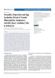
The activities of the two enzymes are presented in (Tables 1-3). The protein concentrations of Tilapia zillii, Hepstus odea and Sarothodelon galileaus gills were estimated to be 5244.37 ± 391.09, 1490.08 ± 105.58 and 1166.59 ± 368.07 respectively; while in their flesh, it was estimated to be 4431.94 ± 56.82, 1606.89 ± 142.94 and 799.47 ± 1.81 respectively, also the protein concentration in the gut was found to be 4084.26 ± 1285.08, 456.49 ± 67.63 and 1530.19 ± 214.32 respectively. From these results, Tilapia zillii has the highest protein concentration, followed by Sarothodelon galileaus and Hepstus odea. The results also showed that both enzymes have a statistically high specific activity in Hepstus odea in all the tissues, followed by Sarothodelon galileaus and Tilapia zillii. Moreover from table 3, the tissue with the highest protein concentration is the gill, then the flesh and the gut, although the differences in their protein concentrations were statistically insignificant. The gill also showed the highest MST specific activity statistically, afterwards the flesh and the gut, and then rhodanese specific activity was however revealed to be high in the gut than other tissues, but not statistically significantly different from the other tissues.
DISCUSSION
Fish and aquatic invertebrates are particularly sensitive to cyanide exposure. Free cyanide was reported to be the primary toxic agent in the aquatic environment. Environmentally relevant exposures to cyanide ions can cause stress, increase in mortality and place an appreciable metabolic load on fishes and other aquatic organisms [7]. Sulphurtransferases are widely distributed enzymes of prokaryotes and eukaryotes [20]. The enzymes catalyze the transfer of sulphane sulphur from a donor molecule, such as thiosulphate or 3-mercaptopyruvate, to a nucleophilic acceptor, such as cyanide or mercaptoethanol.
2/4
Falode et al. (2014) Email:
Central Table 1: Protein concentration and enzyme activity assays in different tissues of the three fishes. TISSUE
Protein Concentration
Rhodanase Specific Activity
MST Specific Activity
TZ GILLS
5244.37 ± 391.09
0.09 ± 0.00
0.07 ± 0.00
SG GILLS
1166.59 ± 368.07
0.17 ± 0.02
0.09 ± 0.01
HO GILLS
1490.08 ± 105.58
TZ FLESH
0.19 ± 0.07
4431.94 ± 56.82
HO FLESH
1606.89 ± 142.94
TZ GUT
4084.26 ± 1285.08
SG GUT
1530.19 ± 214.32
p Value