Paul Scherrer Institut, 5232 Villigen, Switzerland (E.P.); ETH, Wolfgang-Pauli-Street 10, 8093 Zurich, Switzerland (T.E.B.); and EMPA, Lerchenfeldstrasse 5, ...
Self-Consistent Algorithm for Calibrating Spectrometers to Picometer Accuracy over the Entire Wavelength Range EDITH PERRET,* TOBIAS E. BALMER, and MANFRED HEUBERGER Paul Scherrer Institut, 5232 Villigen, Switzerland (E.P.); ETH, Wolfgang-Pauli-Street 10, 8093 Zurich, Switzerland (T.E.B.); and EMPA, Lerchenfeldstrasse 5, 9014 St. Gallen, Switzerland (M.H.)
Spectrometer calibration accuracies are of high importance for a wide range of applications. Typically, one calibrates the spectrometer with a calibration lamp, providing distinct and well-defined calibration lines. However, for small spectral ranges, where only two calibration lines are present, the calibration becomes inaccurate. We present a high-precision nonlinear wavelength calibration method, which is based on two or more reference lines from a calibration lamp. The additional key element introduced is a Fabry–Perot multilayer structure that yields multiple sharp transmission maxima of similar intensity over the full spectrometer range under broad-band illumination (e.g., white-light source). An iterative algorithm is put forward to obtain a self-consistent calibration of picometer precision over the full spectrometer range. In regions distant from calibration lines the accuracy is enhanced by at least a factor of two compared to conventional methods. Index Headings: Spectrometer calibration; Charge-coupled device; CCD detection.
INTRODUCTION Optical spectrometers are used today in spectroscopic, interferometric, or astronomic applications.1–3 Detectors of this type are calibrated by various methods.4–6 For IR and laser absorption measurements it is common to use the sample cell for spectrometer calibration. The cell acts as a simple Fabry–Perot etalon due to the interferences arising between the cell walls. The Fabry–Perot spectrum is then superimposed by the absorption spectrum of the sample (gas or fluid). Limitations of this method lie in the small peak width of the absorption spectrum. In the field of white-light interferometry or Raman spectroscopy one could also use an absorption spectrum of a solid or liquid for calibration. More often the spectrum from a calibration source, e.g., mercury lamp, is used. The calibration line positions are measured on the position detector. Together with the accurately known wavelengths of these lines, a quadratic or higher order polynomial is fitted into the data, where the detector pixel index represents the independent variable. We shall call the calibration methods using a polynomial of second and third degree the conventional calibration method. The precision of this conventional calibration method is limited, particularly, when it is later used to interpolate or extrapolate in a wavelength range distant from the calibration lines. Especially, in miniature spectrometers the light dispersion is strongly nonlinear and therefore the conventional calibrations become less accurate. Some calibration lamps emit distinct lines at different intensities. This can be challenging for the limited dynamic range of the positional detector (e.g., charge-coupled device (CCD)), also leading to a limitation in calibration precision due to the weaker lines. For high-precision Received 14 April 2010; accepted 27 July 2010. * Author to whom correspondence should be sent. E-mail: edith.perret@ psi.ch.
Volume 64, Number 10, 2010
calibration, an ideal reference lamp would therefore emit many equidistant calibration lines with similar peak intensities. Such a precise calibration standard is not readily available. The key element introduced here to solve those problems is a Fabry–Perot reference filter (FRF) with multiple sharp transmission fringes. A FRF is an optical multilayer structure consisting of a (dielectric) spacer layer with a semi-transparent mirror on each face. If the thickness of the spacer layer is exactly known, the peak positions can be calculated from the known optical constants of the multiple layers and the calibration can be done without using a conventional calibration lamp. However, the spacer layer thickness may be subjected to variations (e.g., thermal expansion). In this case it is useful to determine the exact spacer layer thickness simultaneously during the calibration procedure in combination with a calibration lamp. In this article, we describe an algorithm and the required optical setup to solve this problem with high accuracy. The resulting high-precision calibration of spectrometers (HICALOS) is elucidated. We demonstrate how mercury calibration lines together with the FRF can be used to achieve higher calibration accuracies than those of conventional calibration methods.
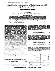
EXPERIMENTAL We describe calibrations performed under realistic experimental conditions. The experimental setup is illustrated in Fig. 1a. At first, two calibration lines of a mercury-argon calibration lamp (CAL-2000 Ocean Optics) are detected to obtain an initial linear calibration of the spectrometer. Reference lines (RL) of a mercury-argon lamp are useful as wavelength standards at the level of 0.1 picometer.7 A high-precision peak localization for these two calibration lines is of high importance since they act as anchoring points for the subsequent self-consistent calibration. Therefore, a stable optical setup and optimized peak detector settings are required. In our setup, mainly two strong RLs at 436 nm (labeled with RL3) and 546 nm (RL4) were used. Secondly, a halogen lamp, which produces white light, illuminates the FRF standard. The transmitted light is collected by a glass fiber and guided to the spectrometer. Glass fibers are used to ensure well-defined in-coupling conditions. Changes in illumination angles or aperture can lead to wavelength shifts and should be avoided. Miniature spectrometers 2000 labeled as m-S1, m-S2 from Ocean Optics (OO), and a half-meter spectrometer from Acton Research Corporation (ARC) labeled as l-S3 were used to investigate the HICALOS performance. The numerical aperture of the Ocean Optics USB 2000 is 0.22 NA according to the SMA 905 single-strand optical fiber connection. A 50-lm entrance slit was used to further reduce NA in the grating direction. The specifications of the spectrometers are given in Table I.
0003-7028/10/6410-1139$2.00/0 Ó 2010 Society for Applied Spectroscopy
APPLIED SPECTROSCOPY
1139
TABLE I. Spectrometer specifications.
FIG. 1. (a) Setup for interferometry experiments: (1) rough calibration with mercury lamp, (2) calibration with a FRF. (b) Sketch of the light path in a spectrometer, where d is the groove spacing, a is the angle of incidence, and b is the angle of diffraction.
The transmission fringes of the FRF close to the borders of the spectrometer are smeared and often show lower intensity. Therefore, only the fringes with high intensity located in the central spectrometer pixel region are taken for further calibration. We shall call this region HICALOS range, which was 25–2020 pixels for m-S1, 400–1600 pixels for m-S2, and 500–4000 pixels for l-S3. We experimentally tested the reliability and accuracy of the HICALOS algorithm. The accuracy of the HICALOS was compared with that of a conventional calibration. The reference lines RL3: 436 nm and RL7: 697 nm were used as anchors for the HICALOS calibration and an FRF with a spacer layer thickness of 15.6 lm was used. A polynomial of eighth (8th) degree was fitted to the data, which consisted of wavelengths against pixel positions. For the conventional calibration, all detected reference lines (RL1–8) were used. However, for many applications only a number � 4 of unequally distributed reference lines are available. We investigated the influence of polynomial degrees on the HICALOS fit accuracy. The spectrometers m-S2 and l-S3 were calibrated with two different FRFs using the HICALOS method. Polynomials of second through seventh degrees were fitted to the data. Root-mean-squared errors were calculated for each fit according to Eq. 2 and averaged over ten independent measurements. The spectrometer curvatures were analyzed and compared to the theoretical grating equation at fixed pixel positions.
THE FABRY–PEROT REFERENCE FILTER When white light is transmitted through an optically layered structure (e.g., etalon, Fabry–Perot), multiple intensity maxima
1140
Volume 64, Number 10, 2010
Labels
m-S1
m-S2
l-S3
Spectrometer type Number of pixels Pixel size [lm] 3 [lm] Groove density [mm�1] Spectral range [nm]
OO USB 2000þ 2048 14 3 200 1200 400–730
OO USB 2000 2048 14 3 200 1800 415–615
ARC 5000 737 300 382–611
at constructive wavelength are observed in the spectrum. Such a transmission spectrum can be described by the multilayer matrix method (MLM).8,9 Therein each optical layer is represented by a complex 2 3 2 matrix. The characteristic matrix of a stack of layers can be readily calculated by a multiplication of the single matrices. The transmittance of the multilayer can then directly be obtained from its characteristic matrix.8 The width of the interference maxima directly depends on the finesse of the interferometer at that wavelength. For use as a FRF, the etalon should produce a high number of peaks in the wavelength range of interest. Because we are focusing on applications in the visual range, the thickness of the dielectric spacer layer(s) will be of the order of a few micrometers. To achieve a high finesse, we use terminal semi-transparent metal (e.g., Ag or Al) mirrors on the outer surfaces of the spacer layer(s). Alternatively, dielectric etalons could be used for this purpose. For high accuracy, the FRF should be of absolutely uniform optical thickness. One way to achieve this is to use a thin sheet of mica as a spacer layer, which can be produced ˚ ) and uniform with atomically smooth surfaces (better than 1 A thickness in the required range over many cm2 areas. The outer surface of the mica sheet is coated with a silver layer 20–50 nm thick. Note that cleaved mica has an, a priori, unknown thickness, resulting in the additional free variable ‘‘spacer layer thickness’’ in this problem. The spacer layer thickness can be determined from the calibrated transmission spectrum using the fast spectral correlation (FSC) method. This method is based on a simplified correlation function between two spectra, one measured and the other theoretically calculated.10 Muscovite Mica was purchased from Spruce Pine Mica USA and was cleaved into thin sheets in a laminar flow cabinet to avoid dust. The mica sheets were cut into small pieces (area ’ 1 cm2) and aligned onto a clean mica support according to the optical axis of the mica crystal.11 Next, the mica support was mounted in the thermal evaporation system. The chamber was evacuated to a pressure of 10�6 mbar. A total silver thickness ˚ /s. The FRF, composed of 40 nm was deposited at a rate of 1 A of two silvered mica sheets with crossed optical axes at 908 (Fig. 1a), was then glued onto glass substrates. The optical axes are crossed in order to cancel birefringence effects that would otherwise lead to double peaks in the transmission spectrum. The glass substrates were cleaned with acetone and spin coated with a thin (, 10 lm) layer of epoxy resin (Shell Chemicals USA), previously dissolved in chloroform (20 mg/mL). Gluing onto the substrates was done on a heating plate at 140 8C, i.e., above the melting point of the casted glue.
THEORY For convenience, we want to limit our theoretical discussion to grating spectrometers in the visual range of the spectrum, although the results will be more generally valid. To measure a spectrum, a spectrometer can be operated as a scanning
monochromator, with a photosensitive detector at the exit slit, while the grating is rotated by a calibrated amount, or, with an array detector in the focal plane (no exit slit). In the latter case, the calibration consists of assigning the correct wavelength to each pixel position in the array. This second variant requires no moving parts and is usually more simple and of higher stability. Approximating the Grating Equation. The grating equation12 describes the dispersion of light in a grating spectrometer: sinðaÞ þ sinðbÞ ¼ nk=d
ð1Þ
where d represents the groove spacing, a is the angle of incidence, b is the angle of diffraction, n is the diffraction order, and k is the wavelength. The light path in a spectrometer is illustrated in Fig. 1b. The fact that the parameters of the grating equation are different and often not specified for a given spectrometer makes the grating equation less practical for calibration purposes. It is more convenient to use a polynomial as an approximation of the grating equation as, e.g., derived by Holy.13 Equally distributed reference lines and higher-order polynomial calibrations result in better calibration accuracies than those of conventional calibrations. In order to use a higher polynomial degree a certain number of reference lines is needed, which is, however, limited in some applications and therefore reduces the choice of the polynomial degree. Here, the additional calibration lines of the FRF come into play. The polynomial degree for the HICALOS calibration should not be higher than half of the number of calibration lines to avoid polynomial over-swinging. The choice of the optimal polynomial degrees will be discussed in more detail later. Self-Consistent Calibration Algorithm. In order to solve for the calibration with an unknown FRF spacer layer thickness, we can invoke a self-consistent algorithm to simultaneously determine the spacer layer thickness and the polynomial coefficients of the calibration function. It will be shown that this problem has a unique solution. A flow diagram of the proposed algorithm is depicted in Fig. 2. At first, two lines of a calibration lamp are measured and their peak positions on the CCD are determined. Two lines towards the upper and lower ends of the spectral range are best suited to obtain a first linear approximation of the calibration. Secondly, the FRF is illuminated with a white-light source and the spectrum is recorded. It is important to keep the optical paths in registry while changing the light sources. The use of pluggable optic fibers can be useful here. The setup is schematically shown in Fig. 1a. The FRF peak positions are determined in units of pixel and the wavelength of each peak is then approximated using the linear calibration. This step essentially determines the FRF interference orders. The numerical tool used for this calculus is fast spectral correlation (FSC),10 which uses the (approximate) measured interference fringe maxima wavelengths to obtain a fitted solution for the FRF spacer layer thickness. After an approximate value of the FRF spacer layer thickness Dinit is determined, the thickness is varied in the model in small steps from Dinit � DD to Dinit þ DD. For each thickness, the theoretical FRF peak wavelengths are calculated and together with the previously measured pixel positions used to produce a new calibration. The best polynomial fit is determined iteratively. As an argument to identify the best fit, a root mean
FIG. 2. Flow diagram: iterative calibration.
squared error (RMSE) is defined as follows: 2 6 6 1 RMSE ¼ 6 6NþM 4X
N þM X
ki
i¼1
31=2
7 7 meas 2 7 ki ðkth � k Þ i i 7 5
ð2Þ
i¼1
The wavelengths kth i correspond to the theoretical locations of represent the measured positions, the maxima and the kmeas i iteratively transformed into wavelength by using the best polynomial calibration. N denotes the number of FRF peaks considered and M denotes the number of mercury-argon lamp peaks used. The (two) calibration lines are used as anchoring points. Therefore, a weighting factor ki is introduced; with typical values 10–100 for the calibration lines and unity for the FRF peaks. Minimizing the RMSE value allows one to iteratively obtain the self-consistent spacer layer thickness. The best polynomial fit at the self-consistent spacer layer thickness is the final calibration curve. The self-consistency of the algorithm is limited by the following three restrictions. First, the algorithm works only for gratings where the nonlinearity in the grating equation is smaller than the spacing between the FRF peaks. Second, the algorithm needs at least one known reference line or an approximate calibration of the spectrometer. The experimental HICALOS accuracy shall be determined by calculating the wavelength differences between the selfconsistent calibration curve and independent wavelengths of mercury-argon lines: HICALOS � kHg=Ar Dkexp Hg=Ar ¼ k
ð3Þ
RESULTS AND DISCUSSION In the first part of this section we test HICALOS with simulated data. In the second part, we present experimental data from real spectrometer calibrations. We will use simulated spectra with intentionally added noise
APPLIED SPECTROSCOPY
1141
FIG. 3. Stability of HICALOS algorithm: The average RMSE (black line) out of 200 simulations with peak noise with a standard deviation of 0.05 pixel is plotted against simulated spacer layer thickness. The initial spacer layer thickness was set to 9.2 lm. The bars indicate the standard deviations. The RMSE for a noise-free simulation (dashed line) is plotted against spacer layer thickness for reference (simulated data).
to quantify systematic and stochastic errors of the selfconsistent algorithm resulting from peak-detection noise. The determined calibration using HICALOS will then be compared to the ‘‘true’’ calibration, which was assumed at the start of the simulations. For this comparison, we evaluate the calibration errors at selected equidistant pixel locations along a virtual CCD detector. The HICALOS accuracy is then given by the differences between the self-consistent solutions kHICALOS and the ‘‘true’’ wavelengths ktrue:
ð5Þ
FIG. 4. (a) Simulation: Bottom: FRF and mercury-argon spectra. Center: Curvature j with respect to a linear calibration through the first and last pixel. Top: Simulated HICALOS accuracy Dksim. Average of 100 simulations (black curve), snapshot (dotted curve), and standard deviation of the calibration error (grey band). (b) Experimental accuracy test using spectrometer m-S1: Bottom: Mercury-argon and HICALOS spectra. Center: Curvature j of the determined conventional calibration. Top: Experimental accuracy Dkexp Hg=Ar of conventional (black triangles) and HICALOS calibration (open circles). For the latter, standard deviations are indicated by bars for eight subsequent measurements.
Using the multilayer matrix (MLM) method,8,9 the transmission spectrum of one virtual FRF was simulated as measured by the virtual spectrometer. We presumed a nonlinear calibration function for the virtual spectrometer such that it resembled the actual nonlinear calibration of a real spectrometer used later in the experimental part. The virtual FRF used for the simulation consisted of a mica sheet of 9.2 lm homogeneous thickness, coated by a 40-nm silver layer on both surfaces. The peak positions in pixel units are determined exactly and scrambled with Gaussian noise of realistic standard deviation, rnoise ¼ 0.05 pixel, and finally transformed into wavelength units, using the self-consistent calibration resulting from the iterative algorithm. The procedure is the same as that illustrated in Fig. 2, except for the addition of Gaussian noise to the peak positions. The HICALOS RMSE, simulated accuracy, and curvature were calculated according to Eqs. 2–5. The RMSE values as a function of spacer layer thickness for applied Gaussian noise and without noise are shown in Fig. 3.
The black curve represents the average error value out of 200 simulated experiments with individual Gaussian noise. The obtained standard deviations are given with black bars. The dashed line represents the simulated calibration without Gaussian noise. This result demonstrates that the algorithm has a unique solution and yields the correct spacer layer thickness of 9.2 lm (at minimum RMSE value), which proves the reliability of the iterative calibration method. The value of the minimum RMSE value was found to be well below 1 pm, which is by itself a measure of quality of the determined calibration curve. The determined accuracy of the HICALOS is shown at the top of Fig. 4a for the simulation. The black curve represents the average obtained from 100 different calibrations with noise, the grey band depicts the standard deviation of the calibration error, and the dotted curve illustrates a selected single result. The error band reflects the overall accuracy of the algorithm, which is found to be 62 pm
Dksim ¼ kHICALOS � ktrue
ð4Þ
The wavelength difference between the HICALOS calibration and a simple linear calibration is used to better visualize the curvature, j, of the calibration curve: j ¼ kHICALOS � klin
1142
Volume 64, Number 10, 2010
in the case of realistic pixel noise of 0.05. This demonstrates that the nonlinearity of the calibration curve can be found by HICALOS with picometer precision. The curvature j of the ‘‘true’’ calibration curve that was used for the simulations is added in the center of the figure. The FRF and calibration lamp spectra are shown at the bottom of the figure. Now, we also present the results from real experimental data. The m-S1 spectrometer was calibrated using both the HICALOS method and the conventional method to compare the two methods. The results are plotted in Fig. 4b. As before, the wavelength deviation Dkexp Hg=Ar (top) reflects how well the calibration function reproduces the measured reference wave˚ lengths. It is found that the calibration accuracy Dkexp Hg=Ar is 0.4 A for the conventional calibration method (black triangles). For comparison, the HICALOS calibration resulted in an accuracy ˚ , as shown in Fig. 4b (top, open circles). The better than 0.2 A strength of the HICALOS lies in uniform high number density of peaks, which leads to the same quality of the calibration, independent of number (if . 1) and separation of Hg/Ar reference lines. In addition, the high number of HICALOS peaks allows one to detect nonlinearities due to grating distortions or refractive index dispersion in the spacer material.14 For example, we also used this method to accurately determine the wavelength dependence of the mica refractive index (not described in this article). Further experiments were done with two different spectrometers m-S2 and l-S3 to test the HICALOS algorithm. Two FRFs of distinct spacer layer thicknesses were used for the HICALOS calibration. Both filters lead to similar results. Polynomials of second, third, and seventh degree were used for fitting. Figure 5 depicts the curvature j as a function of pixel number for the two spectrometers (a) m-S2 and (b) l-S3. Only results from the fitting using a polynomial of third degree are shown. The non-shaded spectral range corresponds to the HICALOS range. Note that the curvature of l-S3 in Fig. 5b is more than one order of magnitude smaller than the one for the miniature spectrometer in Fig. 5a, as can be seen by the different scaling. The insets of Fig. 5 depict the average RMSE values for a polynomial fit of third degree for the two spectrometers (a) ˚ m-S2 and (b) l-S3. The minimum RMSE values are below 1 A for both calibrations. Calibrations of polynomials of seventh degree lead for both filters and both spectrometers to the lowest RMSE values (not shown). For the m-S2 spectrometer the minimum RMSE value is 40 pm and for the l-S3 spectrometer it is 60 pm. The minimum RMSE value for the m-S2 calibrations decreases significantly with increasing polynomial degree. In contrast, the minimum RMSE value is not considerably influenced by the polynomial degree for the l-S3 spectrometer. While the self-consistent mica spacer layer thickness varies ˚ ngstro¨ ms with increasing during iteration by several A polynomial degree for a miniature spectrometer, the same ˚ ngstro¨m for the large spectrometer l-S3, varies only within 1 A which shows the better initial calibration obtained by the latter. The following factors have been found to influence the accuracy of the HICALOS calibration: peak detector precision, optics setup stability, polynomial degrees, the choice of spacer layer thickness, and optical constants used for calculation. These factors will be discussed in detail now. The peak detection error was determined experimentally. Peak variations depend highly on the stability of the setup. The standard deviation of mercury-argon line positions for independent
FIG. 5. Experimental test calibrations: Curvature j for the average of ten measurements against the pixel number for (a) m-S2 and (b) l-S3 spectrometer (for third polynomial degree of fitting). The linear calibration reference is based on the evaluated wavelengths at 500, 1500 pixel for m-S2 and 1250, 3750 pixel for l-S3. The insets show RMSE averaged over ten measurements as a function of ‘‘spacer layer thickness’’ for the calibration of (a) m-S2 and (b) l-S3 spectrometer. The used reference lines for a rough linear spectrometer calibration were RL3: 436 nm and RL4: 546 nm.
measurements using a stable setup is typically 0.05 pixel for the miniature spectrometer m-S2. The standard deviation of the FRF peak positions was found to be in the same range. This indicates that the FRF spectrum can be readily measured with the same precision as conventional calibration lines. In addition, the influence of the light in-coupling angle on the detected pixel line positions was investigated. We observed ˚ ) due to a 658 shift in peak shifts up to 1 pixel (Dk ’ 1A mercury-argon incident angle. It is therefore paramount to use a stable optical setup, where, e.g., the white light is directed to the FRF by a fiber optic and further transmitted to the spectrometer by a second fiber optic. The mercury-argon source is connected to the same first optical fiber as for the
APPLIED SPECTROSCOPY
1143
white-light experiment but without the FRF. Such a stable setup was used for the experimental results presented in Fig. 4b. The polynomial degree used in the HICALOS algorithm significantly influences calibration accuracies of highly curved miniature spectrometers. High polynomial orders yield the lowest minimum RMSE values, i.e., more accurate calibration curves for a given spectrometer. The gain in accuracy of HICALOS is highest for miniature spectrometers with large spectral range. The large focal length l-S3 spectrometer shows a negligible effect of the polynomial degree on the accuracy. This result indicates that higher-order polynomials are useful with spectrometers of small focal length. We note that a higher polynomial would also better account for local optical distortions, if present, regardless of focal length. On the other hand, high-order polynomials tend to oscillate between the anchoring points. The optimal polynomial degree ultimately depends on the number of FRF peaks and, therefore, on the spacer layer thickness. As an empirical rule the optimal polynomial degree lies around the number of FRF peaks N divided by two. When comparing the minimum RMSE values obtained for different FRFs, we find comparable minimum RMSE values for both spectrometers m-S2 and l-S3 at high polynomial degrees. This finding indicates that for these spectrometers the difference in spacer layer thickness is not strongly influencing the calibration accuracy. The RMSE values of miniature and large spectrometers are clearly distinct. We also note that a different setup was used for the half-meter spectrometer (no glass fibers, direct illumination). The higher RMSE value obtained for l-S3 could result from this different setup. It is noted that the calibrations obtained for different FRFs coincide within some picometers. This result confirms the reliability of the algorithm for a high precision calibration.
CONCLUSION A calibration reference that complements the available lines of a calibration lamp leads to higher calibration accuracy over the entire spectral range. A suitable Fabry–Perot filter (FRF) offers the advantage of nearly equidistant lines at tuneable pitch and nearly equal intensity. Due to the higher density of FRF peaks, polynomial fits of higher order can be used to achieve more accurate calibrations. The strength of the HICALOS algorithm lies in calibrating strongly nonlinear miniature spectrometers; in particular this is true for spectral ranges where only a few reference lines are
1144
Volume 64, Number 10, 2010
present. In this case the conventional method has clear disadvantages because higher-order polynomials cannot be fitted. The HICALOS method allows calibrating miniature spectrometers to a level comparable to long-focal-length spectrometers. Another aspect is that the presented HICALOS algorithm used only two reference peaks from a conventional calibration lamp. One could also detect all available mercury peaks in a spectral range and interpolate them with the FRF spectrum. In this case the RMSE line weighting of the mercury peaks should be lowered. Our simulations have demonstrated the robust self-consistency of the algorithm as well as its precision in the picometer regime. Accuracy tests by measurements revealed HICALOS calibration accuracies ˚ , which is at least two times better than that for below 0.2 A the considered conventional calibration, regardless of the spectrometer focal length and reference line distribution. In this work, only calibrations in the visible white-light region were performed. The HICALOS calibration would also work in other wavelength regions as long as a broad-band light source is present. The light source as well as the ideal FRF design depend on the spectral range. Fabry–Perot structures as discussed in this paper are not yet commercially available. In order to assess the full potential of the new calibration method, experiments with spectrometers of different spectral ranges, variable broad light sources, and variable filter designs are necessary in the future. ACKNOWLEDGMENT We thank Ocean Optics for their support. 1. C. Dorrer, J. Opt. Soc. Am. B 16, 7 (1999). 2. A. W. Fountain, T. J. Vickers and C. K. Mann, Appl. Spectrosc. 52, 462 (1998). 3. H. O. Hamaguchi, Appl. Spectrosc. Rev. 24, 137 (1988). 4. J. Wormhoudt, A. C. Stanton, A. D. Richards, and H. H. Sawin, J. Appl. Phys. 61, 142 (1987). 5. C. H. Tseng, J. F. Ford, C. K. Mann, and T. J. Vickers, Appl. Spectrosc. 47, 1808 (1993). 6. S. T. Wollman and P. W. Bohn, Appl. Spectrosc. 47, 125 (1993). 7. C. J. Sansonetti, M. L. Salit and J. Reader, Appl. Opt. 35, 74 (1996). 8. M. Born and E. Wolf, Principles of Optics (Pergamon Press, Oxford, 1980). 9. M. T. Clarkson, J. Phys. D: Appl. Phys. 22, 475 (1989). 10. M. Heuberger, Rev. Sci. Instrum. 72, 1700 (2001). 11. T. E. Balmer, Ph.D.Thesis ETH, Zurich (2007). 12. M. Hutley, Diffraction Gratings (Techniques of Physics) (Academic Press, New York, 1982). 13. J. A. Holy, Appl. Spectrosc. 58, 1219 (2004). 14. J. N. Israelachvili and G. E. Adams, J. Chem. Soc. Faraday Trans. I 74, 975 (1978).