Computer Methods in Biomechanics and Biomedical Engineering
ISSN: 1025-5842 (Print) 1476-8259 (Online) Journal homepage: http://www.tandfonline.com/loi/gcmb20
Semi-automated algorithm for cortical and trabecular bone separation from CT scans K. Janc , J. Tarasiuk , A. S. Bonnet & P. Lipinski To cite this article: K. Janc , J. Tarasiuk , A. S. Bonnet & P. Lipinski (2011) Semi-automated algorithm for cortical and trabecular bone separation from CT scans, Computer Methods in Biomechanics and Biomedical Engineering, 14:sup1, 217-218, DOI: 10.1080/10255842.2011.595192 To link to this article: http://dx.doi.org/10.1080/10255842.2011.595192
Published online: 24 Aug 2011.
Submit your article to this journal
Article views: 80
View related articles
Citing articles: 2 View citing articles
Full Terms & Conditions of access and use can be found at http://www.tandfonline.com/action/journalInformation?journalCode=gcmb20 Download by: [Universite de Lorraine]
Date: 17 January 2016, At: 03:55
Computer Methods in Biomechanics and Biomedical Engineering Vol. 14, No. S1, August 2011, 217–218
Semi-automated algorithm for cortical and trabecular bone separation from CT scans K. Janca,b, J. Tarasiuka*, A.S. Bonnetb and P. Lipinskib a Faculty of Physics and Applied Computer Science (WFiIS), AGH, University of Science and Technology, al. Mickiewicza 30, 30-059 Krako´w, Poland; bLaboratory of Mechanics, Biomechanics, Polymers and Structures (LaBPS), ENIM, Ile du Saulcy, 57045 Metz, France
Keywords: CT scans; bone recognition; image segmentation; genetic algorithm
Downloaded by [Universite de Lorraine] at 03:55 17 January 2016
1.
Introduction
One of the most challenging problems in modern medical imaging techniques is tissue recognition based on image segmentation procedure. The image segmentation may be described as a process of assigning a type of tissue to each pixel of the medical image. Segmentation belongs to an illposed problem class. Usually, its solution is not unique. Even results of segmentation performed manually by two specialists may slightly differ. The main difficulty in bone tissue recognition is to distinguish between cortical and trabecular parts of bone. It is relatively easy to recognise bone among other tissues, especially in computed tomography images. However, a problem arises when two types of bone have to be discriminated. The method proposed in this work combines both medical expert knowledge and skills with genetic algorithm (GA) procedure. The aim is to achieve, in a semi-automatic process saving human expert time, the best possible image segmentation. Our work follows the idea proposed by Bosco (2001) and Cagnoni et al. (1999). A two-stage procedure with learning and appropriate segmentation phases was developed. The GA approach was applied only for the first phase. In contrast to the concepts mentioned by authors, our subjects of evaluation are image-processing filters instead of segmentation curves.
2.
Methods
Let us assume that a great number of computed tomography (CT) scans are available (a few hundred is a reasonable number). Our goal is first to identify bone and next to separate the trabecular and cortical tissues on each CT scan. As the Hounsfield Units (HU; values from CT scans proportional to tissue density) for bone usually range between 400 and 2000, the standard image-processing
*Corresponding author. Email:
[email protected] ISSN 1025-5842 print/ISSN 1476-8259 online q 2011 Taylor & Francis DOI: 10.1080/10255842.2011.595192 http://www.informaworld.com
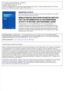
filters based on levels, threshold, and histogram analysis can be applied to distinguish among bone and other tissues. However, there is no strict correspondence between HU and bone type. Indeed, the HU are mainly correlated to tissue apparent density, which can vary from point to point even in the same part of the body, especially for the trabecular bone. Thus, the boundary between trabecular and cortical bones is usually not a single borderline or interface, but rather a little blurred thick line or even region. In the approach presented here, an expert first identifies bone regions in few scans (four or five). Next, the GA searches the set of graphical procedures minimising the differences between its segmentation and the one made by the expert. Afterwards, the set of procedures identified is applied to all CT scans. The important feature of the method proposed in this paper is that GA searches simultaneously the correct image-processing filters, the accurate parameters for these filters as well as the correct order of the filters’ application. The whole procedure, divided into four steps, is illustrated in Figure 1. First, the bone (cortical and trabecular tissues) is selected by GA segmentation from CT scans (Step 1). Next, the mask corresponding to this bone is applied to the scans (Step 2). In Step 3, GA segmentation is applied to extract the trabecular region from the whole bone. The cortical bone is obtained by subtraction of the trabecular tissue from the bone (Step 4). The algorithm was run for a set of few CT scans processed by a human expert. The best combination of image-processing filters and its parameters found by the programme were applied to each scan. All segmented CT scans are treated together. The GA produces only one set of parameters and this set is used for all scans to make segmentation on hundreds of scans now in an automatic way.
K. Janc et al. Step 1
Step 2
Step 3
Step 4 Trabecular Cortical bone bone
218
CT scan
Sub.
GA
Mask
GA
First GA Segmentation
Masking
Second GA Segmentation
Subtraction
Output
Downloaded by [Universite de Lorraine] at 03:55 17 January 2016
Figure 1. Schematic representation of segmentation workflow from one CT scan to three separated regions corresponding to trabecular bone, cortical one and the unused region.
The set of image-processing filters found and its parameters were different in each case. However, for the specific CT machine, it seems that the graphics workflow is repeatable. 4.
Figure 2. Volumetric 3D toothless mandible reconstruction based on GA-treated CT scans. Cortical bone partially removed for better visibility of trabecular bone.
Detailed description of the GA method used to solving the problem is far beyond of this paper and will be published soon in separated work (Janc).
3. Results Several GA parameters and methods were tested in the work. The most important are: size of the population, probability of mutation and type of mutation. Also some specific methods of crossover and individual representations were used. The statistical analysis of obtained segmentation (Janc) shows that for very limited range of GA parameters, it is possible to realise semi-automated procedure to distinguish trabecular and cortical bone with very good results. The regions selected by a human expert and computer programme are almost identical. The method has been successfully tested on three different CT scans: the mandible, skull and knee.
Conclusions
A new approach of the GA used for the segmentation problem has been proposed. The tests prove that the method presented can be successfully applied to distinguish between two very similar CT image regions such as those corresponding to cortical and trabecular bones. The algorithm is very easy to implement. The only time-consuming part is the search for the best imageprocessing filters chain. Once this chain is defined, the correct graphics workflow can be used to treat the remaining scans in a very fast manner. It needs to be emphasised that the method produces well-defined, consistent and disjoint regions for cortical and trabecular bones which result in a very smooth and almost artefactfree 3D reconstruction (see Figure 2). Acknowledgements This work was supported by the Polish Ministry of Science and Higher Education (MNiSW).
References Bosco GL. 2001. A genetic algorithm for image manipulation. In: Paper presented at: Eleventh International Conference on Image Analysis and Processing., Palermo, Italy. Cagnoni S, Dobrzeniecki AB, Poli R, Yanch JC. 1999. Genetic algorithm-based interactive segmentation of 3D medical images. Image Vis Comput. 17:881 – 895. Janc K. Bones elastic properties identification based on the computer tomography measurements [PhD. Thesis]. To be published.