Stimulation of the Human Frontal Eye Fields Modulates Sensitivity of Extrastriate Visual Cortex Juha Silvanto, Nilli Lavie and Vincent Walsh
J Neurophysiol 96:941-945, 2006. First published 19 April 2006; doi: 10.1152/jn.00015.2006 You might find this additional info useful... This article cites 41 articles, 14 of which you can access for free at: http://jn.physiology.org/content/96/2/941.full#ref-list-1 This article has been cited by 29 other HighWire-hosted articles: http://jn.physiology.org/content/96/2/941#cited-by Updated information and services including high resolution figures, can be found at: http://jn.physiology.org/content/96/2/941.full Additional material and information about Journal of Neurophysiology can be found at: http://www.the-aps.org/publications/jn
Journal of Neurophysiology publishes original articles on the function of the nervous system. It is published 12 times a year (monthly) by the American Physiological Society, 9650 Rockville Pike, Bethesda MD 20814-3991. Copyright © 2006 by the American Physiological Society. ISSN: 0022-3077, ESSN: 1522-1598. Visit our website at http://www.the-aps.org/.
Downloaded from http://jn.physiology.org/ by guest on June 3, 2013
This information is current as of June 3, 2013.
J Neurophysiol 96: 941–945, 2006; doi:10.1152/jn.00015.2006.
Report
Stimulation of the Human Frontal Eye Fields Modulates Sensitivity of Extrastriate Visual Cortex Juha Silvanto, Nilli Lavie, and Vincent Walsh Institute of Cognitive Neuroscience and Department of Psychology, University College London, London, United Kingdom. Submitted 5 January 2006; accepted in final form 18 April 2006
INTRODUCTION
The human frontal eye fields (FEFs) are part of a putative control network in which areas in the frontal and parietal lobes are thought to influence the sensitivity of neuronal responses in secondary visual areas (Gitelman et al. 1999; Hopfinger et al. 2000; Giesbrecht et al. 2003). Many studies have examined the parietal component of this network, but fewer have concentrated on the FEF. Early studies of the effects of lesions to the macaque FEF show that this area is required for normal performance on complex tasks such as visual search (Latto and Cowey 1971; Collin et al. 1982), and recording studies have subsequently demonstrated a role for the FEF in visual detection tasks. For example, Thompson and Schall (1999) recorded activity in monkey FEF neurons in a backward masking detection task. Activity of FEF neurons following the target presentation was greater for both hits and false alarms at late latencies, which indicates a role in motor decisions, but at short latencies (beginning around 40 – 60 ms), however, FEF activity was greater following the presentation of a target regardless of subjects’ reports (i.e., for misses as well as hits), which indicates a role in visual registration. Moore and Fallah (2001) found lower luminance detection thresholds following electric stimulation of monkey FEF neurons 50 –175 ms before the onset of a visual stimulus in the motor receptive field of the FEF neuron. It is not clear, Address for reprint requests and other correspondence: J. Silvanto, Institute of Cognitive Neuroscience and Department of Psychology, University College London, 17 Queen Square, London, WC1N 3AR, United Kingdom (E-mail:
[email protected]). www.jn.org
however, whether the improvement in detection was mediated by a direct change in sensitivity of the FEF neuron or by FEF stimulation activating connecting neurons in other areas, such as extrastriate or parietal cortex. A follow-up study of FEF-V4 interactions (Moore and Armstrong 2003) showed that the sensitivity of neurons in extrastriate area V4 was increased if microstimulation was applied to the FEF if the endpoint of the saccade vector of the FEF neuron undergoing stimulation fell within the receptive field of the V4 neuron. This study has been interpreted as evidence of top-down modulation of V4 by FEF and also as supporting the premotor theory of attention (e.g., Smith et al. 2005) because it shows an association between eye movements and covert attention; however, microstimulation was applied for only 50 ms between 200 and 500 ms after visual stimulus onset. In other words, the stimulus had been in the vector field of the FEF neuron for a significant period during which the monkey was attending to the visual stimulus, covering the time period both of visual processing and saccade preparation. Further, the microstimulation was applied only during a period when the monkey was preparing an eye movement to the stimulus. These two aspects of the experiment preclude any dissociation of sensory modulation from eye movement preparation (Juan et al. 2004). To examine the extent to which the findings from nonhuman primate physiology could be applied to the human cortex, Grosbras and Paus (2003), stimulated the human FEFs in a backward masking task and found that TMS applied 40 ms prior to the target onset improved detection sensitivity. As the authors note, however, it is possible that in this study the FEF were rendered more sensitive to incoming information rather than having an effect on the sensitivity of extrastriate visual areas. Moreover, the presentation of a visual stimulus to the retina—and therefore through the geniculostriate pathway— means that one cannot infer a specific effect of FEF stimulation on any particular visual area. More recently, Taylor et al. (2006) applied TMS over the FEF and simultaneously measured occipital visual-evoked potentials (ERPs) while subjects performed a covert orienting task. They recorded from electrodes placed on the same hemisphere as the TMS stimulation and observed that the FEF TMS changed the responses of the visual cortex. They concluded that the “FEF exerts a causal influence over activity in the visual cortex” during voluntary orienting of attention. Taken together, these lines of evidence (Moore and Fallah 2001; Moore and Armstrong 2003; Grosbras and Paus 2003; Smith et al. 2005; Taylor et al. 2006) suggest that visual cortex The costs of publication of this article were defrayed in part by the payment of page charges. The article must therefore be hereby marked “advertisement” in accordance with 18 U.S.C. Section 1734 solely to indicate this fact.
0022-3077/06 $8.00 Copyright © 2006 The American Physiological Society
941
Downloaded from http://jn.physiology.org/ by guest on June 3, 2013
Silvanto, Juha, Nilli Lavie, and Vincent Walsh. Stimulation of the human frontal eye fields modulates sensitivity of extrastriate visual cortex. J Neurophysiol 96: 941–945, 2006; doi:10.1152/jn.00015.2006. The precise role of frontal eye fields (FEF) in vision independent of their role in eye movements remains a matter of debate. One proposal is that the FEF exert top-down influences on the extrastriate visual cortex prior to eye movement preparation. Here we establish, by use of transcranial magnetic stimulation (TMS), that activity in the human FEFs has a direct effect on the sensitivity of extrastriate visual area MT/V5, and that the spatial organization of this top-down effect is lateralized in the human brain. We show that phosphene threshold— the TMS intensity required to elicit a visual perception—for MT/V5 stimulation changes as a function of the delay between the application of TMS over FEF and MT/V5. The effects were specific to the location and time of stimulation. Stimulation of FEF 20 – 40 ms prior to stimulation of MT/V5 decreased the intensity of MT/V5 stimulation required to elicit phosphenes: TMS of the right FEF changed the sensitivity of left and right MT/V5 whereas TMS of the left FEF changed the sensitivity only of the left MT/V5. Thus, the sensitivity of human extrastriate cortex is modulated by activity in the FEF.
Report 942
SILVANTO ET AL.
sensitivity is modulated by FEF. To demonstrate this directly in humans and to explore the hemispheric organization of FEF–sensory cortex interactions, we induced activity in the FEF via transcranial magnetic stimulation (TMS) and measured subjects’ sensitivity to phosphenes induced by TMS over MT/V5. By locally inducing phosphenes with MT/V5 stimulation and hence bypassing the LGN and striate cortex, we were able to obtain a direct measure of the sensitivity of a specific extrastriate visual area – MT/V5. We show that stimulation of FEF 20 – 40 ms prior to stimulation of MT/V5 decreases the intensity of MT/V5 stimulation required to elicit a visual percept and therefore that the sensitivity of human MT/V5 is modulated by activity levels in the FEF. To control for site specificity, we also applied TMS to the vertex and MT/V5 at the same asynchronies as in the FEF condition, and to control for temporal specificity we applied TMS at seven different time points.
Subjects Nine subjects took part in the investigation, eight of whom were naı¨ve to its purpose. Seven subjects were tested in each condition, with five subjects in taking part in both right and left FEF TMS sessions. The subject who was not naive to purpose (J.S.) was naı¨ve to the timing of the TMS pulses, as was the experimenter and all other subjects. The study was approved by the local ethics committee, and subjects gave informed consent. All subjects had previously participated in studies of phosphene perception, the advantage being that their phosphene thresholds (PTs) are stable.
TMS TMS was administered with two Magstim Super Rapid stimulators (Magstim Company, Whitland, UK). The pulses were triggered remotely using a computer that controlled both stimulators, using E-Prime software. Fifty-millimeter figure-eight coils were used over both sites. The stimulation strength was always the same over FEF and Vertex, 65%, an intensity that has been used in many previous FEF studies (e.g., Muggleton et al. 2003; O’Shea et al. 2004) and is known to induce behavioral effects. Subjects were seated on a chair designed for massage so that the subject’s head and body weight were forward and the head rested in a cushioned hole, which allowed the face to peer through. The head is more stable in this arrangement than in a conventional head and chin rest. The coils were fixed in place using “magic arm” (Manfrotto Bassano del Grappa, Italy) coil holders. The subjects’ eyes were covered throughout the experiment, and they were instructed simply to report whether they had perceived a phosphene after each TMS pulse. All subjects reported phosphenes that were restricted to the visual field contralateral to stimulation of V5. To control for temporal specificity, we applied TMS at seven different time points. Specifically, TMS pulses were delivered at FEF/Vertex–MT/V5 stimulation asynchronies between ⫺60 ms (FEF/ Vertex TMS preceding MT/V5 TMS) and ⫹60 ms (MT/V5 TMS preceding FEF/Vertex TMS) in 20-ms steps (Fig. 1C). After each TMS delivery, subjects reported verbally whether or not they had perceived a phosphene. To determine PTs for each TMS condition, the intensity of V5/MT pulse was varied according to a modified binary search (MOBS; Tirell and Owens 1988), an adaptive thresholdfinding algorithm. The TMS intensity was increased or decreased according to the subject’s report on the previous trial. The original upper boundary of the stimulation was 100% of stimulator output, and the lower limit 0%. The number of trials required for setting a threshold depends on the consistency of the subject’s reports and in J Neurophysiol • VOL
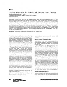
FIG. 1. A and B: transverse and coronal views indicating the location of the left and right FEFs in one subject. C: timeline of an experimental trial. On all trials, a TMS pulse was applied over MT/V5. A second pulse of TMS was applied, depending on the experimental condition, over either FEF or vertex at an SOA between ⫺60 ms (stimulation preceding the MT/V5 pulse) and ⫹60 ms (stimulation postdating the MT/V5 pulse). A trial with a particular combination of stimulation site (FEF; vertex) and SOA (⫺60 ms; ⫺40 ms; ⫺20 ms; 0 ms; ⫹20 ms; ⫹40 ms; ⫹60 ms) was repeated until the PT had been successfully determined for that condition using a modified binary search paradigm. This required between 6 and 15 trials. FEF and vertex were always stimulated with an intensity of 65% of the maximum output of the stimulator.
this experiment was between 6 and 20 trials. Intertrial interval was determined by the subject’s speed of response and was approximately 5 s per trial.
TMS localization TMS was applied over locations that corresponded with the anatomical delineation of left and right FEF (Fig. 1) and left and right MT/V5 (Dumoulin et al. 2000; Watson et al. 1993) by structural magnetic resonance imaging (MRI) in each subject (see Muggleton et al. 2003 and O’Shea et al. 2004 for details). For MT/V5 an additional criterion was the induction of moving phosphenes (Stewart et al. 1999). Vertex was used as an additional control stimulation site to control nonspecific effects of TMS. The stimulation sites were identified on each subject’s T1-weighted MRI scan and coregistered with scalp coordinates. FEF was determined anatomically as lying over the posterior middle frontal gyrus, rostral of the junction of the precentral sulcus, and the superior frontal sulcus (Blanke et al. 2000; Paus 1996). In terms of scalp measurements, the average position of stimulation was 5 cm lateral of the saggital midline and 3– 4 cm anterior of the hand area. This site corresponds well with our previous studies (Muggleton et al. 2003; O’She et al. 2004) and those of others (Leff et al. 2001; Muri et al. 1991). Mean Talairach coordinates were 32, ⫺2, 47 for the right FEF and ⫺32, ⫺2, 46 for the left FEF (Talairach and Tournoux 1988; Paus 1996). V5 can be accurately determined from the production of moving phosphenes (Stewart et al. 1999), and we also determined this using Brainsight coregistration (following Campana et al. 2002, 2005).
Procedure The order of the TMS conditions was intermixed pseudo-randomly, so that a condition in which FEF TMS preceded or followed MT/V5 TMS by a given time window was followed by a condition in which vertex TMS preceded or followed MT/V5 TMS in the same time
96 • AUGUST 2006 •
www.jn.org
Downloaded from http://jn.physiology.org/ by guest on June 3, 2013
METHODS
Report FRONTAL EYE FIELD INTERACTION WITH EXTRASTRIATE CORTEX
RESULTS
The subjects’ PTs in each condition were measured as a percentage of the maximum output of the stimulator unit. To obtain a relative measure of the PTs, the subjects’ absolute thresholds in the FEF and vertex condition were normalized relative to the baseline threshold level in that session. TMS over right FEF The application of TMS to right hemisphere FEF decreased PTs in both right and left MT/V5. Figure 2A shows normalized PTs of right MT/V5 TMS as a function of each TMS condition. For the right MT/V5 TMS, a within-subjects ANOVA with stimulation site (FEF, vertex) and stimulation onset asynchrony (⫺60, ⫺40, ⫺20, 0, ⫹20, ⫹40, ⫹60) as factors indicated a significant interaction between stimulation site and timing (F[1,6] ⫽ 3.549; P ⫽ 0.007; mean square of error [MSE] ⫽ 0.0026). A paired-sample t-test revealed that FEF stimulation applied 20 ms before the MT/V5 pulse significantly lowered the PT in comparison to vertex stimulation at the same time window (t[6] ⫽ 5.012; P ⫽ 0.002; SD ⫽ 0.069; SE ⫽ 0.026). In contrast, there was no statistically significant difference between the FEF and vertex conditions when their stimulation postdated the MT/V5 pulse by 20 ms (t[6] ⫽ 0.323; P ⫽ 0.390; SD ⫽ 0.105; SE ⫽ 0.023). The normalized PTs of left MT/V5 as a function of each right FEF and vertex TMS condition, averaged across the J Neurophysiol • VOL
FIG. 2. Mean of normalized PTs (n ⫽ 7) of MT/V5 in the each experimental condition. All error bars indicate ⫾1 SE. A: effects of rFEF TMS on the sensitivity of ipsilateral visual cortex: TMS of the right FEFs (diamond symbols) lowered the PT of ipsilateral MT/V5, with the peak of this effect occurring when TMS was applied over the FEF 20 ms before MT/V5 was stimulated. TMS over vertex (squares) had no effect. B: effects of rFEF TMS on the sensitivity of contralateral visual cortex: TMS of the right FEFs (diamond symbols) lowered the PT of contralateral MT/V5, with the peak of this effect occurring when TMS was applied over the FEF 40 ms before MT/V5 was stimulated. TMS over Vertex (squares) had no effect. C: Ipsilateral but not contralateral effects of lFEF TMS on cortical sensitivity: TMS applied over the left FEF lowered the PT of the ipislateral MT/V5 (squares) but not contralateral visual cortex (diamonds).
seven participants, are shown in Fig. 2B. For the left MT/V5 TMS a within-subjects ANOVA with stimulation site (FEF, vertex) and stimulation onset asynchrony as factors indicated a significant interaction between stimulation site and time window (F[1,6] ⫽ 3.305; P ⫽ 0.017, MSE ⫽ 0.0047). Pairwise comparisons revealed that FEF stimulation preceding the MT/V5 pulse by 40 ms significantly lowered subjects’ PTs in comparison to vertex stimulation in this time window (t[6] ⫽ 3.01; P ⫽ 0.024; SD ⫽ 0.126; SE ⫽ 0.048). In contrast, the difference between the two conditions was not statistically significant when their stimulation postdated the MT/V5 pulse by 40 ms (t[6] ⫽ 0.795; P ⫽ 0.457; SD ⫽ 0.076; SE ⫽ 0.029). It is apparent in Fig. 2B that the facilitatory effect of right FEF TMS on the activity of left MT/V5 is moderately present at SOAs that precede and postdate the time window of the strongest effect. At these time windows, however, the difference between the FEF and vertex conditions was not statistically significant for (20 ms: (t[6] ⫽ 1.752; P ⫽ 0.130; SD ⫽ 0.143; SE ⫽ 0.054; 60 ms: t[6] ⫽ 1.774; P ⫽ 0.126; SD ⫽ 0.072; SE ⫽ 0.027). TMS over left FEF Figure 2C shows normalized PTs of right and left MT/V5 TMS as a function of each FEF TMS condition. The application of TMS to left FEF decreased PTs in left but not in right MT/V5. A within-subjects ANOVA with phosphene stimulation site (left vs. right MT/V5 TMS) and FEF stimulation onset asynchrony as factors indicated a significant interaction between phosphene stimulation site and SOA (F[1,6] ⫽ 3.402; P ⫽ 0.009; MSE ⫽ 0.0018). A paired-sample t-test revealed that FEF stimulation applied 20 ms before the left MT/V5 pulse significantly lowered the PT in comparison to right MT/V5 TMS at the same time window (t[6] ⫽ 6.148; P ⫽
96 • AUGUST 2006 •
www.jn.org
Downloaded from http://jn.physiology.org/ by guest on June 3, 2013
window, and vice versa. There were three experimental sessions and in each session two conditions were carried out: One session consisted of right FEF–right V5 and vertex–right V5 conditions, another consisted of right FEF–left V5 and vertex– left V5, and another consisted of left FEF–right V5 and left FEF–left V5 conditions. Within and between each session the order of TMS conditions was randomized. The structure of each session was 1) an SOA selected randomly by the software; 2) PTs measured for this SOA for both experimental conditions in that particular session (e.g., right FEF–right V5 and vertex–right V5); 3) the next SOA selected randomly; and 4) again, PTs measured for both experimental conditions for this SOA. The condition that was carried out second for the previous SOA is now carried out first. Whether a given timing started with an FEF–MT/V5 or vertex– MT/V5 condition was counterbalanced. In each condition, the PT was determined once using the binary search paradigm, which takes 6 –20 trials. Because there were seven SOAs (⫺60, ⫺40, ⫺20, 0, 20, 40, 50) and six experimental conditions (right FEF–right V5; vertex–right V5; right FEF–left V5; vertex–left V5; left FEF–right V5; left FEF–left V5), the PT was measured a total of 42 times (14 times in each session), with an average of 12 trials required for each threshold. In addition, the baseline threshold (in which TMS was applied only over left or right MT/V5) was measured a total of 16 times (8 times for each V5)., Baseline thresholds were measured at the beginning and the end of each session, as well as after the fifth and ninth TMS/vertex conditions (as mentioned above, their were 14 threshold conditions in each session). The mean values of the baseline thresholds were 60% and 65% of the maximum stimulator output for left and right MT/V5, respectively. Stimulations of left and right MT/V5 were carried out on separate days. No vertex condition was run in comparison with the left FEF condition based on the prediction from Grosbras and Paus (2003) that there would be left-right field differences in the effects of left FEF stimulation (specifically a null prediction for the left FEF stimulation) and our previous evidence that right and left FEF TMS have different effects on visual processing in the two visual fields (Muggleton et al. 2003).
943
Report 944
SILVANTO ET AL.
0.001; SD ⫽ 0.0387; SE ⫽ 0.0146). In contrast, there was no statistically significant difference between the right MT/V5 and left MT/V5 conditions when their stimulation postdated the MT/V5 pulse by 20 ms (t[6] ⫽ 0.832; P ⫽ 0.437; SD ⫽ 0.10; SE ⫽ 0.376). Again, a moderate facilitatory effect of left FEF TMS on the activity of left MT/V5 is present at stimulation onset asynchronies (SOAs) that precede and postdate the time window of the strongest effect. At these time windows, however, the difference between the FEF and right MT/V5 condition was not statistically significant for the left MT/V5 condition (⫺40 ms: t[6] ⫽ 2.126; P ⫽ 0.078; SD ⫽ 0.103; SE ⫽ 0.039; 0 ms: t[6] ⫽ 2.088; P ⫽ 0.082; SD ⫽ 0.094; SE ⫽ 0.036). DISCUSSION
J Neurophysiol • VOL
96 • AUGUST 2006 •
www.jn.org
Downloaded from http://jn.physiology.org/ by guest on June 3, 2013
Our results clearly show that activity in the human FEF modulates the excitability of extrastriate visual cortex. Together with the findings of Grosbras and Paus (2003), Taylor et al. (2006), Smith et al. (2005), Thompson and Schall (1999), and Bullier (2001), our data suggest that one of the saccadeindependent functions of FEF is to exert top-down modulation over extrastriate visual cortex. Several studies have now shown that some FEF responses are independent of saccade command signals, and it is these visual rather motor FEF neurons that have been associated with top-down control and attention. The content of the top-down control may be either spatial or feature-related. Thompson et al. (2005), for example, have identified spatially selective enhanced activity in visual FEF neurons during a covert spatial attention task and a concurrent suppression of activity in FEF movement neurons. They concluded that “selective activity in FEF visually responsive neurons corresponds to the mental spotlight of attention via modulation of ongoing visual processing.” Other recording studies have shown that some visual FEF neurons’ responses are correlated with the identity of the target, and it is therefore possible that FEF visual neurons are involved in both spatial and object-related processes in selective attention tasks. Bar et al. (2005) have also presented evidence from magnetoencephaolography and functional MRI studies for an early top-down role for FEF in visual object processing. Greater activation in FEF was seen when subjects were presented with objects they successfully recognized than for object they did not. This activity preceded activity in visual object recognition areas by 30 ms (O’Shea et al. 2004; O’Shea and Walsh 2006). Whether this early FEF activity in reflects top-down control processes dedicated to space, object features or both remains to be determined. With regards to how FEF exerts top-down control, it is possible that FEF activity occurs prior to sensory stimulation as opposed to rapid responses to visual stimuli (contrast a possible role for FEF neurons in visual priming: Bichot and Schall 2002). It is important to note that the term “top-down” is used in the temporal context of FEF receiving visual information prior to the extratastriate areas, but it remains entirely possible that top-down processes are mediated by a feed forward anatomical connection from, say, FEF to V4 (e.g., Barone et al. 2000). The FEFs are part of the frontoparietal network involved in many aspect of vision, but, as we noted in the introduction, the role of the FEF has not received the same attention as that of the PPC. To understand the function of the FEF within this network, however, requires a description of the similarities and differences between these two major nodes. One aspect of our
findings reflects the well-established hemispheric asymmetry in attentional functions in humans, namely, that the right hemisphere is more commonly concerned with processing information in both visual fields, whereas the left hemisphere appears to be concerned only with the right visual field (e.g., Mesulam 1981). That right FEF TMS decreased PTs in both visual fields whereas left FEF TMS only decreased PTs in the right visual field is consistent with this and adds to the many examples of similarities between the organization of FEF and PPC in vision and attention. For example, frontal and parietal areas have both been shown to produce neglect when damaged (Keating and Gooley 1988; Mesulam 1999); have both been shown to be important in detecting abrupt changes in the visual field (Beck et al. 2001, 2005; Turatto et al. 2004) and have been shown to be important in visual search (Donner et al. 2000; Nobre et al. 2003). Demonstrating differences between the two nodes, however, is a less common finding, and our result may have implications in this regard. We interpret the effects of FEF. TMS on extrastriate activity to be evidence of a top-down role for FEF because it is consistent with a role for the early visual activity of FEF visual neurons (Murthy et al. 2001). An area involved in control would be expected to be active early and by responding to target features, the FEF could increase the sensitivity of extrastriate neurons to task relevant parameters (e.g., Moore and Armstrong 2003 Moore and Fallah 2001). Our finding may also help to understand why a recent TMS study obtained only equivocal evidence of a role for the PPC in top-down control (Hung et al. 2005): damage to the frontoparietal network may only result in a disruption of top-down modulation if the early input to the FEF is disrupted. Both frontal and parietal nodes may be important for aspects of visual orienting and awareness, and there are reports of early visual responses, attributed to attentional processes in both. Bisley et al. (2005) for example, recently reported rapid responses to visual stimuli of neurons in lateral intraparietal cortex, and Fuggetta et al. (2006) reported that TMS over the PPC after onset of a visual search display can eliminate the N2pc component associated with target detection. This was interpreted as evidence of TMS interfering with spatial processes in the search task. Both FEF and PPC, then, have been associated with spatial top-down processing, but a major difference between them seems to be that the FEFs have also been associated with feature processes. Some physiological properties of the FEF may make it well placed to exert feature-related top-down influence: the FEF responds to visual stimuli very rapidly and is part of what has been called “the fast brain” (Bullier 2001; O’Shea et al. 2004); the organization of FEF-visual cortex connections is retinotopic (Schall et al. 1995); FEF neurons show distinct featurerelated activity (Thompson and Schall 1999) and a preference for stimuli that are relevant as targets (Schall and Hanes 1993); and preactivation of the FEF improves visual detection with the same spatial pattern of hemispheric specialization as found in the current study (Grosbras and Paus 2003) and influences attention-related activity in the visual cortex (Taylor et al. 2006). Further, comparison of the effects of FEF versus PPC stimulation using a partial report task designed to probe topdown control has shown that human FEF is important for target rather than distractor processing (Hung 2005). The pattern of hemispheric asymmetries is an important component of interpreting the role of the FEF in humans and our effects show that
Report FRONTAL EYE FIELD INTERACTION WITH EXTRASTRIATE CORTEX
the right FEF needs approximately 20 ms longer to influence the contralateral than the ipsilateral visual cortex, presumably due to callosal transmission time. In summary, these findings extend and complement the microstimulation studies of nonhuman primate FEF (Moore and Fallah 2001; Moore and Armstrong 2003) and the magnetic stimulation study of Grosbras and Paus (2003) in showing that FEF activity modulates the responses of visual cortex in a manner consistent with a role in top-down control of visual processing. ACKNOWLEDGMENTS
This work was supported by a scholarship from the University College London Graduate School and grants from the United Kingdom Medical Research Council and the Wellcome Trust. V.Walsh. is supported by the Royal Society. REFERENCES
J Neurophysiol • VOL
Hung J, Driver J, and Walsh V. Visual selection and posterior parietal cortex: effects of repetitive transcranial magnetic stimulation on partial report analyzed by Bundesen’s theory of visual attention. J Neurosci 25: 9602–9612, 2005. Juan C-H, Shorter-Jacob SM, and Schall JD. Dissociation of spatial attention and saccade preparation. Proc Natl Acad Sci USA 101: 15541–15544, 2004. Keating EG and Gooley SG. Saccadic disorders caused by cooling the superior colliculus or the frontal eye field or from combined lesions of both structures. Brain Res 438: 247–255, 1988. Latto R and Cowey A. Visual field defects after frontal eye-field lesions in monkeys. Brain Res 30: 1–24, 1971. Leff AP, Scott SK, Rothwell JC, and Wise RJ. The planning and guiding of reading saccades: a repetitive transcranial magnetic stimulation study. Cereb Cortex 11: 918 –923, 2001. Mesulam MM. A cortical network for directed attention and unilateral neglect. Ann Neurol 10: 309 –325, 1981. Mesulam MM. Spatial attention and neglect: parietal, frontal and cingulated contributions to the mental representation and attentional targeting of salient extrapersonal events. Phil Trans R Soc Lond 354: 1325–1346, 1999. Moore T and Armstrong KM. Selective gating of visual signals by microstimulation of frontal cortex. Nature 421: 370 –373, 2003. Moore T and Fallah M. Control of eye movements and spatial attention. Proc Natl Acad Sci USA 98: 1273–1276, 2001. Muggleton NG, Juan C-H, Cowey A, and Walsh V. Human frontal eye fields and visual search. J Neurophysiol 89: 3340 –3343, 2003. Muri RM, Hess CW, and Meienberg O. Transcranial stimulation of the human frontal eye field by magnetic pulses. Exp Brain Res 86: 219 –223, 1991. Murthy A, Thompson KG, and Schall JD. Dynamic dissociation of visual selection from saccade programming in frontal eye field. J Neurophysiol 86: 2634 –2637, 2001. Nobre AC, Coull JT, Walsh V, and Frith CD. Brain activations during visual search: contributions of search efficiency versus feature binding. Neuroimage 18: 91–103, 2003. O’Shea J, Muggleton NG, Cowey A, and Walsh V. Timing of target discrimination in human frontal eye field. J Cog Neurosci 16: 1060 –1067, 2004. O’Shea J and Walsh V. Trickle-down theories of vision. Current Biology 16: R206 –R209, 2006. Orban GA, Van Essen D, van Duffel W. Comparative mapping of higher visual areas in monkeys and humans. Trends Cogn Sci 8: 315–324, 2004. Paus T. Location and function of the human frontal eye fields: a selective review. Neuropsychologia 34: 475– 483, 1996. Rizzolatti G, Riggio L, Dascola I, and Umilta C. Reorienting attention across the horizontal and vertical meridians: evidence in favor of a premotor theory of attention. Neuropsychologia 25: 31– 40, 1987. Schall JD and Hanes DP. Neural basis of saccade target selection in frontal eye field during visual search. Nature 366: 467– 469,1993. Schall JD, Morel A, King DJ, and Bullier J. Topography of visual cortex connections with frontal eye field in macaque: convergence and segregation of processing streams. J Neurosci 15: 4464 – 4487, 1995. Smith DT, Jackson SR, and Rorden C. Transcranial magnetic stimulation of the left human frontal eye fields eliminates the cost of invalid endogenous cues. Neuropsychologia 43: 1288 –1296, 2005. Stewart L, Battelli L, Walsh V, and Cowey A. Motion perception and perceptual learning studied by magnetic stimulation. Electroencephalogr Clin Neurophysiol Suppl 51: 334 –350, 1999. Talairach TJ and Tournoux P. Co-Planar Stereotaxic Atlas of the Human Brain: 3-Dimensional Proportional System—An Approach to Cerebral Imaging (1st ed.) New York: Thieme Medical Publishers, 1988. Taylor PCJ, Nobre AC, and Rushworth MFS. FEF TMS affects visual cortical activity. Cereb Cortex 10.1093/cercor/bhj156, 2006. Thompson KG and Schall JD. The detection of visual signals by macaque frontal eye field during masking. Nat Neurosci 2: 283–288, 1999. Thompson KG, Biscoe KL, and Sato TR. Neuronal basis of covert attention in the frontal eye field. J Neurosci 25: 9479 –9487, 2005. Turatto M, Sandrini M, Miniussi C. The role of the right dorsolateral prefrontal cortex in visual change awareness. Neuroreport 15: 2549 –2552, 2004. Tyrell RA and Owens DA. A rapid technique to assess the resting states of the eye and other threshold phenomena: the modified binary search (MOBS). Behav Res Methods Instrum Comput 20: 137–141, 1998. Watson JD, Myers R, Frackowiak RS, Hajnal JV, Woods RP, Mazziotta JC, Shipp S, and Zeki S. MT/V5of the human brain: evidence from a combined study using positron emission tomography and magnetic resonance imaging. Cereb Cortex 3: 79 –94, 1993.
96 • AUGUST 2006 •
www.jn.org
Downloaded from http://jn.physiology.org/ by guest on June 3, 2013
Bar M, Kassa KS, Ghuman AS, Boshyan J, Schmidt AM, Dale AM, Hamalainen MS, Marinkovic K, Schacter DL, Rosen BR, and Halgren E. Top-down facilitation of visual recognition. Proc Natl Acad Sci USA 103: 449 – 454, 2005. Barone P, Batardiere A, Knoblauch K, and Kennedy H. Laminar distribution of neurons in extrastriate areas projecting to visual areas V1 and V4 correlates with the hierarchical rank and indicates the operation of a distance rule. J Neurosci 20: 3263–3281, 2000. Beck DM, Muggleton N, Walsh V, and Lavie N. Right parietal cortex plays a critical role in change blindness. Cereb Cortex 16: 712–717, 1982. Beck DM, Rees G, Frith CD, and Lavie N. Neural correlates of change detection and change blindness. Nat Neurosci 4: 645– 650, 2001. Bichot N and Schall JD. Priming in macaque frontal cortex during pop-out visual search: feature-based facilitation and location based inhibition of return. J Neurosci 22: 4675– 4685, 2002. Bisley JW, Suresh Krishna B, and Goldberg ME. A rapid and precise on-response in posterior parietal cortex. J Neurosci 24: 1833–1838, 2005. Blanke O, Spinelli L, Thut G, Michel CM, Perrg S, Landis T, and Seeck M. Location of the human frontal eye field as defined by electrical cortical stimulation. Anatomical, functional and electrophysiological characteristics. Neuroreport 11: 1907–1913, 2000. Bullier J. Integrated model of visual processing. Brain Res Rev 36: 96–107, 2001. Campana G, Cowey A, and Walsh V. Priming of motion direction and area MT/V5: a test of perceptual memory. Cereb Cortex 12: 663– 669, 2002. Campana G, Cowey A, and Walsh V. Visual area V5/MT remembers “what” but not “where.” Cereb Cortex Doi: 10.193/cercor/bhj111. 2005. Chambers CD and Mattingley JB. Neurodisruption of selective attention: insights and implications. Trends Cogn Sci 9: 542–550, 2005. Collin NG, Cowey A, Latto R, and Marzi C. The role of frontal eye-fields and superior colliculi in visual search and non-visual search in rhesus monkeys. Behav Brain Res 4: 177–193, 1982. Donner T, Kettermann A, Diesch E, Ostendorf F, Villringer A, and Brandt SA. Involvement of the human frontal eye field and multiple parietal areas in covert visual selection during conjunction search. Eur J Neurosci 9: 3407–3414, 2000. Dumoulin SO, Bittar RG, Kabani NJ, Baker CL, Jr, Le Goualher G, Bruce Pike G, and Evans AC. New anatomical landmark for reliable identification of human area MT/V5: a quantitative analysis of sulcal patterning. Cereb Cortex 10: 454 – 463, 2000. Fuggetta G, Pavone E, Walsh V, Kiss M, and Eimer M. Cortico-cortical interactions in spatial attention: A combined ERP/TMS study. J Neurophysiol 2006. J Neurophysiol (January 25, 2006). doi:10.1152/jn. 01273.2005. Giesbrecht B, Woldorff MG, Song AW, and Mangun GR. Neural mechanisms of top-down control during spatial and feature attention. Neuroimage 19: 496 –512, 2003. Gitelman DR, Nobre AC, Parrish TB, LaBar KS, Kim YH, Meyer JR, and Mesulam M. A large-scale distributed network for covert spatial attention: further anatomical delineation based on stringent behavioural and cognitive controls. Brain 122: 1093–1106, 1999. Grosbras MH and Paus T. Transcranial magnetic stimulation of the human frontal eye field facilitates visual awareness. Eur J Neurosci 18: 3121–3126, 2003. Hopfinger JB, Buonocore MH, and Mangun GR. The neural mechanisms of top-down attentional control. Nat Neurosci 3: 284 –291, 2000. Hung J. Transcranial Magnetic Stimulation Studies on the Control of Visual Selection (unpublished DPhil thesis). Oxford University. 2005.
945