brief communications
Separate but linked functions of conventional myosins modulate adhesion and neurite outgrowth Steven R. Wylie* and Peter D. Chantler*† *Unit of Molecular and Cellular Biology, Royal Veterinary College, University of London, Royal College Street, London NW1 0TU, UK †e-mail:
[email protected]
The potential functional diversity of closely related myosin isoforms found in eukaryotic cells is not yet understood in detail. We have previously provided evidence from functional knockouts of Neuro-2A neuroblastoma cells that myosin IIB is essential for neurite outgrowth1. Here we investigate the role of non-muscle myosin IIA in the same cell line. We show that suppression of myosin IIA transcript and protein expression, brought about through exposure to isoform-specific antisense oligonucleotides, caused a rearrangement of the actin cytoskeleton and loss of cell adhesion. This also led to disruption of focal contacts, as evidenced by coincident reduction in paxillin and vinculin immunofluorescence, but did not diminish transcript expression. All effects were fully reversible. Before myosin IIA antisense-induced detachment, neurite outgrowth remained unaffected. By contrast, antisense oligonucleotides directed against myosin IIB transcripts had no effect on adhesion but severely attenuated neurite outgrowth. We infer that the two main isoforms of neuronal conventional myosin, myosins IIA and IIB, have separate but linked functions during neuronal adhesion and neurite outgrowth.
n understanding of the mechanisms that underlie neuritic outgrowth and growth cone motility, fundamental properties of developing neurons, has been sought ever since the discovery of the neuronal growth cone by Ramón y Cajal, over a century ago2 and such studies in neurons have provided many insights into the wider field of cell motility. Although adhesive contacts are critical for eukaryotic cell motility, a dynamic balance of forces must be struck3, for if adhesion is too strong, movement will cease, whereas if it is too weak the cells will detach. During forward motility, focal contacts are elaborated at the leading edge of the cell in response to Rac- and Rho-dependent signalling pathways4 where they represent sites for signal transduction and the attachment of actin microfilaments5. Focal contacts comprise complex arrangements of cytoskeletal proteins which mediate the adhesive interactions of nerve growth cones, stabilize elongating nerve fibres and facilitate communication between the extracellular matrix and the cell interior through transmembrane receptors: neuronal integrins or RHAMM (receptor for hyaluronic acid-mediated motility) interact with laminin6 and nectins7, or hyaluronan8, respectively. Growth cones are rich in actin microfilaments9, which have the potential to interact with any of the members of the myosin family of molecular motors known to be found in this region, namely myosin I10,11, myosin II1,11–13 and myosin V14. The unconventional myosins—myosin V and myosin I—have been implicated in filipodial extension and restraint of lamellipodial expansion, respectively14. Neuronal cells express two isoforms of conventional myosin: myosin IIA and myosin IIB12. We have shown previously, with the
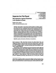
A
88
aid of functional knockouts, that one of these, myosin IIB, is essential for neurite outgrowth1. To test our hypothesis that myosin IIA has a distinctive role in these cells, separate from myosin IIB, we have used an antisense approach to generate isoform-specific functional knockouts of myosin IIA. Upon plating, Neuro-2A cells assumed a rounded phenotype (Fig. 1A, a) before neurite elongation (Fig. 1A, b–d). After 96 h in culture, myosin IIA immunofluorescence was distributed throughout the cytoplasm of untreated Neuro-2A cells (Fig. 1A, b) and cells exposed to sense (Fig. 1A, c and 1B, a) or scrambled (Fig. 1B, e) oligonucleotides. Actin could be seen throughout the cytoplasm of these cells, often concentrated subjacent to the plasma membrane (Fig. 1B, a, e and C, a). By contrast, adherent Neuro-2A cells treated for 96 h with antisense oligonucleotides targeting myosin IIA sequence, not only displayed severe attenuation of myosin IIA expression (Fig. 1A, d and B, c) but also exhibited a disorganized microfilament network with extensive clumping (Fig. 1B, c and C, b). Quantitation of the confocal images shown in Fig. 1B, a, c and e revealed highly significant differences (P < 0.001; one-way ANOVA) in the mean myosin IIA fluorescence intensities between antisense and respective controls (n = 12 cells per comparison) but there were no significant differences in F-actin intensities (P > 0.25). When cells were treated for 96 h with a myosin IIB-directed antisense oligonucleotide, curtailing neurite outgrowth1, but were stained with a primary antibody specific for myosin IIA, no diminution of fluorescence was observed (Fig. 1A, e). Taken together, these results show that the isoform-specific antisense oligonucleotides and myosin IIA antibodies used in these experiments acted specifically, facilitating the selective attenuation of myosin IIA expression and its detection. After recovery for 72 h in oligonucleotide-free medium, myosin IIA immunofluorescence and actin cytoarchitecture were restored (compare Fig. 1B, d and B, b, f). Neurite outgrowth, measured on adherent cells, was not significantly attenuated by antisense oligonucleotides directed against myosin IIA transcripts (Fig. 1D, a), in contrast to the effects of antisense oligonucleotides directed against myosin IIB transcripts reported earlier1 and confirmed in a parallel experiment here (Fig. 1D, b). Mean neurite lengths were comparable following treatment with sense, antisense and scrambled oligonucleotides based on the myosin IIA sequence (Fig. 1D, a). Evidently, myosin IIA is not required for neurite outgrowth per se, in distinct contrast to myosin IIB1. The reverse transcriptase polymerase chain reaction (RT–PCR) revealed that the level of myosin IIA mRNA declined during chronic exposure of Neuro-2A cells to myosin IIA antisense oligonucleotides, demonstrating that the antisense blockade was acting as expected and message did not accumulate (Fig. 2a, lanes 11–13). Following a 72-h period of recovery after removal of the antisense oligonucleotides, the level of myosin IIA mRNA was restored to NATURE CELL BIOLOGY VOL 3 JANUARY 2001 http://cellbio.nature.com
© 2000 Macmillan Magazines Ltd
brief communications A
a
d
c
b
e
50 µm
B
Antisense (AQ3)
Sense (AQ5)
b
c
d
e
f
50 µm 96
50 µm 96
168
C
D
a Neurite length (µm)
20 µm
Myosin IIA oligonucleotides
80 60 40 AQ3 AQ5 AQ3R ACTL
20 0
b
Antisense
h
Myosin IIB oligonucleotides 100 80 60 40 BQ3 BQ5 BQ3R BCTL
20 0
24
Sense
168
96
100
b
Actin
a
50 µm
168
Neurite length (µm)
Actin + Myosin IIA
Myosin IIA
Actin
a
Scrambled (AQ3R)
72
120
Time (h)
168
24
72
120
168
Time (h)
Figure 1 The effect of antisense or control oligonucleotides, derived from myosin IIA or IIB sequences, on the phenotype of cultured mouse Neuro-2A neuroblastoma cells. A, Neuro-2A cells immediately after plating (a) or after 96 h in culture, either untreated (b) or treated with sense (c) or antisense (d, e) oligonucleotides directed against myosin IIA (c, d) or myosin IIB (e) sequences. In all cases rabbit anti-nonmuscle myosin IIA28 was used as primary antibody and detected through addition of fluorescein-conjugated goat anti-rabbit IgG (Sigma) as secondary antibody. B, The effect of chronic exposure to sense, antisense or scrambled oligonucleotides derived from myosin IIA sequence on neurite outgrowth from Neuro-2A cells, before (96 h) and after (168 h) recovery in oligonucleotide-free media. Cells were double stained, using a fluorescein-labelled antibody28 to detect myosin IIA and rhodamine–phalloidin (Molecular Probes) to detect F-actin. C, Images
at higher magnification to illustrate disruption of actin cytoarchitecture by myosin IIA antisense oligonucleotides. Rhodamine–phalloidin (Molecular Probes) staining of Factin viewed in sense-treated (a) and antisense-treated (b) Neuro-2A cells. D, Comparison of treatments with myosin IIA-derived (a) and myosin IIB-derived (b) oligonucleotide sequences on the length of neurite outgrowth. Following transfer to serum-free media, cells were treated, separately, with sense (AQ5, BQ5), antisense (AQ3, BQ3) and scrambled (AQ3R, BQ3R) oligonucleotides. After 96 h exposure, all cells were switched to oligonucleotide-free media (arrow) for a further 72 h. ACTL and BCTL refer to untreated cells. Neurite outgrowth was monitored through DIC optics, images being obtained from a significant number of cells (range: 114–238 cells (IIA); range: 112–292 cells (IIB)) at every time point for each treatment. Standard error of the mean (s.e.m.) is shown at each time point for each sample.
normal (Fig. 2a, lane 14); intensities of the bands seen at 72, 96 and 168 h were 70%, 42% and 97%, respectively, of that at 48 h (Fig. 2a, lanes 11–14). By contrast, myosin IIA mRNA levels remained constant following continuous exposure to, or recovery from, sense (Fig. 2a, lanes 15–18) or scrambled (Fig. 2a, lanes 7–10) oligonucleotides, and in untreated cells (Fig. 2a, lanes 3–6). When mRNAs prepared from these same cells were amplified with primers specific for the myosin IIB isoform, the level of expressed transcript remained constant in all antisense-treated and control cells (Fig. 2b). The same held true for all actin transcripts (Fig. 2c). Although exposure of Neuro-2A cells to myosin IIA antisense oligonucleotides had minimal effect on neurite outgrowth from adherent cells (Fig. 1D, a), this treatment led to cell detachment
(Fig. 3A, a). This response was not observed in untreated cells or in cells treated with sense or scrambled myosin IIA oligonucleotides (Fig. 3A, a). Nor were such changes observed in cells treated with antisense, sense or scrambled oligonucleotides directed at myosin IIB sequence (Fig. 3A, b). Detachment did not lead to cell death: detached cells could be replated, and exhibited neurite outgrowth in serum-free media and cell division in serum-containing media. Before detachment, cell bodies of cells treated with antisense oligonucleotides directed against myosin IIA, but not myosin IIB, sequence underwent significant decreases in diameter (for example, diameters of cells treated with oligonucleotides AQ3 or BQ3 (see Methods) for 96 h were 15.4 µm (s.d. = 1.5; n = 82) and 17.6 µm (s.d. = 1.0; n = 56), respectively. AQ3-treated cells exhibited a
NATURE CELL BIOLOGY VOL 3 JANUARY 2001 http://cellbio.nature.com
© 2000 Macmillan Magazines Ltd
89
brief communications significant (P < 0.05) decrease in diameter of some 13%; these were assessed for all treatments by one-way ANOVA and the Tukey–Kramer multiple comparisons test. Dissociated cells, replated for 96 h, displayed complete restoration of normal cell body diameters (17.3 µm; s.d. = 1.7; n = 122) (P < 0.0001; unpaired ttest) and neurite lengths. Cessation of antisense treatment halted further cell detachment (Fig. 3A, a). Because antisense oligonucleotides directed against myosin IIA sequence brought about loss of cell adhesion, we examined the effect of these myosin IIA oligonucleotide regimes on focal contact formation by immunolocalization of the focal contact proteins, paxillin and vinculin. Neuro-2A cells exhibit focal contacts throughout the cell body, shaft and growth cone; within focal contacts, paxillin and vinculin staining coincided (compare Fig. 3B, a–c). When cells were exposed for 96 h to antisense oligonucleotides derived from myosin IIA sequence, both paxillin and vinculin immunofluorescence were diminished relative to sense and scrambled controls (Fig. 3C, a–c), although some punctate vinculin formations remained (Fig. 3C, b). Quantitation of the confocal images shown in Fig. 3C revealed highly significant (P < 0.001; oneway ANOVA) differences in the mean paxillin and vinculin fluorescence intensities between antisense (n = 6) and respective controls (each n = 11). Furthermore, the paxillin/F-actin ratios were significantly different between antisense- and sense-treated cells (P < 0.0005; unpaired t-test; each n = 12 (data not shown)). Following a 72-h period of recovery in oligonucleotide-free media, vinculin and paxillin immunofluorescence intensities, together with cell body diameters, were restored to control levels. RT–PCR examination of Neuro-2A cells exposed for 96 h to myosin IIA scrambled, antisense or sense oligonucleotides, using amplification primers specific for paxillin and vinculin as well as for myosin IIA, myosin IIB, β-actin and glyceraldehyde-3-phosphate dehydrogenase (G3PDH; signal control), indicated that levels of paxillin (α, β and γ isoforms) and vinculin transcripts remained unchanged irrespective of the nature of the myosin IIA oligonucleotide regime (Fig. 3D). Neuritic outgrowth is complex, involving propulsion and retraction, adhesion and detachment, the growth cone responding to external guidance cues by tentative advance through the extracellular matrix in vivo, or over the substratum in vitro. Adhesion to the extracellular matrix requires formation of focal contacts; these have been observed in chick sensory neuronal growth cones adherent to fibronectin-treated substrata7, growth cones of chick retinal neurons adherent to laminin surfaces6 and in primary spinal neurons and cells from the mouse neuroblastoma cell line NG108-15 adherent to hyaluronan8. Conventional myosin II isoforms have been reported to have an important role in cellular adhesion5: microinjection of either anti-myosin II antibodies15 or myosin II mimetic peptides16 modulates cell rounding and the interaction between integrin receptors and the extracellular matrix. Furthermore, the addition of compounds that inhibit the activity of myosin II results in impairment of neuronal growth cone motility13 and has been shown to attenuate Rhoinduced stress fibre and focal adhesion formation in fibroblasts17. Two main isoforms of myosin II, A and B, are found in neuronal cells12, leading to speculation as to whether these two isoforms have differing functions. A differential localization of the two isoforms is observed within the growth cone, myosin IIB being found in the peripheral (P) domain and adjacent to the central (C) domain where it has a more peripheral distribution than myosin IIA, which is located mainly within the C domain and does not extend into the marginal zone12. Within locomoting cells, myosin II isoforms appear to become localized by mechanisms intrinsic to their structures18. Both myosin II isoforms have been observed within stress fibres, but myosin IIA predominates19, the IIB isoform only accumulating with time18. We have shown directly that myosin IIB drives neurite outgrowth1. Nevertheless, there is an observed correlation between phospho90
a
Myosin IIA primers ACTL
1
2
3
4
5
AQ3R
6
7
b
10
11
2
3
4
5
AQ3R
6
7
8
c
12
13
14
15
16
17
18
19
20
21
22
AQ3
9
10
11
12
13
18
19
20
21
22
18
19
20
21
22
AQ5
14
15
16
17
Actin primers ACTL
1
9
AQ5
Myosin IIB primers ACTL
1
8
AQ3
2
3
4
5
AQ3R
6
7
8
9
AQ3
10
11
12
13
AQ5
14
15
16
17
Figure 2 Demonstration by RT–PCR that the expression of myosin IIA transcripts in cultured mouse Neuro-2A neuroblastoma cells is attenuated specifically through application of antisense oligonucleotides derived from myosin IIA sequence. a, Myosin IIA primers; b, myosin IIB primers; c, actin primers. Within each gel, lanes 3–6, untreated controls (ACTL); lanes 7–10, cells treated with scrambled (AQ3R) oligonucleotides; lanes 11–14, cells treated with antisense (AQ3) oligonucleotides; and lanes 15–18, cells treated with sense (AQ5) oligonucleotides. Markers are seen in lanes 1 and 19 of all gels. Negative controls (lane 20, total RNA as template, to test for genomic DNA contamination; lane 21, an absence of template, to test for systematic DNA contamination) and a positive control (lane 22, G3PDH, to test for the integrity of reverse transcriptase and Taq polymerase, using kit primers and human placental RNA (Clontech)) are included. In a–c, RNA samples were collected from ~ 3 × 10⁵ Neuro-2A cells at 0 h (lane 2), 48 h (lanes 3, 7, 11, 15), 72 h (lanes 4, 8, 12, 16), 96 h (lanes 5, 9, 13, 17) and 168 h (lanes 6, 10, 14, 18) after plating, as described earlier1. Cells were exposed to oligonucleotides for 96 h followed by 72 h recovery in fresh oligonucleotide-free medium. Consequently, lanes with material from 48, 72 and 96 h are obtained during a period of chronic oligonucleotide exposure; lanes with material from 168 h are obtained following 72 h of recovery from exposure.
rylation of myosin regulatory light chain (RLC) and lysophosphatidate-stimulated, Rho-mediated, neurite retraction 17,20–22; neurite outgrowth can be elicited in the presence of inhibitors of myosin II light chain phosphorylation20,21. Furthermore, inhibition of RLC phosphorylation curtails formation of Rho-induced stress fibres and focal adhesions in fibroblasts17. To resolve these issues we hypothesize that despite their conserved structures, myosins IIA and IIB perform different tasks during adhesion and neurite retraction as compared with neurite outgrowth, and may be subject to distinct forms of regulation. Our results support a model in which the two isoforms of conventional myosin, IIA and IIB, have distinct yet complementary functions during neurite outgrowth. We have demonstrated the presence of focal contacts along the ventral surface of mouse Neuro-2A neuroblastoma cells adherent to fibronectin-treated substrata (Fig. 3B) and have shown them to express the focal contact proteins, paxillin and vinculin (Fig. 3B, C). Significantly, isoform-specific knockout of myosin IIA expression (Figs 1 and 2) leads to a rearrangement of the actin cytoskeleton (Fig. 1C) and cell detachment (Fig. 3A), concomitant with a decline in paxillin and vinculin immunofluorescence (Fig. 3C). Furthermore, antisense knockout of myosin IIA alters the shape of the cell body; a NATURE CELL BIOLOGY VOL 3 JANUARY 2001 http://cellbio.nature.com
© 2000 Macmillan Magazines Ltd
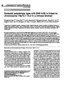
brief communications a
Myosin IIA oligonucleotides
b 500
450
450
400 350 300
ACTL AQ3R AQ5 AQ3
250
B
Myosin IIB oligonucleotides
500
Cell density (no. per mm2)
Cell density (no. per mm2)
A
a Paxillin
400 350
b
300
BCTL BQ3R BQ5 BQ3
250
Vinculin
200
200 0
24
48
72
96
120
144
168
0
24
Time (h)
48
72
96
120 144
168
Time (h)
c
C
a Paxillin + vinculin
Sense (AQ5)
20 µm
D b Myosin IIB Myosin IIA
Paxillin
Vinculin
β-Actin
G3PDH
Antisense (AQ3)
c Scrambled (AQ3R) 50 µm
Paxillin
Vinculin
Paxillin + vinculin
1
2
3
4
5
6
7
8
9
10 11 12 13 14 15 16 17 18 19 20 21 22
Figure 3 The effect of antisense or control oligonucleotides, derived from myosin IIA or IIB sequences, on Neuro-2A neuroblastoma cell adhesion and focal contact formation. A, Effect of oligonucleotide treatment on adhesion to the substratum (adherent cell density per square millimetre): comparison of control or antisense oligonucleotides derived from either myosin IIA (a) or myosin IIB (b) sequence. Untreated cells (ACTL, BCTL) and cells treated with sense (AQ5, BQ5), antisense (AQ3, BQ3), or scrambled (AQ3R, BQ3R) oligonucleotides were grown in culture for 96 h, after which time (arrow) cells were placed into oligonucleotide-free media for a further 72 h. The s.e.m. is shown for every time point. B, Focal contacts found on the ventral surface of a control Neuro-2A cell, throughout the cell body, neuritic shaft and growth cone. High-power image obtained from six confocal slices (~ 3 µm total thickness) at the base of a confocal stack. Cells were stained, as described in Methods, with primary antibodies directed against paxillin and vinculin after 96 h in culture. C, Effect of treatment with sense, antisense or scrambled oligonucleotides derived from myosin IIA sequence on paxillin and vinculin expression within Neuro-2A neuroblastoma cells. Confocal images were obtained in all cases. Cells were double stained for paxillin and vinculin, as described in Methods. Neuro-2A cells after 96 h of chronic exposure to sense (AQ5) (a), anti-
sense (AQ3) (b) and scrambled (AQ3R) (c) oligonucleotides derived from myosin IIA sequence. The default confocal slice thickness was set at ~ 12 µm. D, RT–PCR demonstration that myosin IIA antisense blockade of Neuro-2A cells leads to a decline in myosin IIA transcripts but not in transcripts of myosin IIB, paxillin, vinculin, actin or G3PDH. Neuro-2A cells were exposed for 96 h to scrambled, antisense and sense oligonucleotides derived from myosin IIA sequence (lanes 2 to 19 are arranged in ordered groups of three, respectively). Primers were for myosin IIB (lanes 2–4); myosin IIA (lanes 5–7); paxillin (lanes 8–10, 21); vinculin (lanes 11–13, 21); β-actin (lanes 14–16); G3PDH (lanes 17–19). A negative control in the absence of template is seen in lane 22. Markers are seen in lanes 1 and 20. Note the three paxillin amplification products in lanes 8–10; in order of increasing size we assign these to the alternatively spliced α, β and γ isoforms of paxillin. Whereas the α and β isoforms are well known in both mouse and human neurons, the γ isoform, seen in human material, has not been observed before in mouse tissues29. Lane 21 contains the combined paxillin and vinculin amplification products derived from Neuro2A cells at the time of plating (zero time sample); note that paxillin and vinculin expression is much reduced in these rounded cells (see Fig. 1A,a) compared with adherent cells extending processes at 96 h (see lanes 8–13 and Figs 1A,c and 3B).
13% decrease in cell body diameter was observed, whereas antisense blockade of myosin IIB was without effect (Fig. 3A). Before antisense oligonucleotide-induced detachment, myosin IIA antisense blockade appeared to have no obvious effect on the rate of increase of neurite length (Fig. 1D), in direct contrast to the almost complete cessation of neurite elongation brought about by myosin IIB antisense blockade (ref. 1 and Fig. 1D). Consequently, it would appear that the two isoforms of conventional myosin found in neuronal cells are both required for neuritic outgrowth but in different and characteristic ways. It is possible that the presence of
myosin IIA exerts some form of mechanochemical control over the incorporation of paxillin and vinculin into focal contact sites; our RT–PCR results (Fig. 3D) show that such control is independent of transcriptional regulation. We correlate myosin IIA with the myosin isoform present within mini-sarcomeres (stress fibres) which terminate in focal contacts5,17, and suggest that myosin IIA is responsible for tension generation at these sites, which is required during active adhesion. Furthermore, myosin-driven neurite retraction21,22 is also likely to be powered through myosin IIA action, regulated through RLC
NATURE CELL BIOLOGY VOL 3 JANUARY 2001 http://cellbio.nature.com
© 2000 Macmillan Magazines Ltd
91
brief communications phosphorylation1. By contrast, we attribute the pulling force23,24 exerted by growth cones to myosin IIB, which drives neurite outgrowth and is regulated independently of light chain phosphorylation1,20. Recently, evidence has been obtained25 which suggests that focal contacts link to the actin cytoskeleton by means of a molecular clutch; focal contacts are propelled in a centripetal manner in stationary cells but are immotile in locomoting cells. Further, retrograde actin flow, a myosin II-dependent process, is inversely proportional to the rate of growth cone advance13. If myosin IIA is responsible for this centripetal movement of actin and of focal contacts, one prediction would be that the selective elimination of myosin IIB could allow myosin IIA action to predominate, leading to neurite retraction and cell rounding. This prediction has been borne out by experiment1 (see Fig. 1A, e). Furthermore, following the specific ablation of myosin IIA, tension within stress fibres can no longer be maintained, thereby compromising functional focal contacts and leading to the cellular detachment observed here (Fig. 3A). Note that Neuro-2A cell detachment takes place throughout the myosin IIA antisense blockade. By definition, however, neurite length measurements are made on the population of cells remaining attached at each time point, before their subsequent detachment. As long as these cells retain functional myosin IIA and can remain attached during the myosin IIA antisense blockade, myosin IIB action will remain unaffected, facilitating neurite outgrowth, as observed (Fig. 1D). Consequently, during neurite outgrowth, the actions of these separate but linked functions of myosins IIA and IIB achieve a dynamic balance of directional forces3. Presumably, the prevailing balance of forces is determined through antagonistic Rho and Rac signalling pathways4,17,21,22,26, which modulate both myosin lightchain and heavy-chain phosphorylation patterns22,26, allowing the selective control of myosin IIA and myosin IIB activity postulated earlier1. Our experiments raise the possibility that homologous conventional myosin isoforms may have related functions during the motility of non-neuronal cells.
Methods Cell culture and cytochemistry. Neuro-2A cells were plated (0.8 × 10⁵ per well in Nunc four-well plates) onto fibronectin-modified (10 µg ml–1), polylysinated (0.1 mg ml–1) coverslips and passaged in the presence of serum; neurite outgrowth was initiated by replating cells in serum-free media, as described11. Before cytochemistry, a brief (10 s) extraction step27 in cytoskeletal buffer (10 mM MES pH 6.1, 138 mM KCl, 3 mM MgCl2, 2 mM EGTA) supplemented with 0.32 M sucrose, 0.1% Triton X-100 and 1 µg ml–1 phalloidin (Calbiochem), was followed by fixation for 30 min in cytoskeletal buffer supplemented with 0.32 M sucrose and 4% formaldehyde. Following permeabilization (10 min in PBS plus 0.5% Triton X-100), cells were rinsed in PBS and then immersed in 2% de-complemented horse serum in PBS for 20 min to block nonspecific sites before antibody treatment. Incubation with primary and secondary antibodies was for 2 h and 45 min, respectively. Between incubations, several brief (30 s) rinses in 1% horse serum/PBS + 0.5% Triton X-100 were performed, the final rinse lasting for 5 min. Following exposure to antibodies and further extensive rinses before a final wash in PBS alone, cells were mounted in a solution of glycerol:PBS (containing 2.5% DABCO (Sigma)) (vol:vol = 9:1) to prevent fluorescence quenching, before image acquisition. Non-muscle myosin IIA was detected by indirect immunofluorescence using polyclonal rabbit anti-nonmuscle myosin IIA as primary antibody28 (courtesy of Primal de Lanerolle, University of Chicago) (1:1000 dilution) and fluorescein-conjugated goat anti-rabbit IgG (Sigma) (1:50 dilution) as secondary antibody. Paxillin was detected by indirect immunofluorescence using a primary monoclonal mouse anti-paxillin (Affiniti Research Products) (1:100 dilution) and a secondary rhodamine-conjugated goat anti-mouse IgG (Sigma) (1:60 dilution). Vinculin was detected by indirect immunofluorescence using a primary monoclonal mouse anti-vinculin (Sigma) (1:50 dilution) and a secondary fluorescein-conjugated goat anti-mouse IgG (Sigma) (1:50 dilution). Filamentous actin was detected using rhodamine–phalloidin (Molecular Probes) added at a concentration of 165 nM simultaneously with the secondary antibody. All antibodies were diluted in 1% horse serum/PBS including 0.1% Triton-X100.
Antisense treatment protocols. Sequence information for myosin IIA, available from gene data banks, was used to design isoform-specific antisense and sense (control) oligonucleotides. These were synthesized either to correspond to (in the case of sense oligos) or be complementary to (in the case of antisense oligos) regions within isoform-specific sequence located towards the 5′ end of the myosin IIA transcripts, which encodes the amino-acid sequence NPILEA (single-letter amino acid notation) in AQ5 (AQ3 is the inverse complement of AQ5) (accession number U31463). Scrambled (control) oligonucleotides corresponded in base composition to those of the antisense oligonucleotides, except that their sequences were scrambled and did not match any other entry in the GenBank or EMBL databases. The sequences used were: AQ5 (sense), 5′-CAACCCTATCCTAGAGGCCT-3′; AQ3 (antisense), 5′-AGGCCTCTAGGATA-
92
GGGTTG-3′; AQ3R (scrambled), 5′-CTAAGTGACTAGTGGTGGGC-3′. Oligonucleotides derived from myosin IIB sequence, described previously 1, were: BQ5 (sense), 5′-GCAGATCCAATTCTGGAATCA-3′; BQ3 (antisense), 5′-TGATTCCAGAATTGGATCTGC-3′; BQ3R (scrambled), 5′-GGCTACGATGACAGCTATTTT-3′. BQ3 and BQ5 are derived from an isoform-specific stretch within the 5′ coding region of myosin IIB transcripts (accession number U15766). Neuro-2A cells in culture were transferred to serum-free media and treated, separately, with sense, antisense or scrambled oligonucleotides derived from myosin IIA or myosin IIB sequences. Initial incubations took place with 50 µM oligonucleotide while subsequent additions, every 12 h, were at 25 µM (ref. 1). At appropriate times, cells were fixed and stained with antibodies before examination by confocal laser scanning microscopy.
RT–PCR. RT–PCR was performed according to standard procedures using 42 cycles of amplification (94 °C for 45 s; 62 °C for 1 min; 72 °C for 2 min and a final 7 min extension at 72 °C). Primers used for myosin IIA were 5′-TCCTGGCTATAAGTCACCATG-3′ (upstream primer; from sequence U31463, terminating at the ATG start site) and AQ3 (downstream primer), giving rise to a 680-bp amplification product. For myosin IIB, primers were 5′-CATCTACAACCCTGCCACTCA-3′ (upstream primer: a 21-mer from sequence U34303, located 60 bases downstream from the ATG start site) and BQ3 (downstream primer), giving rise to a 780-bp amplification product. Primers used for paxillin were 5′-ATCTACCACCTCCCACATCT-3′ (upstream primer corresponding to an exon upstream of the αβγ splice sites; sequence provided courtesy of Yuichi Mazaki and Hisataka Sabe) and 5′-CTAGCTTGTTCAGGTCGGAC-3′ (downstream primer)29. Primers used for vinculin were 5′-CGCGATGCCAGTGTTTCATAC-3′ (upstream primer, including the ATG start site) and 5′-ATGGCTTCAGTGTCCTTGCTG-3′ (downstream primer)30. Primers for actin were 5′-TGTGATGGTGGGAATGGGTCAG-3′ (upstream primer) and 5′-TTTGATGTCACGCACGATTTCC-3′ (downstream primer) for mouse β-actin (Stratagene), giving rise to a 514-bp amplification product. Primers for G3PDH were 5′-TGAAGGTCGGTGTGAACGGATTTGGC-3′ (upstream primer) and 5′-CATGTAGGCCATGAGGTCCACCAC-3′ (downstream primer) for mouse G3PDH (Clontech), giving rise to a 983-bp amplification product.
Confocal laser scanning and DIC microscopy. Confocal fluorescence microscopy was performed using a Zeiss LSM 510 confocal laser scanning microscope equipped with a 40× Fluar oil immersion objective (N.A. 1.3). Argon (λex = 488 nm) and He-Ne (λex = 543 nm) lasers were switched between excitation wavelengths for fluorescein and rhodamine fluorescence, which were detected through either band-pass (505–550 nm) or long-pass (LP560 nm) filters, respectively. Neurite outgrowth was monitored through differential interference contrast (DIC) microscopy using a Zeiss Axiovert 135 inverted microscope. Images, recorded at each time point, were digitized before neurite length measurement using Kontron KS-300 software. Statistical analysis was performed using Instat2 and consisted of parametric one-way analysis of variance (ANOVA) including Tukey–Kramer multiple comparisons test and the unpaired t-test. RECEIVED 1 JUNE 2000; REVISED 14 AUGUST 2000; ACCEPTED 20 SEPTEMBER 2000; PUBLISHED 8 DECEMBER 2000.
1. Wylie, S. R., Wu, P-J., Patel, H. & Chantler, P. D. Proc. Natl Acad. Sci. USA 95, 12967–12972 (1998). 2. Letourneau, P. C., Kater, S. B. & Macagno, E. R. (eds) The Nerve Growth Cone (Raven Press, New York, 1991). 3. Palecek, S. P., Loftus, J. C., Ginsberg, M. H., Lauffenburger, D. A. & Horwitz, A. F. Nature 385, 537–540 (1997); see also Erratum, Nature 388, 210 (1997). 4. Hall, A. Science 279, 509–514 (1998). 5. Burridge, K. & Chrzanowska-Wodnicka, M. Annu. Rev. Cell Dev. Biol. 12, 463–519 (1996). 6. de Curtis, I. & Malanchini, B. Exp. Cell Res. 230, 233–243 (1997). 7. Gomez, T. M., Roche, F. K. & Letourneau, P. C. J. Neurobiol. 29, 18–34 (1996). 8. Nagy, J. I., Hacking, J., Frankenstein, U. N. & Turley, E. A. J. Neurosci. 15, 241–252 (1995). 9. Lewis, A. K. & Bridgman, P. C. J. Cell Biol. 119, 1219–1243 (1992). 10. Lewis, A. K. & Bridgman, P. C. Cell Motil. Cytoskel. 33, 130–150 (1996). 11. Miller, M., Bower, E., Levitt, P., Li, D. & Chantler, P. D. Neuron 8, 25–44 (1992). 12. Rochlin, W., Itoh, K., Adelstein, R. S. & Bridgman, P. C. J. Cell Sci. 108, 3661–3670 (1995). 13. Lin, C. H., Espreafico, E. M., Mooseker, M. S. & Forscher, P. Neuron 16, 769–782 (1996). 14. Wang, F-S., Wolenski, J. S., Cheney, R. E., Mooseker, M. S. & Jay, D. J. Science 273, 660–663 (1996). 15. Honer, B., Citi, S., Kendrick-Jones, J. & Jockusch, B. M. J. Cell Biol. 107, 2181–2189 (1988). 16. Sims, J. R., Karp, S. & Ingber, D. E. J. Cell Sci. 103, 1215–1222 (1992). 17. Chrzanowska-Wodnicka, M. & Burridge, K. J. Cell Biol. 133, 1403–1415 (1996). 18. Kolega, J. J. Cell Sci. 111, 2085–2095 (1998). 19. Maupin, P., Phillips, C. L., Adelstein, R. S. & Pollard, T. D. J. Cell Sci. 107, 3077–3090 (1994). 20. Jalink, K. et al. J. Cell Biol. 126, 801–810 (1994). 21. Hirose, M. et al. J. Cell Biol. 141, 1625–1636 (1998). 22. Amano, M. K. et al. Genes Cells 3, 177–188 (1998). 23. Bray, D. Dev. Biol. 102, 379–389 (1984). 24. Lamoureux, P., Buxbaum, R. E. & Heidemann, S. R. Nature 340, 159–162 (1989). 25. Smilenov, L. B., Mikhailov, A., Pelham, R. J. Jr, Marcantonio, E. E. & Gundersen, G. G. Science 286, 1172–1174 (1999). 26. van Leeuwen, F. N., van Delft, S., Kain, H. E., van der Kammen, R. A. & Collard, J. G. Nature Cell Biol. 1, 242–248 (1999). 27. Cramer, L. P. & Mitchison, T. J. J. Cell Biol. 131, 179–189 (1995). 28. de Lanerolle, P., Gogas, G., Li, X. & Schluns, K. J. Biol. Chem. 268, 16883–16886 (1993). 29. Mazaki, Y., Uchida, H., Hino, O., Hashimoto, S. & Sabe, H. J. Biol. Chem. 273, 22435–22441 (1998). 30. Alatortsev, V. E., Kramerova, I. A., Frolov, M. V., Lavrov, S. A. & Westphal, E. D. FEBS Lett. 413, 197–201 (1997).
ACKNOWLEDGEMENTS We thank S. Fletcher for assistance and P. de Lanerolle for supplying the anti-myosin IIA antibody. We thank G. Dunn and W. Gratzer for their comments on the manuscript. This work was supported by grants from the Wellcome Trust and the BBSRC to P.D.C. Correspondence and requests for materials to P.D.C.
NATURE CELL BIOLOGY VOL 3 JANUARY 2001 http://cellbio.nature.com
© 2000 Macmillan Magazines Ltd