Arlette Cohen, Benoit Robert, and Margaret Buckingham. From the ..... B. C. D. E. F. G. Myosin Heavy Chain mRNAs in Developing Mouse Skeletal Muscle.
T H EJOURNAL OF BIOLOGICAL CHEMISTRY Vol258, No. 22, issue of November 25, pp. 13867-13874.1983 Prrnted in U S A
Sequential Accumulation of mRNAs Encoding Different Myosin Heavy Chain Isoforms during Skeletal Muscle Development in Vivo Detected with a Recombinant Plasmid IdentifiedAs Coding for an Adult Fast Myosin Heavy Chain from Mouse Skeletal Muscle* (Received for publication, April 29, 1983)
Andre Weydert, Philippe Daubas, Mario CaravattiSt, Adrian Minty, Gabriele BugaiskyB, Arlette Cohen, Benoit Robert, and Margaret Buckingham From the Pasteur Institute. DeDartmentof Molecular Biology, 75724 Paris Ceden 15, France and §Hoffman LaRoche, Dia/F.E., Bcitiment 619, CH-4002 Basel, Switzerland
In rat skeletal muscle, the sequential appearance of embryonic, neonatal, and adult myosin heavy chain proteins has been described (2). Such multigene families are a characteristic feature of metazoan organisms. Understanding the control of gene expression within an ubiquitous protein family such as themyosin heavy chains should provide some insight into the genomic strategies employed to resolve the problem of constructing a complex organism withmany differentiated tissues, starting from one cell. Recently, cloning techniques have allowed the studyof myosin heavy chain isoforms at the mRNA and gene level, in nematode (3), Drosophila (4), quail (5), chicken (6-9), rabbit (lo), and rat (11-16). Mainly, the COOH-terminal portion of the myosin heavy chain has been investigated this exists as a two-stranded a-helicalcoiled coil (17) in the native myosin molecule and is subject to severe structural constraints which are reflected in its amino acid sequence, nucleic acid sequence, and restriction enzyme pattern. Thegeneral picture emerging from differentlaboratories shows a myosin heavy chain mRNA length of about 7 kb,’ containing regions of homology interspersed with diverged regions when different sequences are compared, and one or several myosin heavy chain genes of20-30 kb length with many intervening sequences (3-16). In Drosophila only one genomic sequence is detected and differential splicing at the 3’ end of the gene generates different RNA transcripts (4). The generationof more than one myosin heavy chain protein from the same genomic sequence has also been suggested for the vertebrates, in this case by post-translational processing (18).In order to investigate gene expression during skeletal The myosin heavy chains represent a multigene family of muscle formation and maturation, we have cloned actin (19, contractile proteins of which different isoforms are expressed 20) and myosin (21) cDNA sequences from newborn mouse to studymRNA in different adult muscle and non-muscle tissues and during skeletal muscle. These have already been used accumulation duringmyotube formation (22). We report here striated muscle development (1). Peptide analyses and immunological data would suggest at least 12 distinct myosin the characterizationof a recombinant plasmid containing the heavy chains in mammals, of which 9 are striated isoforms. 3‘ end of the adult skeletalmuscle myosin heavy chain of the mouse, and an analysisof the accumulationof mRNA encoding this and anotherisoform during skeletal muscle develop*This laboratoryissupported by grants from theMinistry of ment in vivo. Industry and Research, the Centre National de la Recherche Scientifique, the Institut National de Santi! la et de laRecherche Mkdicale, EXPERIMENTALPROCEDURES the North Atlantic Treaty Organization, and the Muscular Dystrophy Construction of Recombinant Plasmids Association of America. The costs of publication of this article were defrayed in part by the payment of page charges. This article must Preparation of poly(A)-containing RNAfrom the leg muscles of 5therefore be hereby marked “advertisement” in accordance with 18 10-day-old mice, fractionation of poly(A)-containing RNA, cell-free U.S.C. Section 1734 solely to indicate this fact. translation, and analysis of the translation products, synthesis, and $ Recipient of a grant from the Swiss National Research Foundation. Present address, Pasteur Institute, Department of Molecular Biology, 75724 Paris Cedex 15, France. The abbreviations used are: kb, kilobase; SDS, sodium dodecyl 7 Supported by the Pasteur-Weizmann exchange program. sulfate.
13867
Downloaded from www.jbc.org by guest, on July 21, 2011
In order to study developmental transitions of myosin heavy chain gene expression, we have cloned from newborn mouse skeletal muscle a recombinant plasmid (plasmid MHC 32) that contains an insertion coding for the COOH-terminal portion of an adult fast myosin heavychain isoform of mouse skeletal muscle. By Northern blots and dot blots, it has been shown that the MHC 32 sequence reveals a broad cross-hybridization with RNA from different mammalian striated muscletissues. Southernblotswith mouse genomic DNA show only one homologous gene, but cross-hybridization at lower stringency to seven to eight different bands, some containing multiple genomic fragments, among which are probably the genes encoding the different striated muscle isoforms. S1 protection experiments with RNA from mouse skeletal muscle before and after birth demonstrate that plasmid MHC 32 is homologous to a major mRNA species of adult skeletal muscle. This adult mRNA is a predominant sequencewithin 5-6 days after birth. It begins to accumulate at 1-3 days; at the 18th day fetal stage, another major mRNA species is detected as partially homologous with the adult MHC 32 sequence. This fetal myosin heavy chain mRNA is still predominant at 1-3 days after birth, but is rapidly (by 5-6 days) replaced by the adult MHC sequence. There is thus a rapid transition after birth from fetal to adult muscle skeletal myosin heavy chain mRNA sequences.
13868
Myosin Heavy ChainmRNAs in Developing Mouse Skeletal Muscle
sizing of single-stranded cDNA and double-stranded cDNA, tailing and annealing of cDNA and pBR322 DNA, transformation of Escherichia coli C 600, in situ hybridization, and extraction of plasmid DNA have been described previously in Minty et al. (19). All manipulations with recombinant bacteria were carried out in a category 2 containment laboratory as stipulated by the French Commission de Classement des Recombinaisons GBnitiques in uitro. Restriction Mapping, Isolation of Restriction Fragments, Labeling, and Sequencing
Preparation of Poly(A)-containing RNA, Northern Blots, and Dot Blots Poly(A)-containing RNA was isolated from various sources by binding to oligo(dT)-cellulose, as described in Minty et al. (19) and titrated with [3H]poly(U) following Bishop et al. (27). The RNA was denatured with 1 M glyoxal, 50% (v/v) dimethyl sulfoxide, 10 mM sodium phosphate, pH 7, a t 50 "C for 1 h as described in Caravatti et al. (22)andtheneither fractionatedon 1.25% agarose gels and transferred to diazobenzyloxymethyl paper according to Alwine et al. (28), or spotted (2 pl) at different concentrations on nitrocellulose filters (Gene Screen, New England Nuclear) previously wetted in 1 X SSC (0.15 M NaCl and 0.015 M sodium citrate) and dried for 0.5 h at 80 "C as described in Minty et al. (20). The spotted RNA was then dried for 2 h a t 80 "C before prehybridization. Preparation of Mouse Genomic DNA and Southern Blotting High molecular weight DNA was prepared from lungs of adult C3H mice by a method suggested by W. Dove.' Excised lungs were immediately placed in liquid nitrogen and crushedusingapestle and mortar, and the powder was resuspended in 5 ml of 1% Sarcosyl, 0.1 mM EDTA. The mixture was incubated at 55 "C for 30 min, digested by 2 mg of proteinase K (Merck), and further incubated for 30 min at 55 "C. The RNA was digested by addition of 1 ml of pancreatic RNase A (Sigma) at 1 mg/ml in 0.01 M potassium acetateand incubation at 37 "C for 30 min. Finally, the DNA was extracted three times using redistilled phenol and chloroform (1:l)and the aqueous phase was dialyzed extensively against the following buffer: 10 rnM Tris, pH8.0,lO mM NaC1,l mM EDTA at 4 "C (30). Five micrograms of DNA were digested with an excess of restriction enzyme, run on 0.7% agarose gels in 40 mM Tris-acetate,pH 7.6, 5 mM sodium acetate, and 1 mM EDTA, and stained using ethidium bromide to check for complete digestion. Then the gel was alkali denatured (0.5 M NaOH, 1.5 M NaCl for 30 min) rinsed, neutralized (with 1 M Tris, pH 7.5, 1.5 M NaCl for 30 min), and transferred to nitrocellulose filters (Schleicher and Schull, BA 85) overnight at 4 "C in 20 X SSC as described by Southern (31). The blots were washed twice in 2 X SSC, dried for 2 h at. 80 "C, and prehybridized. Hybridization of Blots Northern Blots-DNA nick translation (32) of total plasmid DNA with [(u-"P]~CTP(400 Ci/mmol, Amersham), prehybridization, and hybridization at 42 "C in 50% (v/v) deionized formamide (BDH,
W. Dove, personal communication modified from Ref. 29.
Digestion of RNA:DNA Hybrids by SI Nuclease (34, 35)
A restrictionfragment
of the recombinant plasmid MHC 32 (myosin heavy chain clone 32) defined by the HinfI site of fragment 3 (seeFig. 1)and theHue111 site of pBR322 (nucleotides 3,489-3,490) nearest to the PstI site of the insertion was labeled (25) by "filling in" at the HinfI site as described and the labeled anti-messenger strand was isolated (24). An estimated 5 ng of labeled restriction fragment (6,000-12,000 cpm) were hybridized in a volume of 10 pl in sterilized, siliconized capillaries with 875 ng of poly(A)-containing RNA from various sources for 2 h a t 42 "C in 50% (v/v) deionized formamide, 0.9 M NaC1, 50 mM sodium phosphate, pH 7.0, 5 mM EDTA to reach a Rot (noncorrected) of 1.6 X lo-' M X s/liter. The quantity of 875 ng was chosen after a crude assessment of myosin heavy chain mRNA concentration based on several assumptions: namely, that at theprotein level myosin heavy chain molecules are 3 to 4 times less abundant than actin and that the same holds at the mRNA level (36). The hybridization buffer (containing 0.9 M NaCl and 50% formamide) chosen was very similar to the Northern and Southern hybridization buffers. The reaction was stopped in ice-cold water, the sealed capillaries were broken, the contents were expelled into 100 g1 of s1 buffer (0.26 M NaC1, 0.1 M potassium acetate, 1 mM ZnClp, and 100 pg/ml denatured calf thymus DNA (37) digested for 1 h at 37 "C with 100 units of S1 nuclease (Miles)) unless otherwise indicated, and analyzed on 6% acrylamide-urea gels (26) as described in Minty et al. (20). RESULTS
Identification of the Recombinant Plasmid MHC32 Coding for Myosin Heavy Chain-Recombinant plasmids were constructed,containing DNA insertionscomplementaryto poly(A) RNA isolated from leg muscle of newborn mice (510 days). Among these, two actin sequences were characterized (19, 20). One of the recombinant plasmids, plasmid 32, hybridized with a muscle mRNA (38, 39) which on in vitro translation gave a series of polypeptides (results not shown) resembling incomplete products described for myosin heavy chain (8, 11, 12). On Northern blots (28), this plasmid was shown to hybridize to RNA accumulated in myotubes, but not in myoblasts of the mouse myogenic cell line T984-C1 10 (40) and toRNA accumulated in muscle. The mRNAsize of 6900 described nucleotides (22) corresponds approximately to that for myosin heavy chain in other systems ( 6 , l l ) . Furthermore, the DNA of this plasmid, restricted by PstI, separated on a 1.5% agarose gel and blotted on nitrocellulose (31), crosshybridized (results not shown)with a labeled probe identified as encoding a rat myosin heavy chain (kindly provided by U. Nudel, Weizmann Institute) (12). DNA sequencing wascarried out to establish unequivocally that the recombinant plasmid 32 contains a myosin heavy
Downloaded from www.jbc.org by guest, on July 21, 2011
Restriction enzymes were obtained from Bethesda Research Laboratories, P-L Biochemicals, and Boehringer (Mannheim) and used following the instructions suggested by the manufacturers. Recombinant plasmid DNA was restricted by various enzymes. Separation of fragments on 5% acrylamide gels, extraction, and 5' end labeling by the exchange reaction (23) were carried out asdescribed by Maxam and Gilbert (24) using [-Y-~'P]ATP(3000 Ci/mmol, Amersham); 3' end labeling of the DNA molecule was carried out as described in Robert et al. (21) using [a-'*P]cordycepin 5"triphosphate (3000 Ci/ mmol, Amersham). The HinfI site was labeled by "filling in," using [a-"P]dATP (3000 Ci/mmol, Amersham) and the Klenow fragment of DNA polymerase I (New England Biolabs) as described by Challberg and Englund (25). The labeled DNA molecules were subjected to degradation as described by Maxam and Gilbert with the following modifications: the G + A reaction was performed using 25 pl of 88% formic acid for 10 min at 20 "C,before stopping the reaction. The C+ T reaction and the C reaction were performed using hydrazinium hydroxide (Merck, Schuchardt) for 30 and 35 min, respectively. The fragments were analyzed in thin (0.35 mm) 8 and 6% polyacrylamide gels as described by Sanger and Coulson (26) and autoradiographed at -70 "C with NS2T (Kodak) and XAR (Kodak) films.
Chemicals Ltd., England)in NPE buffer (0.9 M NaCl, 50 mM sodium phosphate, pH 7.0,5 mM EDTA) andwashing (40%, v/v, NPE buffer a t room temperature followed by 2%, v/v, NPE buffer at 45 "C) were carried out exactly as described in Caravatti etal. (22). Dot Blots and Southern Blots-Radioactive probes were prepared by nick translation (32) of 200 ng of electrophoretically purified recombinant plasmid fragments 1and 3 of plasmid MHC 32 (see Fig. 1) using [ ( u - ~ ' P ] ~ C T P a n d [ L U - ~ ' P ] (400 ~ T T Ci/mmol, P Amersham) to a specific activity of 2.5 X lo8 cpm/pg. Blots were prehybridized for 2 h at 42 "C in 5-20 ml of 50% (v/v) deionized formamide, 5 X SSPE (0.9 M NaCl, 50 mM sodium phosphate, pH 7.7, 5 r n M EDTA, 0.1% (w/v) SDS, 10 X Denhardt's solution, i.e.0.2% bovine serum albumin(Sigma,fraction V), 0.2% polyvinylpyrrolidone (Sigma), 0.2% Ficoll 400 (Pharmacia), 0.1 mg/ml denatured, sonicated salmon sperm DNA (28,33). Hybridization was carried out overnight at 42 "C in the same buffer withthe radioactive probe. Blots were washed with 2 X SSC, 0.1% SDS (5 X 10 min with 200 ml). The final wash was in 0.1 x SSC, 0.1% SDS at temperatures indicated in the figures. The blots were autoradiographed using XAR (Kodak) films. After hybridization of dot blots, melting curves were performed as follows: the hybridizing region of the filter was cut out and washed at increasing temperatures in 0.1 X SSC, 0.1% SDS, and the eluted radioactivity was counted.
13869
Myosin HeavyChain mRNAs in Developing Mouse Skeletal Muscle 1'1
P s t l - P s t Fl r a g m e n t
AGA AAC CAC CTC AGA G T T G T G GAG T C C A T 6 CAG AGC ACG C T G GAC G C C A r g A s n H i s L e u A r g V a l V a l G l u Ser M e t G l n S e r T h r L e u Asp A l a GCC GCC A T 6 A T G GCG GAG GAG C T G AAG AAG GAG CAG A l a A l a Met Met A l a G l u G l u Leu Lys Lys G l u G l n
GAG............
Glu
GAG ACC AGC GCC CAC X T G GAG CGG A T G AAG AAG AAC A T G GAG CAG ACC A s p T h r S e r A l a H i s L e u G l u A r g n e t L y S L y s Arn net G l u G l n T h r GTG AAG GXC CTG V a l Lys
-
Le"
P s t I - P s t Fl r a g m e n t
N.2
CAS CAC C G T C T G GAG GAG GCT GAG G l n H i s A r g Leu A s p G l u A l a G l u
............AGG Arg .
I,?
GAG C T T GAA L A C
G l u Leu G l u A s " "
CGT AAG CAC GAG CGC AGA G T G AAG GAA CTC A C C TAG CAG ACC GAG G i i A r g Lys H I S G l u A r g A r g V a l Lys G l u L E U T h r Tyr G l n T h r G l u G 1 u "
"
"
"
"
"
"
"
I"
IL
GAC CGC AAG AAC G T G C T G AGG CTG A s p A r g L y s A t " V a l L e u A r g Leu . .
-
-
-
-
-
P s t l - P s t lF r a g m e n t
-
and N.4
N.3
&A "
I
I
T T G GTG GAG AAA CTA CAG ACT AAA GTG AAA G C C TAC AAG Leu V a A l sp L y s L e u G l n T h r L y s V a l L y s A l a T y rL y s
-
-
-
-
-
-
Ser
-
-
.
-
-
I
I
-
Ala
AGA Arg
-
." I"
CAG GCT GAG GAG GCT GAG G l n A l s Glu G l u A l a .
-
-
-
-
T C C RAT GTC AAC
GAA CAA
G l u G l uG l n
-
-
-
Ser Arn V a l A s " (rle)
-
-
-
Ser
AAG T T C LIS Phe
-
-
?*
C G T AAG A T C CAG CAC GAG CTG GAG GAA GCC GAG GAG CGG G C T GAC A i C A r g Lys I l e G l n H i s G l u Leu G l u G l u A l a G l u G l u A r g A l a Asp I l e Le" . + . .
-
GCG AVl a l
I
-
I
I
ACT AAA G T CA T A Thr L y s V a lI l e (Ser)
-
-
-
-
-
-
-
-
-
GTC AAC AAG CTG CGG GTG AAG 4 % " L y s L e u AVr ag l
AGC CCA GAG GTT CAC L y s Ser P Gr Vgl ua l His (Asp)
BAA TAA TCC ATC AGC GAR Ser G l u G l u
TTT
_ " _ " -
-
AGG AGA AAT CAC AAA ATG TGA CGT
C G G A A AT A A T C T T GC A GA T A T T T GC A AT C T
-
-
-
CTGTTGAGAGGT
-
-
GAG
-
TCT
T T GT C AC T GT C CT G A T T AT C A
A69
FIG. 2. Partial nucleotide sequence of the insertion of plasmid 32. The restriction fragments of plasmid MHC 32 (see Fig. 1) were end labeled and sequencedaccording to Maxam and Gilbert (24). Fragments 1 and 2 havebeensequencedonlyon one strand whereas fragments 3 and 4have largely beensequenced on both strands (see Fig. 1 for sequencing strategy). The nucleotide sequence and the derived amino acid sequence are written from 5'-P to 3"OH and from NH, to COOH but the numbering of amino acidresidues is from the COOH terminus toward the headof the molecule. . . . . ., an undetermined portion of the sequence; the oblique lines in fragments 3 and 4 correspondto PstI, HaeIII, and Hinfr restrictionsites. Comparison with the amino acid sequence of adult rabbit skeletal muscle myosin heavy chain (41) is presented by lining up the amino acids to residue 116. - - -, identicalamino acids;differences are indicated. +, a supplementary Glu (position29). Residues in brackets (7, 10, 45, 112) reflect microheterogeneity (41).
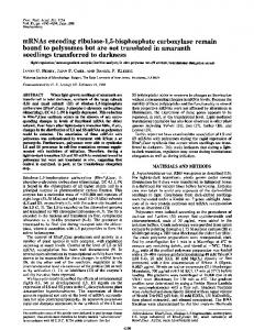
origins (in the same concentration range of 6.25, 12.5, and 25 ng) was denatured and spotted onto nitrocellulose filters. The results with 12.5 ng of poly(A) RNA from fetal liver( A ) ,adult heart ventricles ( E ) , adultfast (G) and slow (soleus) ( C ) , newborn (1-3 days) ( E ) ,and 5-6 days (F) and fetal (18 days) (D)skeletal muscle are shown in Fig. 3. Hybridization using PLt I PYUH PSt I ~ r t ~w n f I pItl pStl insert fragment 3 as a probe, after a 45 "C wash is seen with Pvul P*"W SSt I Bgll P r v l nseul all the RNA samplesexcept thefetal liver control RNA, although it is most intense with the 5-6 day newborn and "1" .... adult skeletal muscle RNA. To distinguish the homologous EcoRle e c BglI isoform corresponding to the MHC 32 sequence, blots were washed at higher stringency since two factors, namely ho- 0 0.2 Kb FIG. 1. Restriction map and sequence strategy for the in- mology and concentration of the MHC RNA sequences, are important in determining the degree of hybridization. Hybridsertion of plasmid 32. EcoRI and BglI arerestrictionsitesin pBR322 allowing orientation of the cloned sequences. The cleavage ization with skeletal muscle RNA from 1-3-day-old, 5-6-daysites for PstI, PuuII, SstI, BglI, HaeIII, and Hinfr are indicated. The old, and adult mice persists after a 55 "C wash. After a 65 "C four different PstI-PstI restriction fragments have been numbered 1 wash, two major spots remain: those withskeletal muscle to 4: fragment 1 (about 633 nucleotides long), fragment 2 (about 213 RNA from 5-6-day-old and adult mice. The newborn skeletal nucleotides long), fragment 3 (about 303 nucleotides long), and fragment 4 (about 103 nucleotides long). The arrows indicate sequence muscle RNA from I-3-day-old mice only hybridizes faintly, reflectingamuch lower concentration of the homologous strategy. The labeling was carried out as described under "ExperiRNA. Melting curves at increasing temperatures of the two mental Procedures." - - -, pBR322; -, coding;-, noncoding;.--., spots with 5-6-day newborn skeletal muscle RNA and with poly(A).
i ' 1 ~ 7 ' 7 ~ 1
r
l
-
.i
",
,
Downloaded from www.jbc.org by guest, on July 21, 2011
chain sequence. The total length of the insertion is about 1250 nucleotidescovering 17% of the mRNAlength. The restriction map of the plasmid revealed many PstI and PuuII sites in the insertion, corresponding to Leu-Gln and Gln-Leu, respectively, as noted by several authors (13,41). Fourdifferent PstI-PstI fragments were identified and their order was assigned by partial sequencing (Fig.1). The insertionincluded deoxycytidine residues (18 at the 5' end and 16at the 3' end) addedduringthetailing reaction. The codingcapacityis approximately 345 amino acids corresponding to the COOHterminal of the molecule. The 3"noncoding region contains 108 nucleotides and includes the polyadenylation signal AAATAAA 23 nucleotides upstream from the poly(A) which is 69 nucleotides long (Figs. 1 and 2). Comparison of the derived amino acid sequence withthe partial rabbit adult fast skeletal muscle myosin heavy chain aminoacid sequence publishedby Elzinga and TNS(41) confirms that the recombinant plasmid, MHC 32, contains an insertion coding for myosin heavy chain: the last 19 carboxyl-terminal amino acids are identical but some substitutions (positions 37, 63, 102, and 108 from the COOH end), mostly of a conservative nature, are seen. Identification of the Isoform Encoded by the Insertion of Plasmid MHC 32 and Its Use to Detect Developmental Transitions of Myosin Heavy Chain mRNAs in Skeletal MuscleSince the myosin heavy chains are encoded by a multigene family, the question arose as to which of 11-13 isoforms (for review, see Ref. 1) the plasmid MHC 32 encodes, especially with reference to the differentisoforms of fetal, neonatal, and adult fast skeletal muscle (2). Because the cloning was carried out with skeletal muscle RNA from 5-10-day-old mice, we suspected thatthe plasmid MHC 32 might code for the neonatal or the adult isoform. Northern blots (results not shown) with RNA from different sources showed hybridization with an mRNA of 6900 nucleotides in striated muscles (skeletal and cardiac) of fetal and adult mice. Given a 3'noncoding sequence of 108 nucleotides and a poly(A) stretch of about 200 nucleotides, this would suggest a rather long 5'noncodingsequence,assuminga length for the coding sequence of about 6000 nucleotides or even less (8). Weaker hybridization with RNA from myotubes from the mouse myogenic cell line T 984 C1 10 suggests a lower homology with the mRNA for the MHCembisoform expressed in early myotubes of these cultures (22). Cross-species hybridization is seen with rat and human muscle. In order to obtaina more quantitative assessmentof crosshybridization with RNA from various sources, dot blots were carriedout (22). Poly(A)-containing RNA fromdifferent
13870
Myosin Heavy ChainmRNAs in Developing Mouse Skeletal Muscle sequence (Figs. 1 and 4). After labeling the HinfI site by “filling in” as described under “Experimental Procedures,” part of the preparation was subjected to Maxam and Gilbert degradation and another part of it was used for S1 nuclease protection experiments. The homologous mRNA should protect a 240-nucleotide fragment, whereas hybridization to heterologousmRNA should only protectpartial fragments stretching from the labeled end to thesite of sequence diver-
A B C
379
-
”
257, 241‘
D
1724
E
G 64-
45”
55”
65”
FIG.3. Dot blots of poly(A)-containingRNA from different mouse tissues. Dot blots of poly(A)-containing RNA from different mouse tissues were hybridized to the purified nick-translated fragment 3 of plasmid MHC 32 (containing the 3”noncoding portion). The specific activity was 2.5 X 10” cpm/pg. The quantity of poly(A) RNA for all the samples was 12.5 ng.The temperatures onthe abscissa correspond to the washing temperatures of the blot in 0.1 X SSC, 0.1% SDS. The exposure time was 24 h. Lane A, RNA of fetal liver; lane R, RNA of adult cardiac ventricle; lane C, RNA of adult soleus; lane D, RNA of fetal skeletal muscle (18 days); Lane E, RNA of newborn skeletal muscle (1-3 days); laneF, RNA of newborn skeletal muscle (5-6 days); lane C, RNA of adult skeletalmuscle (1-2 months).
adult skeletal muscle RNA did not show a significant difference (results not shown). These results suggest that an RNA homologous to the plasmic MHC 32 insertion is present in these two RNApreparations. S1 protection experiments were undertaken in order to discriminate between the two contributing factors of homology and concentration. The S1 protection experiments were designed to take advantage of the fact that some regions of the myosin heavy chain nucleotide sequence are conserved so that different isoforms can cross-hybridize, while other regions such as the 3‘ end of the coding region and the 3”noncoding region are diverged and will not be protected against S1 nuclease digestion except by the homologous mRNA. First, such an experiment should clearly show if the plasmid MHC 32 is coding for a newborn or an adult sequence. Second, S1 protection experiments with this probe should permit the detection of other partially homologous myosin heavy chain transcripts accumulated during muscle development. We therefore isolated a single-stranded anti-messenger sequence between the HinfI site in fragment 3 and a Hue111 site in pBR322. This fragment contains the last 21 amino acids of the coding sequence, the 108 nucleotides of the 3”noncoding sequence, the poly(T), the poly(dG) tailing, and the neighboringpBR322
454 384
FIG.4. Protection of the HinfI-Hue111restriction fragment from S1 nuclease digestion by RNAs from different mouse tissues. The HinfI site, a t the 3’ end of the coding sequence of the myosin heavy chain, was end labeled as described under “Experimental Procedures.” The HaeIII restriction site is in the pBR sequence. The isolated anti-mRNA contains the last21 COOH-terminal amino acids (coding), the 3’ end noncoding region, the poly(T), the poly(dG) tailing, and theneighboring pBR322 sequence (up to the HaeIII site) as indicated. Part of the preparation was sequenced (lanes C, C-A, AC, C-T, C ) and part of the preparation was hybridized to 875 ng of poly(A) RNA from various sources. After S1 digestion, the protected fragments were analyzed on a 6% acrylamide-urea gel. The numbering of the nucleotides is given assuming the same structure of the HinfI site as in Umeda et al. (7). All samples were treated with 100 units of S1 nuclease, except sample 2 which was treated with 200 units. All muscle RNA samples were from C3H mice. The results with RNA from newborn (1-3 days) skeletal muscle were obtained in a separate experiment under similar conditions. Lane I, RNA from adult skeletal muscle; lane 2, RNA from newborn (5-6 days) skeletal muscle; lane 3, RNA from newborn (5-6 days) skeletal muscle; lane 4, RNA from newborn (1-3 days) skeletalmuscle; lane 5,RNA from fetal (18days) skeletal muscle; lane 6, RNA from adult cardiac ventricle; lane 7, RNA from adult brain; lane8, control without RNA.
Downloaded from www.jbc.org by guest, on July 21, 2011
F
Myosin Heavy Chain
mRNAs in Developing Mouse Skeletal Muscle
172-
1 2 3 FIG.5. Analysis of the HinfI-HaeIII-labeled fragments protected from SI nuclease digestion by hybridization with different RNA preparations (see Fig. 4) on a more highly resolving 6%acrylamide urea gel. Lanes I , 2,and 3 are the same as in Fig. 4.
relative intensities are not the same, e.g. two bands which on a more resolutive gel (results not shown) correspond to nu-
*** **
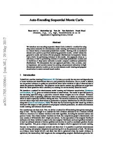
cleotide positions 113-115 and 117-118 (CGTTCTTTG) which are AT-rich sites. We conclude that the plasmid MHC 32 insertion is homologous to an mRNAfrom adult skeletal muscle and that myosin heavy chain mRNA of this isoform accumulates immediately after birth and is already the major RNA species in skeletal muscle from 5-6-day-old mice.In late fetal skeletal muscle and in the same tissue immediately after birth (1-3 days), the major species of mRNA is only partially homologous to the MHC 32 probe. No additional major mRNA species was identifiable in newborn mice, distinct from the fetal and adult isoform. The Myosin Heavy Chain Multigene Family: Detection of Multiple Genomic Fragments by the MHC 32 Sequence and Its Assignment to One Homologous Gene-Hybridization of purified nick-translated fragments of plasmid MHC 32 (fragments 1 and 3on Fig. 1)to a blot (31) of mouse genomic DNA restricted by EcoRIwhichdoes not cut in the MHC 32 sequence, permitted the detection after washing at low stringency (50 “C) of a number of bands corresponding to crosshybridizing genomic sequences in this multigene family (see Fig. 6A). An EcoRI digest of mouse (C3H) genomic DNA revealed seven (possibly eight or more given the intensity of the 4.5kilobase band) different restriction fragments (Fig. 6A, lane a) of variable intensities and ranging from 0.9 up to 10.6 kb when fragment 1 of the MHC 32 insertion (entirely coding) was used as a probe. Two supplementary bands appeared at 13 and 7 kb when fragment 3 of the insertion (containing most of the 3”noncoding region) was used as a probe (Fig. 6A, lane d ) . Already at low stringency with this more specific probe, the homologous gene hybridizes most strongly. Three main bands (8, 5.5, and 4.5 kb) still persist at 70 “C with fragment 1 as a probe (see lane b, Fig. 6A) showinga substantial homology to this part of the MHC 32 sequence. After a stringent wash at 75 “C with 0.1 x SSC and 0.1% SDS, only one band of 8 kb remains with fragment 1 as a probe (see lane c) and an additional fainter band of 7 kb with fragment 3 as a probe (see lane f ) . We conclude that the restriction fragments of 8-kb and7-kb length contain the 3‘ end of the adult fast skeletal muscle gene of mouse, homologous to the MHC 32 sequence. It is very unlikely that the adult fast skeletal muscle gene is interrupted by multiple intervening sequences containing
Downloaded from www.jbc.org by guest, on July 21, 2011
gence expected to lie at the end of the coding sequence (see “Discussion”) and in the 3”noncoding sequence. The results shown in Fig. 4 demonstrate essentially the same protection pattern for both adult skeletal muscleRNA (lane I ) and newborn (5-6 days) skeletal muscle (lanes 2 and 3 ) . A minor fraction of the labeled fragments is protected up to theend of the poly(A), probably including the tailing (more than 240 nucleotides) but themajority of the protected sequences reach only to theend of the 3”noncoding region showing two main series of bands. The hybridization of the poly(A:T) sequence is probably not sufficiently stable to confer a protection from S1 nuclease digestion. Numerous minor bands are seen below the two major protected series of bands. Most of these probably correspond to local regions of instability in the hybrid since they tendto fall in AT-rich stretches (see “Discussion”). A more highly resolving6% acrylamide gel (Fig. 5) shows that each series of protected fragments includes several bands: the upper (longer) bands correspond to nucleotides 167, 168, and 169, the lower (shorter) bands correspond to nucleotides 159, 160, and 162 counted from the HinfI site (see Fig. 4). This hybridization pattern is slightly affected by doubling the S1 concentration (lane 2): the enzyme seems to trim away the upper series of bands. The end of the 3”noncoding sequence of plasmid MHC 32 reads ATA ATT TTG CAA TCT A69 and the sequence lying betweenthe two main series of bands reads ATT TTGC. This heptanucleotide is also present in the same position in a rat cardiac myosinheavy chain cDNA clone (ATTTTGCCTGCASO) (13). The sequence of this rat cDNA, apart from the additional 11 nucleotides, is identical with another rat cardiac myosin heavy chain cDNA clone, suggesting heterogeneity in the polyadenylation site of this mRNA (13). The two series of bands seen in the experiments reported here with adult skeletal muscle mRNA may be due to a similar phenomenon. An alternative explanation is that there is someinstability of the hybrid in this region, since the upper series of bands is diminished with increasing S1 concentration. In contrast, hybridization with fetal skeletal muscle RNA and with adult cardiac ventricular RNA results in much shorter protected fragments (lunes 5 and 6, Fig. 4): fetal skeletal muscle RNA protects a sequencewhich stops at nucleotide 45 (from the HinfI site) corresponding to the 7th amino acid (i.e. Thr) from the COOH terminus but several other bands are present at nucleotides 46, 43, and 41. These bands (45, 46, 43, and 41) are also seen after hybridization with adult and newborn (5-6 days) skeletal muscle RNA (see “Discussion”). RNA from adult cardiac ventricle protects an even shorter fragment: there are several bands ranging from nucleotide 32 to 38 (from the HinfI site) and corresponding to the 11th (Arg), 10th (Glu), and 9thamino acid (Val) from the COOH terminus and two additional bands at nucleotides 24 and 25 corresponding to the 14th (Val) and 13th amino acid (Lys) (see Ref. 13). Hybridization with RNA from brain (lane 7, Fig. 4) shows a band migrating in the same position as the control without RNA (lane 8). Since RNA from 5-6day-old skeletal muscle already contains the mRNA for the adult isoform, RNA frommuscle of 1-3-day-old micewas isolated in order to look for the mRNA encodingthe neonatal isoform. The result is shown in Fig. 4, lane 4. Most of the protected fragments form bands at a position expected for the mRNA from fetal skeletal muscle, but a minor fraction of the labeled HinfI-HaeIII fragment is protected up to the end of the 3”noncoding region, giving the same pattern with two major series of bands as that seen after hybridization with adult skeletal muscle RNA. There are minor bands which seem to be superimposable on those obtained after hybridization with adult and newborn (5-6days) RNA although their
13871
13872 3
Myosin Heavy Chain mRNAs in Developing Mouse Skeletal Muscle 1
3
23.7-
-23.7
is detected (Fig. 6B, c and f ) . Since other related genomic sequences are very homologous, the washing conditions are critical in these experiments. The relative intensity of the other bands in relation to the remining one diminishes with increasing stringency.We therefore conclude that there is one gene corresponding tothisadultfastskeletal myosin sequence, and that it is probably interrupted by a n intervening sequence containing an EcoRI site in the region covered by fragment 3. DISCUSSION
We have cloned and characterized a recombinant plasmid (MHC32)thatcontainsaninsertion codingfor the 3'terminalpart of a myosin heavy chain.The sequence is homologous with an mRNA present in adult mouse skeletal muscle, and we conclude that the proteinencoded is an adult fast skeletal muscle isoform. This recombinant plasmid has been used to demonstrate the sequential transition from fetal 0 to adult myosin heavy chain mRNAs during skeletal muscle development in vivo in the mouse. Comparison of the derived amino acid sequence with that described for an adult skeletalmuscle myosin heavy chain in 62.3 2-36 the rabbit (41) shows some differences in the last 116 amino -2-0 2.0" acidresidues at the COOH terminus (Fig. 2). There is an additional Glu residue at position 28 or 29 (counting from the COOH-terminal amino acid), which is absent in the published protein sequence. Residues 37 (Ile + Leu), 42 (HwIII site, probably Ser + Ala), 63 (Thr + Ser orAla), 102 (Ile + Val), and 108 (Asn + Ser) are different. These changes may be a b c d e f a b c d e f species specific whereas another series of positions found to be variable in rabbit and termed microheterogeneous (41)may A B be isoform specific, one combination of these residues being FIG. 6. Southern blots of restricted mouse genomic DNA. Southern blots of restricted mouse genomic DNA were hybridized to found in the mouse plasmid MHC 32: residue7 (Thr), 10 plasmid MHC 32 restriction fragments 1 and 3 (see Fig. 1). Mouse (Glu), 45 (Val), and 112 (Asn). When the MHC 32 sequence genomic DNA wasdigested by EcoRI (A, lanes a to f ), BclI ( E , lanes iscompared to a cloned ratcardiac myosin heavy chain a to c), &/I1 (E, lanes d to f ) . The digested DNA was run on a 0.7% sequence (13),several notable variant positions, already proagarose gel, blotted on nitrocellulose, and hybridized to nick-trans- posed as species specific in the comparison with the rabbit, lated probes of fragments 1 (entirely coding; A , a-e) and 3 (213 coding and 113 noncoding;A, d-f, and B, a-f ) and washed at thetemperatures can be identified 37 (Ile + Val), 63 (Thr+ Leu), 102 (Ile + Ala), and 108 (Asn + Ala). It is striking that the last 10 indicated: 50,70,75 ( A , a-c, and d-f), and 60 "C; 75 and 77 "C ( E , aamino acidsat theCOOH terminus are highly diverged (70% c, and d-f). The specific activity of the probes was 1-3 X 10' cpm/pg. Size markers usedwere X DNA cut by Hind111 and indicated in in amino acids and43% in nucleotide sequence) between the kilobases. C3H mice were used in all digestions except those with adult mouse skeletal muscle and rat cardiac (13) sequences, BglII where the results with BALB/c are shown. An identical pattern compared with amuch lower divergence (6-17% in amino of bands was given with C3H DNA. acid and 12-24% in nucleotide sequence) in the remainderof this region. Comparing the mouse MHC 32 sequence with a myosin heavy chain cloned from embryonic chicken skeletal EcoRI sites giving rise to the7-8 bands at 50 "C, first because muscle (6, 7), we also observe a high divergence for the 10 we used two probes (insertion fragments 1 and 3 which are COOH-terminal amino acids (50%divergence in aminoacids over 200 nucleotides apart in the cDNAsequence of plasmid and 47% divergence in nucleotide sequence). It is not clear MHC 32) revealing several common bands (compare lanes a whether thedivergence in the COOH-terminalsequence is of and d for EcoRI digestion) and second because under high functional significance, or is permissible in the nonhelical terminal tailof the molecule where the structural constraints stringency conditions (75 "C) most of the bands disappear, demonstrating that they are nothomologous and correspond of the rod are relaxed (17). Apart from the polyadenylation signal and a TT"tetrato othermyosin heavy chain-related sequences. At high stringency (75 "C), one homologous sequence is seen after digestion nucleotide a few nucleotides before the poly(A) present in (13) sequences,the 3"noncoding with EcoRI and hybridization to fragment1. The single band chicken (6,7) and rat cardiac seen afterEcoRI digestionwith fragment1 (also after BanHI, sequence is diverged. However, the chicken skeletal muscle resultsnotshown) would suggest thatthereis only one 3"noncoding sequence does showa striking homology (AGAAGAAATCAhomologous gene; however, the two fragments seen at high ATTGCACAAAATGTGA inchickenand stringency with fragment 3 after EcoRI digestion introduce CAAAATGTGA in mouse) with a region in the middle of the some ambiguity into this interpretation. Genomic DNA was 3"noncoding sequence of the mouse skeletal muscle sequence. thequasiconserved nucleotherefore digested with two other enzymes, BclI and BglII and It is tempting to speculate that 18 theSouthernblots were hybridized withfragment 3 and tides in the mouse may be important in the transcriptional washed under increasing stringency (Fig. 6B). At low strin- regulation, metabolism, or translation of myosin heavy chain gency, a series of bands of differing intensities are seen(Fig. mRNA in skeletal muscle. A similar sequence is not seen in 6B, a and d ) , whereas at higher temperatures only one band other skeletalmuscle mRNAs, such as that for mouse skeletal
8-
Downloaded from www.jbc.org by guest, on July 21, 2011
Myosin Heavy Chain mRNAs in Developing Mouse
:’ S. Alonso, personal communication. B. Robert, personal communication.
13873
transcripts in the adult skeletal muscle, and possibly also of some cardiac heavy chain mRNA species. The use of homologous fetal and cardiac probes should clarify this point. As far as the major bands are concerned,twoseries of protected fragments are seen with RNA from adult skeletal muscle: they correspond probably to slightly different sitesof polyadenylation on the same mRNA (see “Results” and Ref. 13). It seems unlikely that two mRNAs encoding different (adult or neonatal) myosin heavy chains are detected, since the 3”noncoding sequence of the mRNAs is identical except for the last 12 nucleotides and on Southern blots only one homologous gene isdetected. From proteindata,there is evidence to suggest that two or three differentmyosin heavy chains are present in adult fast skeletal muscle (43). The amino acidsequence in the COOH-terminal region of the molecule shows microheterogeneity (41), and onemight have expected to detect other major species with some homology in the COOH-coding region. For the technical reasons discussed, suchfurther speciesmay not havebeendetected. Alternatively, it has been proposed that additional isoforms may be generated by post-translational processing, in which case they would not be detectable in the experiments reported here (18). In developing skeletal muscle,asecondmajor partially protected typeof fragment is detected in addition to the two series of adult skeletal muscle. This is the major sequence seen in 18-day fetalmuscle. It is a doublet, and may represent two closely related sequences or result from some terminal digestion of the fragment assuggested for the series of bands seen with adult muscleRNA. By 1-3 days after birth, the adult sequence, although a minor species, is accumulating and by 5-6 days, it is the major component. These results were unexpected in view of the clearly defined transitions at theprotein level of accumulated myosin heavy chain isoforms in the rat, from fetal (MHC,,,,) to newborn (MHC,,,, from 1 day before birth to 13 days after birth) and to adult proteins (2). Noevidence was obtained with the mouse probe for a major component specific to newborn skeletal muscle. We cannot exclude the possibility that such a neonatal sequence is not detected by our probe. It is also possible that the neonatal isoform is generated by post-translational modifications.However, thisseems unlikely, andthedataon protein accumulation in the rat and mRNA accumulation in the mouse can be reconciled if the messenger sequence seen in late fetal muscle (18 days) codes for the neonatal myosin heavy chain protein. There is someevidence that skeletal muscle development proceeds more rapidlyin themouse than in the rat,5 with an earlier accumulation after birth of the adult isoform in mouse (7th day) than in rat (13thday). It is thereforenot inconceivable, assumingthatthere is a lag period during which accumulated myosin heavy chain reaches its maximum level (10-20% of total protein (44)), that such a relationship exists. The sequential transitions in the accumulation of mRNAs coding for different myosin heavy chain isoforms during skeletal muscle development in vivo clearly differ from the situation for the alkali myosin light chain and actin multigene families. In fetal skeletal muscle (17-20 days), the mRNAs for the adult alkalilight chains are already accumulated (21). It is not clear whether the mRNA for the foetal light chain, LCl,,h, is still present at this stage, or whether this mRNA accumulatesfirst a t earlierstages of muscle development, followed by that for the adult isoforms, or whether the fetal and adult mRNAs co-accumulate.However, the timing of accumulation of the adult myosin light chain mRNAs clearly differs from that of the adult myosin heavy chain mRNAs.
’ R. Whalen and G. Butler-Browne, personal communication.
Downloaded from www.jbc.org by guest, on July 21, 2011
muscle actin3 ormyosin alkali light chainsMLC1/3.4 Detection of messengersfor other isoforms, particularly developmental isoforms, with plasmid MHC 32 raises some interesting questions. The experiments with RNA from different tissues would suggest that the sequence cross-hybridizes with mRNAs for other striated muscle isoforms, namely those in slow skeletal muscle, fetal skeletal muscle, and probably several in cardiac muscle. In view of the low concentration of myosin heavy chain mRNA in non-muscle tissues, it is not possible to draw any conclusion about the sequence homology of this isoform. From Southern blotting experiments with mouse genomic DNA, we conclude that the MHC 32 sequencedetects 7-8 bands of differentlength as also reported for chick (9) and rat (12-16) DNA. Several of the bands shown in Fig. 6 (e.g. the 4.5-kb band in the EcoRI digest, or the 5.5-kb band in the BclI digest) are very intense a t low stringency, suggesting multiple myosin heavy chainrelated sequences. It is not known whether these are functional genes, or pseudogenes, or partial reduplications in the 3‘ region (for a detailed discussion of this point, see Ref. 30). Such multiple myosin heavy chain-related sequences of the same restriction fragment length may result from a recent amplification event in evolution. Only one genomic sequence is homologous to the adult skeletal muscle probe, suggesting only one gene for this adultisoform. Interpretation of the S1 protection experiments requires some discussion before the results canbe assessed. With this approach, it is possible to distinguish different types of myosin heavy chain mRNAs and their relative amounts, since not only homologous mRNAsaredetectablebut also mRNAs with partial homology expected, with the probeused, to lie in the more conserved region of the coding sequence proximal to the 5”labeled end. If this more conserved region has in fact diverged throughout its length, or only in a few nucleotides corresponding to the5’ end-labeling site, theprobe will not detect such mRNA species. This will also be the case, under the conditions used here, if the mRNA species to be detectedis at avery low concentration. The only way to exclude this possibility is by the use of other regions of the fast adultcoding sequence,or of homologous probes. A second important point concerns the significance of the fragments which are partially protected from S1 digestion. The interpretation is straightforwardwhentheyareintensebands,or series of bands, the latter probably resulting from some instability at the extremity of the hybrid molecule and consequent S1 activity. The difficulty arises when minor bands are present. This is the case for hybridization with mRNA from adult and newborn (5-6 days) skeletal muscle, where numerous bands appear below the two majorseries of protected fragments. These minor bandsseem to be the product of two phenomena: an artifactual one, reflecting the technical conditions of the experiment (conditionsof hybridization and S1 digestion resulting in minor nicking of the sequence in ATrich regions) and a real one, reflecting the presence of minor isomorphic mRNA species. Both their number and preferential location in AT-rich regions would suggest that most of the minor bands seen with the adult andnewborn (5-6 days) skeletal muscle RNA are probably artifacts. With l-3-day newborn muscle RNA, a few distinct minor bands are seen above the major fetal fragments and bellow the adult bands. Other minor bands seen with adult and newborn (5-6 days) mRNA samples correspond to the positions of major bands seen with the fetal and cardiac RNA. This may indicate the continuing presenceof low levels of fetal myosin heavy chain
Skeletal Muscle
13874
Myosin Heavy ChainmRNAs in Developing Mouse Skeletal Muscle
B. (1980) Proc. Natl. Acad. Sei. U. S. A . 77,5749-5753 12. Nudel, U., Katcoff, D., Carmon, Y., Zevin-Sonkin, D., Levi, Z., Shaul, Y., Shani, M. & Yaffe, D. (1980) Nucleic Acids Res. 8, 2133-2146 13. Mahdavi, V., Periasamy, M. & Nadal-Ginard, B. (1982) Nature (Lond.)297,659-664 14. Nguyen, H. T., Gubits, R. M., Wydro, R. M. & Nadal-Ginard, B. (1982) Proc. Natl. Acad. Sci. U. S. A . 79, 5230-5234 15. Czosnek, H., Nudel, U., Shani, M., Barker, P. E., Pravtcheva, D. D., Ruddle, F. H. & Yaffk, D. (1981) EMBO J . 1, 1299-1305 16. Wydro, R. M., Nguyen, H. T., Gubits, R. M. & Nadal-Ginard B. (1983) J . Biol. Chem. 258, 670-678 17. MeLachlan, A. D. & Karn, J. (1982) Nature (Lond.) 299, 226231 18. Bandman, E., Matsuda, R. & Strohman, R. C. (1982) Cell 29, 645-650 19. Minty, A. J., Caravatti, M., Robert, B., Cohen, A., Daubas, P., Weydert, A., Gros,F. & Buckingham, M. E. (1981) J. Biol. Chem. 256,1008-1014 20. Minty, A. J., Alonso, S., Caravatti, M. & Buckingham, M. E. (1982) Cell 30, 185-192 21. Robert, B., Weydert,A,,Caravatti, M., Minty, A,, Cohen, A., Daubas, P., Gros, F. & Buckingham, M. E. (1982) Proc. Natl. Acad. Sei. U. S. A . 79, 2437-2441 22. Caravatti, M., Minty, A,, Robert, B., Montarras, D., Weydert, A., Cohen, A,, Daubas, P. & Buckingham, M. (1982) J. Mol. Biol. 160,59-76 23. Berkner, K. L. & Folk, W. R. (1977) J. Biol. Chem. 252, 3176Acknowledgments-We would like to thank Odette Jaffrezou for 3 184 manyhours of dedicated dissection. We also thank Paul Barton, 24. Maxam, A. M. & Gilbert; W. (1980) Methods Enzymol. 65,499Serge Alonso, Robert Whalen, and Gill Butler-Browne for helpful 560 discussion. We are especially grateful t o Francois Gros for his gen25. Challberg, M. D. & Englund, P. T. (1980) Methods Enzymol. 65, erous support and constant encouragement. We would like to thank 39-43 M. Elzinga for sending us data on the rabbit skeletal myosin heavy 26. Sanger F. & Coulson, A. R. (1978) FEBS Lett. 87,107-110 chain sequence priorto publication, and P. K. Umeda for a preprint 27. Bishop, J. O., Rosbash, M. & Evans, D. (1974) J. Mol. Biol. 85, on S1 protection experiments with myosin heavy chain sequences in 75-86 developing chick muscle. We very much appreciated the helpfulness 28. Alwine, J. C., Kemp, D. J., Parker, B. A,, Reiser, J., Renart, J. of U. Nudel (Weizmann Institute) in sending us the rat p82 clone. Stark, G. R. & Wahl, G. M. (1979) Methods Enzymol. 68,220242 Addendum-After submission of this manuscript, we were able to 29. Blin,N. & Stafford, D. W. (1976) NucleicAcidsRes. 3, 23032308 compare the DNA sequence of pMHC 32 with new sequence data 30.Minty, A. J., Alonso, S., Guenet, J . L. & Buckingham, M. E. (46) on the 3’ ends of myosin heavy chain cDNAs from rat skeletal (1983) J . Mol. B i d . 167, 77-103 muscle. Oneof the two adult sequences is very homologous to plasmid 31. Southern, E. M. (1975) J. Mol. Biol. 98, 503-517 MHC 32. 32. Rigby, P. W. J., Dieckmann, M., Rhodes, C. & Berg, P. (1977) J . REFERENCES Mol. Biol. 113,237-251 33.Denhardt,D. T. (1966) Biochem.Biophys.Res.Commun. 23, 1. Buckingham, M. E. & Minty, A. J . (1983) in Eukaryotic Genes: 641-646 Their Structure, Actiuity, and Regulation (MacLean,N., Gregory, S. & Flavell, R., eds) Buttenvorths, London, in press 34. Berk, A. J. & Sharp, P. A. (1977) Cell 12, 721-732 35.Weaver, R. F. & Weissmann,C.(1979) NucleicAcidsRes. 7, 2. Whalen, R. G., Sell, S. M., Butler-Browne, G. S., Schwartz, K., 1175-1193 Bouveret, P.& Pinset-Harstrom, I. (1981) Nature (Lond.)292, 36. Potter, J . D. (1974) Arch. Biochem. Biophys. 162, 436-442 805-809 37. Vogt, V. M. (1980) Methods Enzymol. 65, 248-255 :3, MacLeod, A.R., Karn, J. & Brenner, S. (1981) Nature (Lond.) 38. Stark, C. R. & Williams, J . G. (1979) Nucleic Acids Res. 6, 19529 1,386-390 203 4. Rozek, C. E. & Davidson, N. (1983) Cell 32, 23-24 5. Hastings, K. E. M. & Emerson, C. P., Jr. (1982)Proc. Natl. Acad. 39. Smith, D. F., Searle, P. F. & Williams, J . G. (1979) Nucleic Acids Res. 6,487-506 Sei. U. S. A. 79, 1553-1557 6. Umeda, P. K., Sinha, A. M., Jakovcic, S., Merten, S., Hsu, H. J., 40. Jakob, H., Buckingham, M. E., Cohen, A., Dupont, L., Fiszman, M. & Jacob, F. (1978) Exp. Cell Res. 114, 403-408 Subramanian, K. N., Zak, R. & Rabinowitz, M. (1981) Proc. 41. Elzinga, M. & Trus, B. (1980) in Methods in Peptide and Protein Natl. Acad. Sci. U. S. A. 78, 2843-2847 Sequence Analysis (Birr, C., ed) pp.213-224, Elsevier, Amster7. Umeda, P. K., Kavinsky, C., Sinha, A. M., Hsu, H. J., Jakovcic, dam S. & Rabinowitz, M. (1983) in Muscle Deueloprnent: Molecular and Cellular Control (Pearson, M. L. & Epstein, H. F., eds) pp. 42. Yaffe, D. (1968) Curr. Top. Deu. Biol. 4, 37-77 43. Zweig, S. E. (1981) J . Biol. Chem. 256, 11847-11853 169-176, Cold Spring Harbor Laboratory,New York 8. Jakowlew, S. B., Khandekar, P., Datta, K., Arnold, H.-H., Narula,44. Squire, J. M. (1981) in T h e Structural Basis of Muscular Contraction (Squire, J., ed) pp.265-380, Plenum Press, New York S. K. & Siddiqui, M. A. 0. (1982) J . Mol. Biol. 156, 673-682 9. Robbins, J., Freyer, G. A,, Chisholm, D. & Gilliam, T. C. (1982) 45. Fritsch, E. F., Lawn, R. M. & Maniatis, T. (1980) Cell 19, 959972 J. Rid. Chem. 257,549-556 H. T., Periasamy, 10. Sinha, A. M., Umeda, P. K., Kavinsky, C. J., Rajamanickam, C., 46. Nadal-Ginard, B., Medford, R. M., Nguyen, M.,Wydro, R. M.,Hornig,D.,Gubits,R.,Garfinkel, L. I., Hsu, H. J., Jakovcic, S. & Rabinowitz, M. (1982) Proc. Natl. Weiczorek,D.,Bekesi, E. & Mahdavi, V. (1982)in Muscle Acad. Sci. U. S. A . 79,5847-5851 Deuelopment: Molecular and Cellular Control (Pearson, M. L. 11. Medford, R. M., Wydro, R. M., Nguyen, H. T. & Nadal-Ginard, (Ir Epstein, H. F., eds) pp. 143-168, Cold Spring Harbor Laboratory, NY ti P. Barton, personal communication.
The actins also present another pattern of accumulation. During skeletal muscle development, fetal actin gene transcripts co-accumulate with the skeletal muscle actin mRNAs. In 18-day fetal muscle, both mRNAs are present (approximately 60:40, adult skeleta1:fetal actin) and the fetal transcripts persist innewborn (5-10 days) muscle (approximately 80:20, adult ske1etal:fetal actin) (20). Thus, both in the coaccumulation andin the timing of developmental transitions for fetal and adult mRNAs, the actin multigene family differs from the myosin heavy chain multigene family. At the level of mRNA accumulation, there seems to be no coordination between the developmental transitions, suggesting a specific developmental regulation foreach multigene family. The transcriptional activity of these different genes during development remains to be investigated. The organization of actin and myosin genesis also different. Striated muscle actin genes are not linked (15,30), whereas myosin heavy chain genes are on the same chromosome in the mouse (15) and in humans appear to be clustered.6 The sequential expression of skeletal muscle myosin heavy chain genes during development may be reflected in the spatial organization of these genes as is the case for the globins (45) where embryonic, fetal, and adult sequences are aligned sequentially in the mammalian genome.
Downloaded from www.jbc.org by guest, on July 21, 2011