only by either a 2- or 0-nt overhand on the 3â²end (primer-free). These methods ... protocols. To address these problems ..... a central random domain of 27 nt for.
Short Technical Reports Minimal primer and primer-free SELEX protocols for selection of aptamers from random DNA libraries Weihua Pan, Ping Xin, and Gary A. Clawson Gittlen Cancer Research Foundation, Hershey Medical Center, Departments of Pathology, Biochemistry & Molecular Biology, College of Medicine, Pennsylvania State University, Hershey, PA, USA BioTechniques 44:351-360 (March 2008) doi 10.2144/000112689
Standard systematic evolution of ligands by exponential enrichment (SELEX) protocols require libraries that contain two primers, one on each side of a central random domain, which allow amplification of target-bound sequences via PCR or RT-PCR. However, these primer sequences cause nonspecific binding by their nature (generally adding about 20 nt on each end of the random sequence of about 30–40 nt), and can result in large numbers of falsepositive binding sequences and/or interfere with good binding random sequences. Here, we have developed two DNA-based methods that reduce and/or eliminate the primer sequences from the target-binding step, thus reducing or eliminating the interference caused by the primer sequences. In these methods, the starting selection libraries contain a central random sequence that is: (i) flanked by only 2 nt on each side (minimal primer); or (ii) flanked only by either a 2- or 0-nt overhand on the 3′end (primer-free). These methods allow primer regeneration and re-elimination after and before selection, are fast and simple, and don’t require any chemical modifications for selection in a variety of conditions. Further, the selection rounds are performed with DNA oligomers, which are generally employed as end product aptamers.
INTRODUCTION The identification of tumorspecific molecular markers for cancer diagnostics and the targeting of tumorspecific moieties/pathways for developing nontoxic and effective anticancer
therapies are important goals. Efficient targeting of cancer cells will depend upon development of molecular probes suitable for in vivo applications, probes that are endowed with the required affinity, specificity, and favorable pharmacokinetic properties.
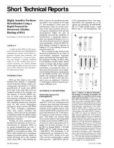
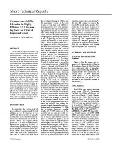
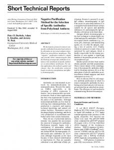
Aptamers are highly structured oligonucleotides (DNA or RNA) that can bind to targets with affinities comparable to antibodies. They are identified through an in vitro selection process called systematic evolution of ligands by exponential enrichment (SELEX; (1,2)) to recognize a wide variety of targets, from small molecules to proteins, and from cultured cells to in vivo imaging targets (3–11). These oligonucleotides have properties that are well suited for in vivo diagnostic and/or therapeutic applications: besides good specificity and affinity, they are poorly immunogenic, and their relatively small size can result in facile penetration of tissues. The SELEX technology can now accept chemically modified nucleotides for improved stability in biological fluids (12). Less than two decades after the first applications of the technique, several lead compounds are currently in clinical trials, and the first aptamer drug, Macugen, was approved by the U.S. Food and Drug Administration (FDA) in 2004 (see Reference 13). However, aptamers that are identified through the standard SELEX process usually comprise ∼70–80 nt, since they are typically selected from nucleic acid libraries with ∼30–40-nt-long randomized regions plus fixed primer sites of ∼20 nt on each side. There are substantive problems with these primer sequences (which generally comprise ∼50% of the library sequences) in that they may comprise a portion of the Figure 1. The minimal primer (MP) DNA aptamer selection protocol. The library oligonucleotides are annealed together and subjected to PCR amplification to yield a double-stranded DNA library (Step A). The random region from the MP single-stranded DNA library is prepared by Nt.BstNBI /NotI digestion and subsequent gel purification. Top-right panel (Step B) shows the digested PCR products after PAGE on a 10% gel under denaturing conditions (the +31-fragment is indicated to the right). After selection (Step C), the selected library is hybridized with its 5′-end bridge-pair and 3′-end bridge-pair, and ligated to regenerate the previously removed primer regions (Step D). The ligated products are then reamplified by PCR. Step E (bottom right) shows the products reamplified after 5, 10, 15, 20, 25, and 30 PCR cycles, after separation by PAGE on 8% gels under nondenaturing conditions. Migration of PhiX174/HinfI markers is shown to the left, and the position of the 64 bp fragment is indicated. The selected products can then be used for additional rounds of selection.
Vol. 44 ı No. 3 ı 2008
www.biotechniques.com ı BioTechniques ı 351
Short Technical Reports
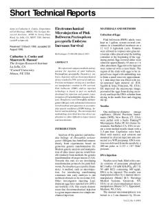
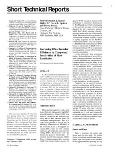
Figure 2. The PF1 DNA aptamer selection protocol. The library oligonucleotides are annealed together and subjected to PCR amplification to yield a double-stranded DNA library (Step A). The 5′-end primer-free (PF) random region from the PF single-stranded DNA library is prepared by Nt.BstNBI/Nt.BbvCI digestions (cleavage sites are marked with red arrowheads) and gel purification, and the self-bridge (black arrow) is also isolated by gel purification. After selection (Step C), the selected sequences (designated pS30) are hybridized with their corresponding oligomers and the self-bridge and ligated to regenerate the previously removed primer regions. At this stage, primers are introduced that contain an Sp6 promoter at the 3′-end (Step D). This is then used to transcribe RNA containing the selected regions (Step E). Finally, the RNA is then exclusively reamplified (the template DNA is digested) using RT-PCR (Step F), and the selected sublibrary is ready for the next round of selection.
selected binding sequences, and/or they may base-pair with the central random fragments to form structures that are selected as sites for target binding, which often compromises the significance of results from standard SELEX protocols. To address these problems using RNA libraries, primer sequences have been blocked by complementary oligonucleotides or switched to different sequences midway during the rounds of SELEX (14), or have been trimmed to only 6 and 4 nt (15), or to 9 and 7 nt (16). Wen and Gray (17) designed a primer-free genomic SELEX method, in which the primer sequences were completely removed from the library before selection and were then regenerated to allow amplification of the selected genomic fragments. However, to employ the technique, a unique genomic library had to be constructed, a very elaborate procedure, and there is presumably very limited diversity in the genomic library. While this protocol might be adaptable for use with a random region (in place of the genomic segments), the reliance on a linear extension step to regenerate primers would likely prove problematic. Alternatively, efforts to circumvent problems caused by fixed primer sequences using high efficiency partitioning are met with problems regarding PCR amplification (18). Here we have developed two methods, minimal primer (MP) selection (termed primer-bridge PCR) and primer-free (PF) selection (termed 352 ı BioTechniques ı www.biotechniques.com
self-bridge RT-PCR), which significantly simplify SELEX procedures and effectively eliminate primerinterference problems. Here we show that the protocols (the MP protocol and two closely related PF protocols) work in a straightforward manner. The central random regions of the libraries were purified without extraneous flanking sequences, and were bound to a melanoma cell line. The bound sequences were obtained, reunited with flanking sequences, and reamplified to generate a selected sublibrary. Successive rounds of selection increased library binding and increased selectivity for melanoma cells versus binding to nontarget fibroblasts. MATERIALS AND METHODS Oligonucleotides for MP Selection The random library for the MP selection protocol was (L7-N27) = GAA CAG GAT TAG CGG CCG C-(N)27-TGA TTC GAC TCT AGA GCG, which contains 427 (1.80 × 10¹⁶) different sequences. The amplification primers were as follows: 5′-end primer (L7-P1): CGC TCT AGA GTC GAA TCA; 3′-end primer (L7-P2): GAA CAG GAT TAG CGG CC; 5′-end bridge pair (5′-end top-strand, 5TS = CGC TCT AGA GTC GAA T and 5′end bottom-strand, 5BS = TGA TTC GAC TCT AGA GCG); and 3′-end bridge pair (3′-end top-strand, 3TS =
pGGC CGC TAA TCC TGT TC and 3′-end bottom-strand, 3BS = GAA CAG GAT TAG CGG CCG C). Oligonucleotides for the PF Selection The random library for the PF protocols was (L8-N30) = GTA TAC CTG CAG CTG AGG-(N)30-ATT CGT CTC TAG AGC GCA, which contains 430 (1.15 × 10¹⁸) different sequences. The amplification primers were as follows: 5′-end primer (L8-P1): TGC GCT CTA GAG TCG AAT; and 3′-end primer (L8-P2): GTA TAC CTG CAG CTG AGG. Sp6 promoter top-strand for the PF1 and PF2 protocols were Sp6TS-1 = pTCA GCT GCA GGT ATA CTA TAG TGT CAC CTA AAT AAG CTT GGG or Sp6TS-2 = pCCT CAG CTG CAG GTA TAC TAT AGT GTC ACC TAA ATA AGC TTG GG, respectively. The bottom-strand primer was Sp6BS = CCC AAG CTT ATT TAG GTG ACA CTA TAG TAT ACC TGC AGC TGA GG for both protocols. All of the oligonucleotides were HPLC-purified and were purchased from Integrated DNA Technologies, Inc. (Coralville, IA, USA). Cell Lines and Culture Selection The human melanoma cell line SKMEL-31 (HTB-73; ATCC, Rockville, MD, USA) was used for selection, whereas a normal skin fibroblast cell line Detroit 551 (CCL-110, ATCC) was used for counter-selection. The Vol. 44 ı No. 3 ı 2008
Short Technical Reports Selection Using Cultured Mammalian Cells as Target
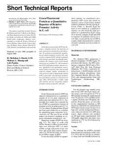
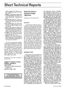
Figure 3. The PF2 DNA aptamer selection protocol. The library oligonucleotides are annealed together and subjected to PCR amplification to yield the double-stranded DNA library (Step A). The primer-free (PF) random region from the PF single-stranded DNA library is prepared by Nt.BstNBI/BspMI digestions (cut sites are denoted with arrowheads) and gel purification. For the PF2 protocol, the self-bridge is isolated either from the PF1 protocol and/or by single digestion of the starting library with Nt.BstNBI (Step B), coupled with gel purification. After selection (Step C), the selected sequences (again designated pS30) are hybridized to their corresponding oligomers and the self-bridge, and ligated to regenerate the previously removed primer regions. As in PF1, primers are introduced that contain an Sp6 promoter at the 3′-end (Step D). This is then used to transcribe RNA containing the selected regions (Step E). The RNA is then exclusively reamplified using RT-PCR (Step F), and the selected sublibrary is ready for the next round of selection.
cells were maintained in Dulbecco’s modified Eagle’s medium containing 12% heat-inactivated bovine calf serum in a humidified incubator at 37°C with 5% CO2. Preparation of Minimal-primer/ Primer-free DNA from Random DNA Libraries 1.6 nmol L7-N27 and L8-N30 (approximately 1015 sequences from each library) were amplified by PCR with 4.8 nmol P1 and 3.2 nmol P2 (respectively) in 1.6-mL reactions as previously reported (19–22). During the PCR amplification, a trace amount of α-32P-dCTP was incorporated in the reaction to radiolabel the dsDNA product. The PCR products were extracted once with an equal volume of Phenol:ChCl3:IAA (Ambion, Austin, TX, USA), precipitated with 250 mM NaCl and 2.5 volumes of ethanol at -70°C, and recovered by centrifugation at 21,000× g. DNA pellets were rinsed once with 70% ethanol and were dried in a Speed Vac (GMI, Ramsey, MN, USA). DNA was cleaved with 400 U of NotI for the MP 354 ı BioTechniques ı www.biotechniques.com
selection, or 400 U of Nt.BbvCI for the PF1 selection, at 37°C overnight in 800 μl reactions. The cleaved PCR products were again extracted and precipitated, rinsed and dried, and were then subjected to a second cleavage with 400 U Nt.BstNBI at 55°C for 4 h in 800 μl reactions. The DNA fragments were separated by PAGE in 10% gels under denaturing (7 M urea in Tris-Borate-EDTA) conditions, and the corresponding gel strips were isolated after identification by autoradiography. The +31-, +32-, and -66-fragments were eluted for 1 h at room temperature in buffer of 20 mM Tris-HCl (pH 7.4) and 250 mM NaCl, and then were heated for 3 min at 85°C before precipitation as described above in this section. All restriction and nicking endonucleases were purchased from New England BioLabs (Ipswich, MA, USA) and reaction conditions followed the manufacturer’s instructions. The reaction volumes for further PCR amplifications and enzymatic digestion were decreased by half for each selection round, eventually reaching a 200 μl volume, which was then used for all subsequent rounds.
Preparation of MP and PF library DNAs. The 32P-labeled MP/PF library DNAs were heated at 85°C for 3 min in 200 μl of 20 mM TRIS-HCl (pH 7.4) and were cooled down to 37°C. MgCl2 was added to a final concentration of 5 mM, and the samples were incubated for 3 min. Prewashing of target cells. Detroit551 cells and SK-MEL-31 cells were plated and allowed to grow for 1 day (about 2 × 10⁶ cells in 60 mm plates), then washed for 10 min in 4 mL Eagle’s Minimum Essential Medium (EMEM) three times. Counter-selection and selection. The structured library DNAs were mixed with 2 mL EMEM and were incubated with the washed Detriot-551 skin fibroblast cells for 30 min (as a counter-selection), then the unbound DNA-EMEM mixture was removed and incubated with the washed SKMEL-31 cells for 30 min (selection). After selection, bound sequences were harvested (as described below in Recovery of SK-MEL-31 cell-bound DNAs (selected DNA)). The counterselection was repeated once for the second round, twice for the third round, and then three times for the rest of the selection rounds. Post-selection rinsing. The DNAbound SK-MEL-31 cells were washed under the same conditions as described in the prewash, except that the incubations were for 20 min for each washing. All of cell washings and selections were performed at 37°C and with gentle rocking. Recovery of SK-MEL-31 cellbound DNAs (selected DNA). The DNA-bound SK-MEL-31 cells were harvested by incubating the cells for 10 min at 37°C within 800 μl TrypLE Express (Gibco, Invitrogen, Carlsbad, CA, USA) before being heated at 85°C for 15 min. Selected DNAs were extracted and precipitated twice as described. Reamplification of the Selected MP/ PF DNA Sequences Ligation. The selected +31- or +32fragments were respectively heated at Vol. 44 ı No. 3 ı 2008
������
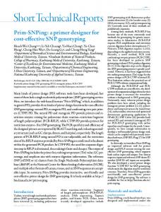
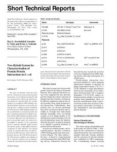
Figure 4. Experimental results/reduction to practice of the primer-free (PF) selection protocols. Designated steps correspond to those depicted in Figures 2 and 3. After digestion of the PF library with restriction endonucleases (as indicated), the digested products were separated by PAGE on a 10% gel under denaturing conditions. The corresponding fragments obtained with the PF1 and PF2 selection protocols are shown (Step B). 32P-labeled RNAs were transcribed using ligated products of 1, 0.5, 0.25, and 0.125 μl from 5 μl reactions, and were separated by PAGE on 6% gels under denaturing conditions (Step E). The RNA preparations were then treated with DNase to removed the unselected random DNA regions, reverse-transcribed into cDNA, and then reamplified for 7, 14, 21, or 28 PCR cycles. Products were separated by PAGE on 8% gels under nondenaturing conditions. The 66 nt fragment represents the full-length product containing the selected pS30 sequences re-embedded within the 5′- and 3′- flanking sequences. Migration of PhiX174/HinfI markers are shown at left. Controls included omission of the reverse transcription (RT) step. A small amount of product in such samples could be seen at high cycle numbers; this can be completely eliminated with a second (or prolonged) DNase digestion step.
85°C for 3 min with 100 pmol oligonucleotide of each primer bridge-pair (5TS and 5BS, 3TS and 3BS in the MP protocol) in 20 μl of 20 mM TRIS-HCl (pH 7.4), or with the -66-fragment and 2 nmol of each primer component for the PF protocol primer regeneration (LS8-P1, Sp6TS-1, and Sp6BS) in 200 μl of 20 mM TRIS-HCl (pH 7.4). The temperature was allowed to slowly return to room temperature within 1 h to allow efficient hybridization. Eight hundred U of T4 DNA ligase (2 × 10⁶ U/mL) and 10× ligase buffer (both from New England BioLabs) were added to bring the reaction volume to 40 mL for the MP protocol, and 8000 U of T4 ligase and 10× ligase buffer were added to bring the reaction volume to 400 μl for the PF protocols; reactions were incubated at 37°C for 1 h. Transcription (and reverse transcription for the PF protocols). The ligated product was extracted once and precipitated, then transcribed into RNA by Sp6 RNA polymerase in 400 μl of reaction (23). The nucleotide fragments (RNA and DNA) were
extracted and precipitated, resuspended in 400 μl, and then digested with 40 U RNase-Free DNase I (New England BioLabs) for 2 h at 37°C. In later experiments, this DNase digestion step was repeated once to completely destroy residual DNA fragments. The remaining RNA was extracted and precipitated in 300 mM NaAc (pH 5.2) and 2.5 volume of ethanol at -70°C.The RNA pellet was harvested by centrifugation and the pellet was rinsed with ethanol, then resuspended in 20 mM Tris-HCl (pH 7.4), heated with 1 nmol L8-P1 at 85°C for 3 min, and then chilled on ice (220 μl reaction volume). Two hundred μl of the annealed RNA/DNA complexes were reverse-transcribed into complementary DNA at 37°C for 1 h using SensiScript Reverse Transcriptase (Qiagen) in 400 μl reactions. As a no-reverse transcription (RT) control, 20 μl of the RNA/DNA complexes were incubated as above but without reverse transcriptase in 40 μl reactions. The reaction volumes of subsequent transcription and reverse-transcription
����������������� ��������������������������� ����������������������������� ������������������������ �������������������������� �������������������������������� �������������������������������� �������������������������������� ������������������������������ ����������������������������� ��������������������������������� ����������������������� �������������� �������������
����������������� ���������������������
Short Technical Reports
reactions were decreased by half for each selection round, eventually reaching 100 μl, a volume that was used for all subsequent rounds. Titration of PCR cycles. Two μl of ligated products from MP selection and 20 μl of reverse-transcribed products from PF selection were prepared for 100 μl PCR reactions. These were divided into 5 aliquots, which were then run for various numbers of PCR cycles. The PCR products were analyzed in an ethidium bromide–stained 8% polyacrylamide TBE-gel (run under native conditions) to determine the optimal number of PCR cycles suitable for large-scale amplification. The volume of MP-ligated product used for pilot-PCR was increased by half for each selection round, eventually reaching 8 μl, a volume that was used for all subsequent rounds. Half of each MP ligated–product and/or PF RT-product was used for preparing 32P-labeled MP/PF selected libraries to determine their binding to the selection targets. Selected DNA aptamers were identified by TOPO TA cloning kit (Invitrogen, Carlsbad, CA, USA) and sequencing as previously described (22). A detailed Online Protocol is available at www.BioTechniques.com. RESULTS AND DISCUSSION Two double-stranded DNA libraries were constructed using PCR with the corresponding oligonucleotides (Figures 1–3, Step A), which contain a central random domain of 27 nt for the MP library, or 30 nt for the PF library, flanked by two primer regions. In each library, the 5′- regions contain an endonuclease “nicking” site for the endonuclease Nt.BstNBI; this enzyme recognizes dsDNA but cleaves only one strand of the DNA substrate. In the 3′-region, the MP library contains a NotI endonuclease restriction site that cleaves dsDNA substrate, while the 3′-region in the PF library contains another “nicking” site for the endonuclease Nt.BbvCI, which also recognizes dsDNA but cleaves only one strand, as well as a BspMI endonuclease restriction site. 358 ı BioTechniques ı www.biotechniques.com
For the MP protocol, the internal 5′-pCA-N27-GC-3′ fragment, 31 nt in length (designated +31-fragment) was generated by NotI/Nt.BstNBI cleavage of the MP dsDNA library followed by gel purification. As shown, this produces the random N27 sequences with 2 nt flanking sequences (“docks”) on each side (Figure 1, Step B). The +31-fragments with the 2 nt docks were gel-purified after the digestion and used in a selection procedure for binding to cell surface markers on melanoma cells (see below in this section). Following selection, the bound +31-fragments were used for primer regeneration (Figure 1, Step D) to allow reamplification of the selected sequences for the next round of selection, and a number of subsequent rounds were performed. Under our modified hybridization:ligation conditions, use of the 2 nt docks resulted in a yield of >90% ligated products, when compared with use of 9 nt docks in this primerbridge approach (data not shown). The hybridization:ligation with 2 nt docks was equivalent to that obtained with 4–8 nt docks, whereas use of 1 nt docks resulted in very low yields of ligated products (