... simplicity of its development and use by non-specialists patient's e.c.g.. ~_~ 12 bit. I m.p.u. from the. --. a.d.. /'/. Motorola bedside. '-I. I monitor. I convertor. 6800.
Med. & Biol. Eng. & Comput., [98l, 19, 497 500
Technical note Simple microprocessor-based system for on-line e.c.g, arrhythmia analysis K e y w o r d s - - A r r h y t h m i a , E.C.G., Microprocessor, Monitoring
9 1 Introduction THIS paper presents a simple microprocessor system for the on-line analysis of electrocardiograms (e.c.g.). The system is
2 Description o f the system
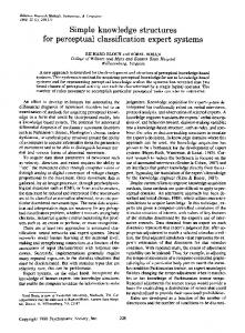
Microprocessor technology was chosen (Fig. 1) because of the simplicity of its development and use by non-specialists
I
patient's e.c.g. from the -bedside monitor
~_~ 12 bit a.d. '-I I convertor
I
m.p.u. Motorola
I
6800
/'/
I I I /
I
I I I P[A
ORS beat
/
I~l~J'-~ J
Fig. 1
Block diagram of system
- - Minimum heart-rate, calculated from the largest R-R interval - - Mean heart-rate, calculated with the exact number of heart beats - - Maximum heart-rate, calculated from the shortest R-R interval - - Number of premature beats, with the same width as the reference QRS - - Number of premature beats, with a width larger than the reference QRS (p.v.b.s) - - Number of missing beats - - A symbolis printed at the end ofthe line if the signal is too noisy or if artefacts are present.
(QRS)ref + 4 ms
(QRS)ref
0140~)118/81/040497 + 04 S01.50/0 (~ IFMBE: 1981 M e d i c a l & Biological Engineering & C o m p u t i n g
artefact
wide premature beat
narrow
normal
1 ,l'(RR)ref: RR I [width (QRS)ref=
missing beat
premQturebeat 0"850 X (~C~)ref~)ref 1:750X(R~)ref R~ interval a QRSwidth artefact
200ms wide premature beat (GRS)ref* 4ms (QRS)ref
narrow premature beGt
normal
II
I i (no calculation otf new references),JillI
0"850x(RR)ref (RR)ref 1750x(RR~rd RRinterval
b Fig. 2
First received lOth March and in final form 26th August 1980
stand by
ORS width
200ms designed to be used in a coronary care unit (c.c.u) to complement the usual monitoring systems already in use. Its main objective is to print out the following five parameters every minute, calculated during each preceding minute:
of learning phase
Classification of the QRSs (a) QRS occurring after a normal QRS or missing beat (b) QRS occurring after a premature beat
J u l y 1981
497
(MOREAU, 1977). Consequently, the information given by this system is not as complete as that given by more sophisticated systems (NYGARDSet al., 1979; VICTORet al., 1978; KNOEBEL et al., 1976; BUSMANNet al., 1975; LEBLANCet al., 1975), but is comprehensive enough to establish a document which helps the cardiologist in following the patient's arrhythmia history and. the medical shifts in transferring the information.
The choice of this technology meant that elaborate algorithms were not used for wave recognition and classification as in larger systems (MURTHY et al., 1979; MACFARLANE, 1971). There was no need for preprocessing or data compression ( C o u M E L et al., 1978; Cox et al., 1968; KORTMAN, 1967) because the memory is used only for storing the information of the last two samples, a few parameters
IDEMTTFICATIOH
HOM: PREHOM: AGF:
DRTE: HFURE:14HO0 CNMMEHTRIPF~:/
FIH~L Y~E
HFIJRET 14H0010 I I I I I l I I I 14HIOT I I I I I I T I I
RYTHHE~ I~0 80
4N
160
200
.@+
./ /
-@§ @-r
--~§
---
---
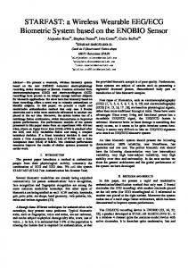
Fig. 3
498
,/ /
./ /
./
./
,.-
./
,/
,/
,/
/
/
/
.~,
,..,-
..-'
/,
/ /.
./ j
./ l
./
/
,/ /,
t
;~ /
,.,
/
4".
/"
5./
,/
,/
@
+,
/ 3/
./
./
/
/
#
/
,/
./
/ /
@§
/DE
./
,/
,/
~§
/
./-
/
/
@+
/
,.-~
/
/
--@
/
,/
,/
.#
,/
/
4)+
-@§
14H20I I I ! I I I
J /
@
@§ --
9 RRYTHMI E ~ @EF4,EL4,PR@
--
,/ 1 /
@ -@§
--
@
~@-I-
,/
/
/
/ /
/
/"
./
./
./
/
./
./
,./
,/
/
/
/
./
/
/
/
/
/
Narrow premature beats Note the sudden variation of the calculated maximum heart-rate when premature beats occur
Medical &
Biological Engineering &
Computing
J u l y 1981
defining the arrhythmia history in the preceding minute and reference values for the normal QRS. A 500 Hz sampling frequency was used for the acquisition of the e.c.g, signal. Recognition and classification of the QRS complexes takes place between sampling. The QRS amplitude must be within the range 100mV/2 V, automatic gain adjustment being made by using the eight most significant bits among the twelve given by the analog/digital converter. This gain adjustment is made during the learning phase (a few seconds at the beginning of monitoring). Wave recognition was made by comparison of the amplitude and slope of the signal with three calculated
thresholds (TAS ascending slope, TDS descending slope, TA amplitude). These thresholds were computed in the learning phase according to the chosen gain and they remain constant thereafter. A sequence of points is considered as defining a QRS complex if they satisfy the following criteria: - - The first five points must have an amplitude and a slope greater than calculated thresholds TA and TAS, respectively. -The last four points must have an amplitude and a slope greater than calculated thresholds TA and TDS, respectively.
IDEHTIFICATIOM
HNM: PRFHOM: RGE:
DATE:19..-"5/80 HFI_.IPF:16HO0
COMHEHTAIRF~:RRYTHMIE COMPLETE
AHAtY~F 0@@@@0@
RYTHME~ 120 80
HFIJRET
16HOOIO I I I I I I I I I 16H101 I I I I I I I I I 16H201
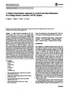
Fig. 4
40
§
@
--
.@
--
§
@
--
@
--
--
@
--
@
--
@
--
--
--
--
t + , +.
.@
--
-t.
+ .4-
,"
/
/65
"
."
.-"71 .,"
.,"
,"
/66.-"
*
i
.." G 4 1
....
..-"
"
-"
+
.,'51 -"
....
+
-'71 -" .,"70 .-"
.-" .-"
+
.
+ +. +,
@.
/67,," .,'67/
," .,"
.-" ."
/64/
.," /
/ .-"
/ 68 .,"
'4+
@
.,'71
+
,."
,,69.~ .,"69 .."
.
@ @
.*57 .-"
-"
@
@ @
*.RRYTHMI E ~ ~EF ~,EL+,PR4, / 4 6 .," / /
-'66/
r
r
200
.--"70 /
@
--
160
/71
+
.-"
.-"71 .,"
§
,"
/
.... .-"
/ .."
,,"
.-"
,--"
,"
Atrial fibrillation Note the wide but symmetric distribution of the calculated maximum and minimum heart rate around the mean heart rate
Medical & Biological Engineering & Computing
July 1981
499
All the points included between the sequences of five and four points must have an amplitude greater than the threshold TA. The QRS width is known from the number of points included in such a sequence. This is not the real width of the QRS, but a representation of the width used only for comparison of the QRS complexes with references. Classification of the complexes is based on two variables, RR interval and QRS width, in accordance with the table in Fig. 2. The references for R-R interval and QRS width are calculated the first time with the first two successive normal complexes (under control of the operator). If the QRS identified at time t is classified as 'normal', the references (calculated at 't - 1') are replaced by new values. This allows the system to follow variations of R.R. interval (e.g. with respiration). If a QRS is classified as a premature beat or as occurring after too long a delay (missing beat) the references are not re-evaluated. The classification of the first QRS complex occurring after a premature beat is somewhat different because of the refractory period, which is not to be interpreted as a missing beat. Minimum and maximum values of the R-R interval used for computing minimum and maximum heart rate are modified when justified. At the end of a minute, the stored values are printed and reinitialised. This system requires 2 k byte of p.r.o.m, and 128 bytes of r.a.m. So far, it has been tested on more than ten different pathological subjects, on periods varying from 20 to 90 min. Over a total monitoring period of eight hours, about 39000 QRS complexes were analysed, and about 200 premature beats recognised. In this test the percentage of false positive p.v.b.s was less than 1% and only four QRS complexes in the 39000 were not recognised. The total number of 'transducer default' was 66, which means that a correct analysis could not be made because the signal was too noisy or had artefacts (due to movements of the patient or breaking of contact). It is important to note that in most of such cases, intervention of the personnel was required as the monitor's alarm was ringing. In this case, if the automatic resetting of the system had not been triggered, it could be done manually by the nurse. Examples of results are presented in Figs. 3 and 4, the symbols EF, EL, PA and DE meaning respectively: narrow premature beat, wide premature beat, missing beat and transducer default.
Acknowledgment--The authors want to thank P. Kerbiriou and M. Beausoleil for the clinical experimentation. P. MORIZET-MAHOUDEAUX C. MOREAU D. MOREAU
Universitb de Compibgne Dbpartement de Gknie Biologique B.P. 233 60206 Compi~gne Cedex France
500
J. J. QUARANTE
Centre Hospitalier de Compibgne Service de Cardiologie 42, rue de Paris 60208 Compiltgne Cedex France
References
BUSSMANN, W. D., VOSWINCKEL, W., AMELING, W. and EFFERTS, S. (1975) Online analysis of ECG arrhytmias with a digital computer. Med. & Biol. Eng., 13, 382-387. COUMEL, P., ATTUEL, P., BERTHON, A. and BARBIER, D. Evaluation quantitative des troubles du rythme cardiaque par ordinateur. Communication B.V.I.7 Biosigma 1978. COX, J. R., NOLLE,F. M., FOZZARD,H. A. and OLIVIER,G. C. (1968) AZTEC, a preprocessing program for real time ECG analysis. IEEE Trans., BME-15, 128-129. KNOEBEL,S. B., LOVELACE,D. E., RASMUSSEN,S. and WASH, S. E. (1976) Computer detection of premature ventricular complexes: a modified approach. Am. J. Cardiology, 38, 440-447. KORTMAN, C. M. (1967) Redundancy reduction. A practical method of data compression. Proc. IEEE, 55, 253-263. LEBLANC, A. R., ROBERGE, F. A. and NADEAU,R. A. (1975) Evaluation of a new ECG measurement program for the detection of rhythm disturbance. Med. & Biol. Eng., 13, 370-381. MACFARLANE,P. W. (1971) ECG wave form identification by digital computer. Cardiovascular Research, 5, 141-146. MOREAU, C., LE BEUX,P., DUCH~NE,J. and DERENNE,J, P. {1977) Utilisation des microprocesseurs e n Instrumentation biom6dicale: Exemple d'application une 6preuve d'exploration fonctionnelle respiratoire. In Medical Computing, 143-150. MURTHY, I. S. N. and RANGARAJ,M. R. (1979) New concepts for PVC detection. IEEE Trans., BME-26, 409-416. NYGARDS, M. E. and HULTING, J. (1979) An automated system for ECG monitoring. Computers & Biomedical Research, 12, 181-202. VICTOR, J., GESL1N, P. and TADEI, A. Syst6me modulaire d'analyse des arythmies cardiaques. Communication A.III.5 Biosigma 78.
Medical & Biological Engineering & Computing
July 1981