Yvette M. van der Linden a,*, Herman ...... der Linden; Hans van Houwelingen, Department of. Medical ... Enschede; Ineke van Mierlo, Daniel Den Hoed Cancer.
Radiotherapy and Oncology 69 (2003) 21–31 www.elsevier.com/locate/radonline
Simple radiographic parameter predicts fracturing in metastatic femoral bone lesions: results from a randomised trial Yvette M. van der Lindena,*, Herman M. Kroonb, Sander P.D.S. Dijkstrac, Judith J. Lokd, Ed M. Noordijka, Jan Willem H. Leere, Corrie A.M. Marijnena, for the Dutch Bone Metastasis Study Group a
Department of Clinical Oncology K1-P, Leiden University Medical Center, Albinusdreef 2 2300 RC Leiden, The Netherlands b Department of Radiology, Leiden University Medical Center, Leiden, The Netherlands c Department of Orthopaedic Surgery, Leiden University Medical Center, Leiden, The Netherlands d Department of Medical Statistics, Leiden University Medical Center, Leiden, The Netherlands e Department of Radiotherapy, St. Radboud Medical Center, Nijmegen, The Netherlands Received 14 January 2003; received in revised form 26 May 2003; accepted 11 June 2003; available online 9 October 2003
Abstract Background and purpose: In the randomised Dutch Bone Metastasis Study on the palliative effect of a single fraction (SF) of 8 Gy versus six fractions of 4 Gy on painful bone metastases, 14 fractures occurred in 102 patients with femoral metastases. Purpose of the present study was to identify lesional risk factors for fracturing and to evaluate the influence of the treatment schedule. Material and methods: Pretreatment radiographs of femoral metastases were collected. Three observers separately measured the lesions and scored radiographic characteristics. Results: Ten fractures occurred after median 7 weeks in 44 SF patients (23%) and four after median 20 weeks in 58 multiple fraction patients (7%) (UV, P ¼ 0:02). In 110 femoral metastases, an axial cortical involvement .30 mm significantly predicted fracturing (MV, P ¼ 0:02). Twelve out of 14 fractured lesions and 40 out of 96 non-fractured metastases had an axial cortical involvement .30 mm (negative predictive value, 97%). When correcting for the axial cortical involvement, the treatment schedule was not predictive anymore (MV, P ¼ 0:07). Conclusions: Fracturing of the femur mostly depended on the amount of axial cortical involvement of the metastasis. We recommend to treat femoral metastases with an axial cortical involvement #30 mm with an SF of 8 Gy for relief of pain. If the axial cortical involvement is . 30 mm, prophylactic surgery should be performed to minimize the risk of pathological fracturing or, if the patient’s condition is limited, irradiation to a higher total dose. q 2003 Published by Elsevier Ireland Ltd. Keywords: Palliation; Bone metastases; Femur; Fracture; Radiation therapy; Prophylactic surgery
1. Introduction Metastases are the most frequent cause of malignant destructive lesions in the skeleton. The majority of bone metastases develop in patients with breast, prostate or lung cancer [35,37]. Most lesions are located in the spine, pelvis and proximal part of the extremities. Besides local or generalized pain, patients can suffer from pathological fracturing, the loss of limb function, compression of nerves or spinal cord from bulging bone or tumour fragments, or * Corresponding author. 0167-8140/$ - see front matter q 2003 Published by Elsevier Ireland Ltd. doi:10.1016/S0167-8140(03)00232-9
systemic conditions such as hypercalciemia. Palliative treatment options are external beam radiotherapy, chemotherapy, hormonal therapy, surgery, systemic administration of radioisotopes, regular infusions with bisphosphonates and the use of analgesics. The choice for a specific type of treatment depends on the symptoms experienced by the patient and on the localization and number of metastases. In addition, the clinician has to consider the performance status and life expectancy of each individual patient. Treatment should be aimed at providing the patient with maximum palliation and minimum discomfort. For a patient with a metastasis in the femur,
22
Y.M. van der Linden et al. / Radiotherapy and Oncology 69 (2003) 21–31
the occurrence of a pathological fracture can be most distressing and causes considerable morbidity. Therefore, prevention of a femoral fracture is an important palliative topic. In the multicenter prospectively randomised Dutch Bone Metastasis Study (DBMS), the palliative effect of a single fraction (SF) of 8 Gy versus six fractions of 4 Gy on painful bone metastases was investigated in 1157 patients. No major differences were found between the treatment schedules in response to pain, treatment side effects, analgesic consumption or overall quality of life [33]. In a subgroup of 102 patients with femoral metastases, 14 fractures occurred during follow-up. The purpose of the present study was to identify lesional risk factors for femoral fracturing and to evaluate the influence of the treatment schedule on the occurrence of a femoral fracture. Therefore, all femoral patients and femoral lesions within the DBMS were reviewed. The objective of the study was to define a practical guideline that can be used in a clinical setting, to decide which femoral lesions should be treated with radiotherapy and which with prophylactic surgery.
2. Material and methods 2.1. Patient selection and follow-up Between March 1996 and September 1998, 1157 Dutch patients with painful bone metastases from solid tumours were randomised between an SF of 8 Gy (n ¼ 579) and six fractions (multiple fractions, MF) of 4 Gy (n ¼ 578) [33]. Purpose of the study was to prove the equal effectiveness of SF versus MF; endpoint of the study was response to pain. Seventeen out of 21 radiotherapy institutes in the Netherlands participated in the trial. Patients had a minimal pain score of 2 on an 11-point scale of 0 (no pain) to 10 (worst imaginable pain) [19]. Already fractured lesions and lesions that were suspected to have a high risk of fracturing by the treating physician were excluded from the trial. The Medical Ethics Committees of all participating institutions approved the study and all patients signed informed consent forms. Registration of patients who did not meet the entry criteria (n ¼ 2927) was performed to identify the reasons for exclusion. After randomisation, intensive follow-up with 13 weekly and afterwards monthly questionnaires on pain using the 11-point scale, treatment side effects, quality of life, and analgesic consumption was carried out to a maximum of 2 years or until death. Data managers in the participating hospitals collected data on all events, such as death, retreatment and occurrence of a fracture or spinal cord compression. In December 1998 the follow-up on survival and fractures of all randomised patients was updated and the study was closed. For the present study all patients with a femoral metastasis within the DBMS were selected.
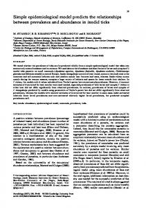
2.2. Scoring of radiographic aspects and measurements of the lesions The pretreatment radiographic imaging material and the radiotherapy simulation films of patients with a femoral metastasis were collected. Radiographs taken no more than 2 weeks prior to randomisation were considered suitable for evaluation. Without knowledge of the treatment schedule or outcome after treatment (i.e. a fracture yes or no), all femoral lesions were separately analysed by three experienced observers (a radiologist, an orthopaedic surgeon and a radiation oncologist). The following radiographic aspects were scored: appearance (predominantly osteolytic or predominantly osteoblastic), feature (solitary lesion, multiple lesions, diffuse lesions in the femur), pattern of bone destruction (geographic, moth-eaten, permeative pattern [21,25]), axial localization (proximal femur, shaft, distal femur) and transverse localization (mostly medial, mostly central, mostly lateral, or a combination of medial, central and/or lateral). In case of discrepant scoring between the observers they re-evaluated the radiographs and reached a consensus. The observers measured on the available conventional radiographs the sizes of the lesions in mm (Fig. 1): largest axial length of the entire lesion (L-lesion), largest transverse extension of the lesion (W-lesion) and largest axial cortical involvement of the lesion (L-cort). They also measured the largest transverse width of the bone (W-tot), the maximal thickness of the cortex without lesional involvement (C-tot) and the maximal thickness of the cortex with lesional involvement (C-lesion). The measurements were summarized and a mean score for the three observers was calculated. In the analysis cut-off points of measurements were chosen to detect differences in fracture risk. In order to compare the lesional measurements correctly, only conventional radiographs were measured. If a lesion was only visible on a CT scan, MRI scan and/or bone scintigram, no measurements were taken, but only the radiographic aspects were scored. In case of lesions with a permeative or moth-eaten pattern of bone destruction, the observers carefully measured the entire lesion. In case of more than one lesion within the radiation field, only the lesion considered at risk of fracturing was measured and scored. In patients with more than one lesion considered at risk of fracturing, both lesions were separately analysed only if their localizations were at a minimal distance of 50 mm. 2.3. Statistical analysis The database was analysed using SPSS 10.0 for Windows (SPSS Inc., Chicago, IL, USA). Spearman’s rank correlation tests were used to analyse the interobserver variability of separate measurements. Fisher’s Exact tests were used to compare proportions for baseline characteristics and baseline scored radiographic aspects. Mann – Whitney tests were used
Y.M. van der Linden et al. / Radiotherapy and Oncology 69 (2003) 21–31
23
Fig. 1. Measurements of metastatic lesions in the femur (in mm): largest axial length of the entire lesion (L-lesion), largest transverse extension of the lesion (W-lesion), largest axial cortical involvement (L-cort). Measurements of the femur (in mm): largest transverse width of the bone (W-tot), maximal thickness of cortex without lesional involvement (C-tot) and maximal thickness of cortex with lesional involvement (C-lesion).
to compare quantitative and ordered variables for baseline characteristics and baseline lesional measurements. Student’s t-tests were used to analyse differences in normally distributed data for baseline characteristics. Predictive values of risk factors were calculated. Kaplan – Meier curves and log rank tests were used for survival analyses (Fig. 2). Log rank tests were used to determine differences in time to fracture with end of follow-up or death considered as censored. Because the endpoint of this study was prevention of fractures, patients who died without a fracture were regarded as being successfully palliated. Therefore, to get a good estimate of the probability of fracture before death for each new patient presenting with a femoral lesion, patients who died without a fracture were not censored but were considered to have an uneventful follow-up until December 1998 (Figs. 3 and 4). A Cox proportional hazards model was used for uni- and multivariate analyses. All reported P-values are based on two-sided tests with P , 0:05 taken to be significant.
3. Results 3.1. Registered and randomised patients Of 2927 registered patients who did not meet the entry criteria, 8% (n ¼ 234) were excluded because of impending or actual fractures. More than 50% of these patients (n ¼ 121) had femoral metastases. The patient characteristics of the registered patients have been reported elsewhere [33]. In short, no major differences were noted between registered and randomised patients for sex, age, Karnofsky
Performance Scale, initial pain score, primary tumour or treatment site. Of the 1157 randomised patients, 110 patients were entered into the trial with a femoral metastasis. When the radiographs were evaluated, eight patients turned out to have an acetabular metastasis and were therefore not included in this analysis. Of the remaining 102 patients, 44 patients (43%) received an SF and 58 patients (57%) received MF. 3.2. Availability of radiographs and number of femoral lesions From the 102 patients with a femoral lesion all available radiographs were collected in hospitals throughout the Netherlands. The radiographs of two MF patients were missing and could therefore not be evaluated. From the remaining 100 patients the following imaging films were available: 91 radiotherapy simulation films (AP direction), 100 conventional radiographs (51 AP radiographs, 49 multidirectional radiographs), six CT, or MRI scans and nine bone scintigrams. The radiotherapy simulation films were checked for geographical misses: all lesions were adequately treated within the radiation fields. In 10 patients the lesion was only visible on a CT scan, MRI scan or bone scintigram. Five SF patients and three MF patients had two lesions considered at risk of fracturing, one MF patient had three lesions. In these nine patients, the lesions were located in different parts of the femur with a minimal distance of 50 mm. All lesions were included in the analysis. In total, in 100 patients with available radiographs, 110 lesions were scored on radiographic aspects and 100 lesions were measured.
24
Y.M. van der Linden et al. / Radiotherapy and Oncology 69 (2003) 21–31
Fig. 2. Survival in 102 patients with femoral metastases treated within the Dutch Bone Metastasis Study for the treatment schedule single fraction of 8 Gy (SF) versus six fractions of 4 Gy (MF).
3.3. Patient characteristics, survival and occurrence of fractures: SF versus MF Between the 44 SF patients and 58 MF patients no major differences in age, sex, pain score at time of randomisation, Karnofsky Performance Scale, or primary tumour were found (Table 1). Median follow-up was 21 months in both groups (range 4– 36 months). Median time from randomisation to the start of radiotherapy was 3 days in SF patients (range 0 –22 days) versus 6 days in MF patients (range 0 – 26 days). In total, nine SF and five MF patients received a second irradiation because of continuing or recurring pain (20 versus 9%, respectively, P ¼ 0:14). Fig. 2 shows the overall survival of the treatment groups. SF and MF patients had a median survival of 33 weeks (95% CI 15– 51 weeks) and 41 weeks (95% CI 30 – 51 weeks), respectively (P ¼ 0:31). During follow-up, 10 fractures (23%) occurred in 44 SF patients versus four fractures (7%) in 58 MF patients (P ¼ 0:02). Fig. 3 shows the probability of a fracture before death in both treatment groups, demonstrating that SF patients had a higher chance to experience a pathological fracture than MF patients. Median time to fracturing after randomisation was 7 weeks in 10 SF patients (range 2 – 29
weeks) and 20 weeks in four MF patients (range 3– 36 weeks). Because SF patients were treated sooner after randomisation than MF patients, the median time to fracturing from the start of radiotherapy was studied additionally: 10 SF patients experienced a fracture at a median of 6 weeks from the day the radiation was delivered (range 2 –29 weeks) compared to median 17 weeks for four MF patients (range 2– 35 weeks). These results suggest that MF postponed the occurrence of a pathological fracture. 3.4. Lesional characteristics and measurements: SF versus MF To evaluate the variability between the observers, Spearman’s rank correlation coefficients for the separate measurements were calculated, ranging from 0.64 to 0.70 for L-lesion, 0.52 – 0.57 for L-cort, 0.65 – 0.74 for W-lesion, 0.71– 0.75 for W-tot, 0.46 –0.65 for C-tot and 0.58 –0.80 for C-lesion. Variability of measuring was acceptable and justified combining the measurements into one outcome. Table 2 lists the lesional characteristics and measurements for SF versus MF lesions. Between the randomisation groups no significant differences in radiographic feature, appearance, pattern of destruction or axial localization of
Y.M. van der Linden et al. / Radiotherapy and Oncology 69 (2003) 21–31
25
Fig. 3. Probability of fracturing in 110 femoral metastases for the treatment schedule single fraction of 8 Gy (SF) versus six fractions of 4 Gy (MF) in patients treated within the Dutch Bone Metastasis Study.
the lesions were noted. The SF lesions were more often a combination of a medial, central and/or lateral located lesion than MF lesions (54 versus 29%, respectively, P ¼ 0:005), suggesting that SF lesions were larger in the transverse plane than MF lesions. The transverse width of both bone and lesion were indeed larger in SF lesions compared to MF lesions (median W-tot 45 versus 33 mm, respectively (P ¼ 0:01), median W-lesion 31 versus 21 mm, respectively (P ¼ 0:003)). SF lesions were located more often proximally in the femur than MF lesions (71 and 62%, respectively) and this perhaps caused the significantly larger W-tot and W-lesion. The axial measurements L-lesion and L-cort and the cortical measurements C-tot and C-lesion were not significantly different between lesions in the two treatment groups. 3.5. Patient characteristics and survival: fractures versus no fractures Between the 14 patients that experienced a fracture and the 88 patients that remained free from fracturing no major differences existed for age, sex, pain score at time of randomisation, Karnofsky Performance Scale or primary tumour. Six fractures were reported in a total of 45 patients
with breast cancer (13%), three in 17 patients with prostate cancer (18%), two in 26 patients with lung cancer (8%) and three in 14 patients with other primary cancers (21%) (P ¼ 0:93). Median follow-up was 21 months for patients with or without a pathological fracture. Median overall survival for patients with a fracture was 44 weeks (95% CI 16 – 71 weeks) and 38 weeks for patients without a fracture (95% CI 25– 51 weeks) (P ¼ 0:93). Patients who remained free from fracturing during follow-up were not retreated more often than patients who developed a fracture (15 versus 21%, respectively, P ¼ 0:44). Therefore, a higher total dose of irradiation due to more retreatment was not a valid explanation for non-fracturing of lesions. 3.6. Lesional characteristics and measurements: fractures versus no fractures In Table 3 the radiographic aspects and lesional measurements are listed for fractured versus non-fractured lesions. Radiographic feature, appearance, pattern of destruction, axial or transverse localization of the lesions were not different between fractured and non-fractured lesions. Most fractured and non-fractured lesions were solitary, osteolytic lesions, located proximally in the femur.
26
Y.M. van der Linden et al. / Radiotherapy and Oncology 69 (2003) 21–31
Fig. 4. Probability of fracturing in 110 femoral metastases for an axial cortical involvement L-cort ,30 mm versus L-cort .30 mm in patients treated within the Dutch Bone Metastasis Study.
Although fractured lesions more often had a permeative radiographic pattern, this was not predictive for fracturing (P ¼ 0:13). Most fractured lesions were located medially (29%) or were a combination of medial, central and/or lateral (57%) (P ¼ 0:36). The largest axial length of the entire lesion, the transverse measurements W-tot and W-lesion and the cortical measurements C-tot and C-lesion were not significantly different between fractured and non-fractured lesions. The axial cortical involvement L-cort was the only parameter in the univariate analysis significantly predictive for fracturing (P ¼ 0:001). Median L-cort for fractured lesions was 42 versus 29 mm for non-fractured lesions. Cut-off points for the axial cortical involvement were chosen to determine a more objective predictor for fracturing. A cut-off point of 30 mm significantly predicted fracturing (P ¼ 0:01, HR 7 (95% CI 1.6– 31.4)). Twelve fractured lesions had L-cort . 30 mm. Fig. 4 shows the probability of fracture for each patient presenting with a lesion with L-cort # 30 mm versus L-cort . 30 mm. In total, 52 lesions had L-cort . 30 mm of which 12 fractured: the positive predictive value of L-cort . 30 mm was limited (23%). No fractures occurred in the 10 lesions that were only visible on a CT scan, MRI scan or bone scintigram. If we consider those 10 lesions to have an L-cort # 30 mm, a total of 58 lesions had L-cort # 30 mm. Only two lesions
Table 1 Characteristics at time of randomisation of 102 patients with a metastatic femoral bone lesion per treatment schedule (single fraction versus multiple fractions) 1 £ 8 Gy (N ¼ 44)
SD
6 £ 4 Gy (N ¼ 58)
SD
Gender Male Female
48% 52%
Age Mean
63.5
10
Pain scorea Mean
6.3
2
6.3
2
Karnofskyb Mean
68%
17
70%
16
Primary tumour Breast Lung Prostate Other
43% 25% 16% 16%
P-value
0.84 45% 55% 0.92 63
14 0.88 0.53 0.97
45% 26% 17% 12%
SD, standard deviation. Pain score at randomisation, minimal score of 2 on an 11-point score from 0 (no pain) to 10 (worst imaginable pain). b Karnofsky Performance Scale is conditional score, ranging from 100% (normal situation, no complaints) to 0% (death). a
Y.M. van der Linden et al. / Radiotherapy and Oncology 69 (2003) 21–31
27
Table 2 Radiographic aspects and lesional measurements of the femoral lesions for the treatment schedules single fraction versus multiple fractions 1 £ 8 Gy (N ¼ 49 lesions)
6 £ 4 Gy (N ¼ 61 lesions)
Radiographic feature Solitary Multiple Diffuse
54% 27% 19%
42% 37% 21%
Radiographic appearance Osteoblastic Osteolytic
14% 86%
7% 93%
Radiographic pattern Geographic Moth-eaten Permeative
23% 70% 6%
37% 52% 11%
Axial localization Proximal Shaft Distal
71% 23% 6%
62% 36% 2%
Transverse localization Medial Central Lateral Combination
23% 19% 4% 54%
18% 26% 26% 29%
Length (median and range in mm)a L-lesion L-cort
53 (17– 251) 35 (7– 155)
46 (14–232) 31 (0–94)
0.12 0.37
Width (median and range in mm)a W-tot W-lesion
45 (25– 81) 31 (7– 59)
33 (22–68) 21 (7–54)
0.01 0.003
Cortex (median and range in mm)a C-tot C-lesion
6 (1–18) 2 (0–9)
6 (1–10) 2 (0–9)
0.44 0.82
P-value 0.44
0.21
0.24
0.19
0.005
Radiographic aspects were scored in 110 lesions in 100 patients of whom radiographs could be collected (conventional radiographs, CT scans, MRI scans and/or bone scintigrams). a Lesional measurements on only conventional radiographs of 100 lesions (in mm) (see Fig. 1): largest axial length of the entire lesion (L-lesion), largest transverse extension of the lesion (W-lesion) and largest axial cortical involvement (L-cort). Measurements of the femur (in mm): largest transverse width of the bone (W-tot), maximal thickness of cortex without lesional involvement (C-tot) and maximal thickness of cortex with lesional involvement (C-lesion).
with L-cort # 30 mm eventually fractured (one SF and one MF lesion). The negative predictive value of L-cort # 30 mm was high (97%). Because 51% AP and only 49% multidirectional radiographs were available for review, we evaluated whether underestimation of L-cort occurred if measured only on AP radiographs. Of the 52 lesions with L-cort . 30 mm, 46% were measured on AP radiographs only. Of the 48 lesions with L-cort # 30 mm, 50% were measured on AP radiographs. The median L-cort was not significantly smaller (32 versus 33 mm for AP versus multidirectional, respectively, P ¼ 0:15), demonstrating that AP films could be used reliably to measure L-cort. Between SF and MF patients the percentage of lesions with L-cort . 30 mm was not different (53 versus 43%, respectively, P ¼ 0:34). Subsequently, we performed a multivariate analysis with the variables that predicted fracturing in the univariate analysis: randomisation
treatment schedule and L-cort . 30 mm. When correcting for L-cort . 30 mm, we could not demonstrate the treatment schedule to be a significant predictor for fracturing (P ¼ 0:07, HR 2.9 (95% CI 0.9 –9.4)). An axial cortical involvement . 30 mm remained predictive for fracturing in the multivariate analysis (P ¼ 0:02, HR 6 (95% CI 1.3 – 27)).
4. Discussion In this study on fracture risks in femoral metastatic lesions, we demonstrated the axial cortical involvement of the lesion to be significantly predictive for fracturing (L-cort . 30 mm, MV-analysis, P ¼ 0:02). Although more fractures occurred after a single dose of 8 Gy, we could not prove the treatment schedule to be predictive in the multivariate analysis when correcting for L-cort (P ¼ 0:07).
28
Y.M. van der Linden et al. / Radiotherapy and Oncology 69 (2003) 21–31
Table 3 Radiographic aspects and lesional measurements of femoral lesions for the outcome fracture yes or no Fracture no (N ¼ 96 lesions)
Fracture yes (N ¼ 14 lesions)
P-value
UVa HR (95% CI)
Radiographic feature Solitary Multiple Diffuse
43% 34% 23%
83% 17% 0%
0.18
1 0.2 (0.1– 1.1) 0.0 (0.0– 7.3)
Radiographic appearance Osteoblastic Osteolytic
12% 88%
0% 100%
0.44
1 29 (0.0– . 100)
Radiographic pattern Geographic Moth-eaten Permeative
34% 60% 6%
22% 57% 21%
0.13
1 1.6 (0.4– 6.1) 4.9 (0.9– 24.5)
Axial localization Proximal Shaft Distal
64% 33% 3%
86% 7% 7%
0.18
1 0.1 (0.0– 1.5) 2.4 (0.3– 18.9)
Transverse localization Medial Central Lateral Combination
19% 25% 17% 37%
29% 7% 7% 57%
0.36
1 0.2 (0.02– 1.8) 0.3 (0.04– 2.8) 1.0 (0.3– 3.3)
Length (median and range in mm)b L-lesion L-cort
48 (14–251) 29 (0–120)
58 (31–229) 42 (27–155)
0.13 0.001
Width (median and range in mm)b W-tot W-lesion
39 (22–81) 23 (7–59)
40 (26–74) 31 (15–52)
0.53 0.22
Cortex (median and range in mm)b C-tot C-lesion
6 (1– 11) 2 (0– 9)
6 (2–18) 1 (0–6)
0.11 0.29
Radiographic aspects were scored in 110 lesions in 100 patients of whom radiographs could be collected (conventional radiographs, CT scans, MRI scans and/or bone scintigrams). a Lesional measurements on only conventional radiographs of 100 lesions (in mm) (see Fig. 1): largest axial length of the entire lesion (L-lesion), largest transverse extension of the lesion (W-lesion) and largest axial cortical involvement (L-cort). Measurements of the femur (in mm): largest transverse width of the bone (W-tot), maximal thickness of cortex without lesional involvement (C-tot) and maximal thickness of cortex with lesional involvement (C-lesion). b UV, univariate analysis, HR, hazard ratio calculated with Cox proportional hazards model, 95% CI, 95% confidence intervals.
Treatments with a palliative intent comprise a large proportion of the total care that is delivered to cancer patients every day [18]. It is therefore necessary that all involved professionals are informed about issues relating to palliative care. However, Barnes et al. showed recently that only 1.3% of all abstracts presented at the annual meetings of the American Society for Therapeutic Radiology and Oncology (ASTRO) during the period 1993 –2000 were devoted to symptom control and palliation [2]. One of the major problems in palliative care is the management of bone metastases. In the treatment of femoral metastases, one has to consider on the one hand prevention of pathological fractures with considerable morbidity, and on the other prevention of unnecessary surgical procedures. Elective surgery on patients with a relatively good performance status has been recommended because it is easier to perform than after pathological fracturing [32]. In addition, it has
been reported that pain is often reduced to a minimum directly after surgery and the ability to walk is usually regained within a few days [11]. Some physicians even advocate preventive osteosynthesis in all patients with proximally located femoral metastases, irrespective of the estimated risk of fracturing. However, we believe it is better to spare a patient with a limited life expectancy an operation with associated morbidity and mortality. Therefore, it is necessary to formulate good criteria for the correct application of surgical and non-surgical treatments for femoral metastases. In the literature, several authors have studied femoral lesions and formulated risk factors for fracturing [4,6 – 8, 10– 13,15,16,20,22 –24,28,31,32,38,39]. Most often mentioned are the size of a lesion . 25 mm, increasing pain, and a circumferential cortical involvement . 50%. However, the majority of these studies were retrospective and used
Y.M. van der Linden et al. / Radiotherapy and Oncology 69 (2003) 21–31
mostly surgical data. Patients in these studies already presented with a femoral fracture or underwent prophylactic fixations. Therefore, no information on the natural course of these lesions without fixation was available. Because most patients have limited life expectancies, it is possible that a large proportion of the lesions would have never progressed into a fracture. The present study has the major advantage that all reviewed femoral patients were randomised into a prospective radiotherapy trial with adequate follow-up on survival and development of fractures. Fourteen of the 102 randomised patients with a femoral lesion developed a pathological fracture during follow-up. We demonstrated the axial cortical involvement of the lesion to be significantly predictive for fracturing (L-cort . 30 mm, MV-analysis, P ¼ 0:02). These results are in line with Menck et al., who studied 69 pathological femoral fractures and concluded that 90% of the fractured lesions had an L-cort . 30 mm [22]. Dijkstra et al. noted a median L-cort of 54 mm in nine actual subtrochanteric fractures (range 38 – 100 mm) [10]. In our study, all other lesional measurements and radiographic aspects, such as the size of the lesion, the amount of transverse cortical involvement or a proximally located, solitary, osteolytic lesion were not predictive for fracturing. The strength of our study lies in the high negative predictive value of L-cort (97%), although the positive predictive power of L-cort was limited (23%). Of all 58 lesions with L-cort , 30 mm, only one SF lesion and one MF lesion progressed into a fracture (3%). The use of L-cort pointed out that, as long as L-cort is # 30 mm, even large lesions could be safely treated with radiotherapy. Although more fractures occurred after a single dose of 8 Gy, we could not prove the treatment schedule to be predictive for fracturing, when correcting for L-cort in the multivariate analysis (P ¼ 0:07). This outcome was probably due to the limited number of events. However, we showed that six fractions of 4 Gy seemed to postpone the occurrence of a fracture. There are some remarks to be made. First, the radiation doses used in the DBMS were chosen for treatment of pain due to metastatic involvement of the bone. Prevention of fracturing by inducing remineralisation of the affected bone was not expected with these limited dose schedules. Therefore, the trial excluded all suspected high risk and already fractured lesions. The study protocol did not provide criteria on high risk lesions because it was believed that the existing risk factors in the literature were perhaps not very accurate in fracture prediction. Instead, only lesions which the treating physician expected to be high risk of fracturing were excluded. Although we did not review the radiographs of the 121 excluded patients, it is most likely that these expected high risk and already fractured lesions were larger than or at least as large as the expected low risk lesions that were randomised into the DBMS. Therefore, we believe that the use of L-cort as a guideline for treatment is applicable to all femoral lesions. Secondly, the scoring and measuring of
29
three observers were combined into one outcome to simulate a multidisciplinary setting in which clinicians join their individual experience. The interobserver variability of the measurements was reasonable and justified this approach. As a result, the length of the axial cortical involvement seemed to be a simple and objective parameter to predict femoral fracturing. Thirdly, only 49% multidirectional radiographs were available for review, as has been reported in earlier studies [32]. One could question the reliability of studying AP radiographs only. We therefore analysed if L-cort was smaller when measured on AP radiographs only. The difference in median L-cort for AP radiographs versus multidirectional radiographs was only 1 mm (P ¼ 0:15). Nonetheless, we advocate the use of multidirectional radiographs to study femoral lesions because AP radiographs provide less detailed information on lesional sizes, extensions and characteristics. In addition, routine use of a CT scan to study the three-dimensional anatomy of bone would be most optimal, but unfortunately, also least practical. Many randomised prospective trials have reported the equal palliative effect of a single dose or short-term radiotherapy compared to more protracted regimens for the treatment of pain due to bone metastases [1,9,14,17,26,29, 33], although some authors disagreed [5,30]. The overall percentage of fracturing in the DBMS, 3% in 1157 eligible patients, was similar to what other radiotherapy trials report (1 – 8%) [1,26,27,29,34,36]. In the femoral subgroup, 14 fractures occurred. Two other trials also reported the percentage of fracturing in subgroups: Uppelschoten et al. observed 8% fractures in pelvic and femoral lesions after a single dose of 6 Gy in a non-randomised prospective study of 170 patients, but they made no distinction between the two sites [36]. Tong et al. studied 1016 patients in a randomised prospective trial with five different treatment schedules [34]. They reported 13% fractures in 96 patients with a metastasis in the long bones, but made no distinction between humeral or femoral sites. In case of a solitary lesion (n ¼ 164) they noticed 18% fracturing after 40.5 Gy and only 4% after a lower dose of 20 Gy (P ¼ 0:02). No explanation was given for this remarkable difference, but it is likely that other factors play a role, such as lesion size. Only a few studies focused on the role of radiotherapy in fracture prevention and mentioned percentages of reossification after radiotherapy [8,20,27]. Cheng et al. [8] retrospectively studied 97 bone metastases in 59 breast cancer patients. For impending fracturing they took criteria that Beals et al. and Harrington suggested for predicting fracturing [4,15]: a lesion . 25 mm in diameter and/or any lesion with a circumferential destruction of the cortex . 50%. Thirty-nine expected impending femoral lesions did not break during follow-up after a radiation dose of 30– 40 Gy. In 11 patients evidence of reossification was visible on serial radiographs. Keene et al. [20] discussed the follow-up radiographs of 35 patients with femoral metastases after
30
Y.M. van der Linden et al. / Radiotherapy and Oncology 69 (2003) 21–31
average doses of 30 Gy (range 15– 60 Gy). They noted progression in 26% of the lesions, no change in 57% and partial healing in 17%. We agree with Bates, who suggested in her review on bone metastases that adequate doses for fracture prevention still have to be found by carrying out prospective studies [3]. In conclusion, we recommend the use of the simple radiographic parameter axial cortical involvement in deciding which treatment to apply on patients with metastases in the femur. In case of a painful femoral lesion with an axial cortical involvement # 30 mm, an SF of 8 Gy can be safely applied. If the axial cortical involvement . 30 mm and the patient’s condition is limited, radiotherapy using MF should be given to postpone the occurrence of a fracture. If the axial cortical involvement . 30 mm, prophylactic surgery should always be considered, accepting a relatively high percentage of surgical interventions to prevent disabling pathological fracturing of the femur.
Appendix A. The Dutch Bone Metastasis Study Group consists of the steering committee (Jan Willem H. Leer; Yvette M. van der Linden; Hans van Houwelingen, Department of Medical Statistics, Leiden University Medical Centre, Leiden; Job Kievit and Wilbert B. van den Hout, Department of Medical Decision Making, Leiden University Medical Centre, Leiden; Hanneke de Haes, Department of Medical Psychology, University of Amsterdam, Amsterdam and Elsbeth Steenland, Department of Clinical Oncology, Leiden University Medical Centre, Leiden) and the coordinators from the participating institutes (Hendrik Martijn, Department of Radiotherapy, Catharina Hospital, Eindhoven; Bing Oei, Dr. Bernard Verbeeten Institute, Tilburg; Ernest Vonk, Department of Radiotherapy, Deventer Hospital, Deventer; Elzbieta M.van der SteenBanasik, Arnhem Radiation Institute, Arnhem; Ruud G.J. Wiggenraad, Department of Radiotherapy, Haaglanden Medical Centre, The Hague; Jaap Hoogenhout, Department of Radiotherapy, St. Radboud Medical Centre, Nijmegen; Carla C. Wa´rla´m-Rodenhuis, Department of Radiotherapy, University Medical Centre Utrecht; Geertjan van Tienhoven, Department of Radiotherapy, University Medical Centre, Amsterdam; Rinus Wanders, Limburg Radiation Institute, Heerlen; Jacqueline Pomp, Department of Radiotherapy, Reinier de Graaf Hospital, Delft; Matthijs van Reijn, Department of Radiotherapy, Twente Hospital, Enschede; Ineke van Mierlo, Daniel Den Hoed Cancer Centre, Rotterdam; Ewald Rutten, Department of Radiotherapy, Medical Centre Alkmaar; Jan Metsaars, Department of Radiotherapy, Leyenburg Hospital, The Hague; Gerrit Botke, Friesland Radiation Institute, Leeuwarden and Ben G. Szabo´, Department of Radiotherapy, University Medical Centre, Groningen).
References [1] 8 Gy single fraction radiotherapy for the treatment of metastatic skeletal pain: randomised comparison with a multifraction schedule over 12 months of patient follow-up. Bone Pain Trial Working Party. Radiother Oncol 1999;52(2):111–21. [2] Barnes EA, Palmer JL, Bruera E. Prevalence of symptom control and palliative care abstracts presented at the Annual Meeting of the American Society for Therapeutic Radiology and Oncology. Int J Radiat Oncol Biol Phys 2002;54(1):211–4. [3] Bates T. A review of local radiotherapy in the treatment of bone metastases and cord compression. Int J Radiat Oncol Biol Phys 1992; 23(1):217–21. [4] Beals RK, Lawton GD, Snell WE. Prophylactic internal fixation of the femur in metastatic breast cancer. Cancer 1971;28(5):1350–4. [5] Blitzer PH. Reanalysis of the RTOG study of the palliation of symptomatic osseous metastasis. Cancer 1985;55(7):1468 –72. [6] Bunting R, Lamont-Havers W, Schweon D, Kliman A. Pathologic fracture risk in rehabilitation of patients with bony metastases. Clin Orthop 1985;192:222– 7. [7] Bunting RW, Shea B. Bone metastasis and rehabilitation. Cancer 2001;92(4 Suppl):1020–8. [8] Cheng DS, Seitz CB, Eyre HJ. Nonoperative management of femoral, humeral, and acetabular metastases in patients with breast carcinoma. Cancer 1980;45(7):1533– 7. [9] Cole DJ. A randomized trial of a single treatment versus conventional fractionation in the palliative radiotherapy of painful bone metastases. Clin Oncol (R Coll Radiol) 1989;1(2): 59–62. [10] Dijkstra PD, Oudkerk M, Wiggers T. Prediction of pathological subtrochanteric fractures due to metastatic lesions. Arch Orthop Trauma Surg 1997;116(4):221–4. [11] Dijkstra S, Wiggers T, van Geel BN, Boxma H. Impending and actual pathological fractures in patients with bone metastases of the long bones. A retrospective study of 233 surgically treated fractures. Eur J Surg 1994;160(10):535–42. [12] Fidler M. Prophylactic internal fixation of secondary neoplastic deposits in long bones. Br Med J 1973;1(5849):341– 3. [13] Fidler M. Incidence of fracture through metastases in long bones. Acta Orthop Scand 1981;52(6):623–7. [14] Gaze MN, Kelly CG, Kerr GR, et al. Pain relief and quality of life following radiotherapy for bone metastases: a randomised trial of two fractionation schedules. Radiother Oncol 1997;45(2): 109 –16. [15] Harrington KD. New trends in the management of lower extremity metastases. Clin Orthop 1982;169:53 –61. [16] Hipp JA, Springfield DS, Hayes WC. Predicting pathologic fracture risk in the management of metastatic bone defects. Clin Orthop 1995; 312:120–35. [17] Hoskin PJ, Price P, Easton D, et al. A prospective randomised trial of 4 Gy or 8 Gy single doses in the treatment of metastatic bone pain. Radiother Oncol 1992;23(2):74–8. [18] Janjan NA. Radiation for bone metastases: conventional techniques and the role of systemic radiopharmaceuticals. Cancer 1997;80(8 Suppl):1628–45. [19] Jensen MP, Karoly P, Braver S. The measurement of clinical pain intensity: a comparison of six methods. Pain 1986;27(1):117– 26. [20] Keene JS, Sellinger DS, McBeath AA, Engber WD. Metastatic breast cancer in the femur. A search for the lesion at risk of fracture. Clin Orthop 1986;203:282– 8. [21] Lodwick GS. The bones and joints, an atlas of tumor radiology. Chicago, IL: Year Book Medical Publishers Inc.; 1971. p. 38–44. [22] Menck H, Schulze S, Larsen E. Metastasis size in pathologic femoral fractures. Acta Orthop Scand 1988;59(2):151– 4. [23] Miller F, Whitehill R. Carcinoma of the breast metastatic to the skeleton. Clin Orthop 1984;184:121–7.
Y.M. van der Linden et al. / Radiotherapy and Oncology 69 (2003) 21–31 [24] Mirels H. Metastatic disease in long bones. A proposed scoring system for diagnosing impending pathologic fractures. Clin Orthop 1989;249:256 –64. [25] Mulder JD, Kroon HM, Schutte HE, Taconis WK. The diagnosis of bone tumours. Radiologic atlas of bone tumours. Amsterdam: Elsevier Publishers; 1993. p. 28 –31. [26] Nielsen OS, Bentzen SM, Sandberg E, Gadeberg CC, Timothy AR. Randomized trial of single dose versus fractionated palliative radiotherapy of bone metastases. Radiother Oncol 1998;47(3):233–40. [27] Oszaran Z, Yalman D, Anacak Y, Esassoloak M, Haydaroglu A. Palliative radiotherapy in bone metastases; results of a randomized trial comparing three fractionation schedules. J BUON 2001;6: 43–8. [28] Parrish FF, Murray JA. Surgical treatment for secondary neoplastic fractures. A retrospective study of ninety-six patients. J Bone Joint Surg Am 1970;52(4):665–86. [29] Price P, Hoskin PJ, Easton D, Austin D, Palmer SG, Yarnold JR. Prospective randomised trial of single and multifraction radiotherapy schedules in the treatment of painful bony metastases. Radiother Oncol 1986;6(4):247–55. [30] Ratanatharathorn V, Powers WE, Moss WT, Perez CA. Bone metastasis: review and critical analysis of random allocation trials of local field treatment. Int J Radiat Oncol Biol Phys 1999;44(1): 1– 18.
31
[31] Snell WE, Beals RK. Femoral metastases and fractures from breast cancer. Surg Gynecol Obstet 1964;119:22–4. [32] Springfield DS. Pathologic fractures. Fractures in adults (Rockwood and Green). Lippincott Williams Wilkins; 2001. p. 557 –83. [33] Steenland E, Leer JW, van Houwelingen H, et al. The effect of a single fraction compared to multiple fractions on painful bone metastases: a global analysis of the Dutch Bone Metastasis Study. Radiother Oncol 1999;52(2):101–9. [34] Tong D, Gillick L, Hendrickson FR. The palliation of symptomatic osseous metastases: final results of the study by the Radiation Therapy Oncology Group. Cancer 1982;50(5):893 –9. [35] Tubiana-Hulin M. Incidence, prevalence and distribution of bone metastases. Bone 1991;12(Suppl 1):S9–S10. [36] Uppelschoten JM, Wanders SL, de Jong JM. Single-dose radiotherapy (6 Gy): palliation in painful bone metastases. Radiother Oncol 1995; 36(3):198– 202. [37] Woodhouse EC, Chuaqui RF, Liotta LA. General mechanisms of metastasis. Cancer 1997;80(8 Suppl):1529–37. [38] Yazawa Y, Frassica FJ, Chao EY, Pritchard DJ, Sim FH, Shives TC. Metastatic bone disease. A study of the surgical treatment of 166 pathologic humeral and femoral fractures. Clin Orthop 1990;251:213–9. [39] Zickel RE, Mouradian WH. Intramedullary fixation of pathological fractures and lesions of the subtrochanteric region of the femur. J Bone Joint Surg Am 1976;58(8):1061– 6.