Proceedings of the IASTED International Conference NANOTECHNOLOGY AND APPLICATIONS (NANA 2008) September 29 - October 1, 2008 Crete, Greece
SINGLE PARTICLE COUNTER USING NANOPORES: MULTIPHYSICS SIMULATIONS AND EXPERIMENTS H. V. B. Achar 1, 3, M. Rossi 1, 2, S. Cavalcanti1, E. Giordano1, E. Sangiorgi1 and M. Tartagni1 1
ARCES, II Facoltà di Ingegneria, Università di Bologna, Sede di Cesena, Cesena, Italy 2 Silicon Biosystems S.p.A., Bologna, Italy 3 Department of Electrical Engineering, Indian Institute of Technology Madras, Chennai, India Email:
[email protected]
ABSTRACT Investigations on micro-fabricated devices for biomedical applications such as biosensors have swiftly advanced in the last few years. The suitability of these devices for such applications has been extensively documented by universities and private laboratories alike. Detection of single molecules is fundamental for enhancing our understanding of delicate biological mechanisms. This would open new promising areas in diagnostics and therapeutics. This paper discusses the simulations and experiments carried out on electric field-induced translocation of polystyrene beads through silicon nitride nanopores. The behavior of the spherical particles suspended in the electrolyte solution to the applied electric field is studied. The use of focusing electrode to focus the charged particles into the centre of the chamber is also discussed. The electric field, the ionic current though the pore, and the translocation time are computed using Femlab simulations and later verified experimentally.
electrophoresis. As the particle of interest passes through the nanopore, ionic current is partially blocked. This blockage or perturbation is measured using commercial systems like Axon patch clamp amplifier. The magnitude and duration of this perturbation is correlated with the particle’s size and charge. This is used to obtain information such as the particle’s length, diameter, charge, mobility, etc., which are useful for conducting basic scientific studies and for practical applications and diagnosis. Researchers have showed that nanopores are a powerful tool for molecular probing. There are some obvious advantages of nanopore measurements compared to fluorescence measurements. Non requirement of labeling of molecules is one advantage. Initially, α-haemolysin was used for nanopore experiments. The drawback of this approach was that the fluidity of the bilayer platform produced instabilities, and therefore led to extremely short functional lifetimes of the pore [1]. An alternative approach is to build apertures on a solid state platform. Different methods such as ionbeam sculpting, focused ion beam (FIB) drill, ion track etching, etc., are currently being used for the fabrication of nanopores. Wide ranges of diameters of pores have been reported in the literatures [2]-[7].
KEY WORDS Biosensors, nanopores, coulter counters, multiphysics simulations.
1. Introduction
In this paper we report on the Femlab simulations and experimental observations of current perturbations caused due to the translocation of carboxylated polystyrene beads through silicon nitride nanopores. The main purpose of this work is to investigate the control of the motion of charged particles in the microfluidic chamber by using the concept of focusing electrode. Using the finite element simulations, we found that electrophoretic motion of a spherical charged particle in an aqueous electrolyte solution is strongly affected by the applied electric potential and the charge of the particle. By modifying the electric field lines inside the chamber using focusing electrode, it was possible for us to obtain the desired direction of particle movement.
Biological particles such as proteins and DNA carry electrical charges and hence they can be made to move due to an applied electric field in an electrolyte solution. The origin of this idea is from the Coulter Counter device invented in the 50’s. The device mainly consists of an aperture or nanopore of size slightly larger than that of the particle of interest. This pore connects two micro chambers filled with electrolyte solution, separated by a thin membrane. Two electrodes are immersed in the electrolyte, one in each chamber, causing the ionic current to flow through the pore. The induced electric field causes the charged particles to move in the direction determined by the field. This phenomenon is known as
615-084
37
Fig. 1: Geometry of the device (not to scale)
Fig. 2: Electric field lines without focusing electrode
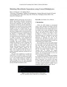
2. Methods, Materials and Experiments The cylindrical nanopore used in the simulation and experiments has a diameter and length of 200 nm each, and is situated in the middle of a silicon nitride (Si3N4) layer of thickness 200 nm. A silicon substrate of thickness 300 µm supports the nitride layer, as shown in figure 1. The entire chip is mounted into a home-made PDMS holder containing two reservoirs for conducting the experiments. 2.1 Simulation Simulations were performed using Comsol Multiphysics package in order to study the electrode arrangement and to analyze the movement of the charged particles inside the microfluidic chambers. The electric field distribution inside the microfluidic chamber was simulated using AC/DC module of Comsol. Figure 2 shows the streamline plot of electric field lines when a potential difference of 100 mV was applied between the electrodes (positive electrode on top). As can be seen from the figure, the electric field lines spread out on either side of the vertical axis in the lower chamber.
Fig. 3: Electric field lines with focusing electrode for simplicity only one nanopore and the associated chambers are shown. The potential at the focusing electrode was set at –20 mV with respect to the ground electrode. The electrophoretic velocity field lines of the charged particles in the microfluidic chambers were then simulated. Electrophoresis is the phenomenon due to which charged particles move in an electrolyte solution under the influence of applied electric field. Electrophoresis has been widely used in characterizing and separating colloidal particles, polyelectrolytes, and macromolecules such as DNA fragments and proteins, manipulating particles in micro / nanofluidic systems, sequencing the genomes of organisms, and forensic analysis, etc.
A negatively charged particle present in the lower chamber will experience Coulomb force acting away from the vertical axis and will tend to move in the sideward direction away from the pore (shown by the arrows). This will result in particles from one chamber drifting into the adjacent chambers; in the case where there are arrays of nanopores arranged in rows and columns, with one nanopore surrounded by four other nanopores, with common upper and lower microfluidic chambers for all. Investigation was done on the approaches of placing a negatively biased focusing electrode surrounding the ground electrode underneath each nanopore in order to focalize the charged particles into their respective nanopores. This arrangement of the electrodes and the resulting electric field lines are shown in figure 3, where
In viscous medium, in the presence of friction and electric field, the movement of charged particle takes place at a constant velocity v, given by: (1)
38
Fig. 5: Current waveform showing bead translocations through the nanopore Fig. 4: Velocity field lines of charged particles
layer which is thin compared to particle radius, the factor 2/3 in the equation can be replaced by unity.
where, µ is the electrophoretic mobility and is the electric field. The mobility of a given particle in the medium is a constant which is the characteristic of that particle. The mobility can be determined by the electric force that the charged particle experiences, balanced by its frictional drag through the medium.
Velocity field of charged particles inside the microfluidic chamber was simulated using Chemical Engineering and AC/DC modules of Comsol Multiphysics. The resultant plot is shown in figure 4. The direction of velocity field lines in the bottom chamber is such that the particles tend to move towards the center of the chamber. This was due to the presence of the focusing electrode surrounding the ground electrode which effectively prevented the dispersion of the particles and avoided crosstalk with neighboring chambers (not shown). With a zeta-potential of –4 mV, positive electrode at 100 mV and focusing electrode at −10 mV, a translocation velocity of 76.85 µm s–1 through the nanopore was obtained. The average pore blockage time or the translocation time through the nanopore was obtained as 2.60 ms.
The electric force is: (2) The frictional force is: (3) where, Q is the charge on the particle, η the viscosity of the solution, r is the particle radius and v is its velocity. From this we can work out the electrophoretic mobility, which is the balance between the electrical force moving the particle and the frictional force slowing it down.
If the bias voltage at the focusing electrode was increased in magnitude, then the focusing effect improved, but at the expense of the velocity of translocation. Some particles even tend to move in the downward direction, instead of passing through the pore. So a focusing electrode which is negatively biased at 10–20 percent of the potential at the upper electrode was found to be ideal.
Many particles, including biological particles, when suspended in an electrolyte, are surrounded by an electric double layer consisting of counter ionic charges. At the surface of the particle there exists stagnant layer in which the liquid is stationary. Outside the stagnant layer, fluid velocity increases with the distance from the surface. Potential at the outer limit of stagnant layer is called zeta potential. Using the zeta potential, we can have an alternate equation for electrophoretic mobility. Hence,
2.2 Experiments After having verified the concepts using simulations, translocation experiments were conducted in the laboratory. Silicon nitride membranes containing one nanopore each of dimensions mentioned earlier were sourced from Applied Nano Structures, Santa Clara, CA, USA and the Polystyrene carboxylated beads of diameter 100 nm were sourced from Polysciences Inc., Warrington, PA, USA.
(4) where, ε is the permittivity of the electrolyte and ζ is the zeta-potential of the charged particle. The mobility of the colloidal particle depends on the thickness of the double layer as compared to the size of the particle. For a double
The membrane was cleaned using deionized water and ethylene prior to mounting it on the custom-made PDMS holder having two chambers and having provisions to fill them with electrolyte solution and the beads. After filling
39
3. Conclusion
the two chambers with KCl solution of known conductivity and pH, two Ag/AgCl electrodes were inserted in the top and bottom chambers. Ionic currents for various applied voltages were measured using the standard Axon Patch clamp 200B amplifier and the Digidata 1322 A/D converter, and hence the resistance of the pore was determined. The resistance values were found to be consistent with the theoretical values with an error of less than 10%.
The report discussed the Femlab simulations of translocation experiments using silicon nitride nanopore and the experimental results confirming the simulated results. The placement of electrodes was considered in the first simulation. With only the ground electrode present in the bottom reservoir, some particles tend to move sideways and away from the pore. With the help of focusing electrode, this problem was overcome. The electrophoretic translocation of charged particles was considered in the next simulation. The velocity and direction of particle motion were studied with and without focusing electrode. The last part of the report discussed the procedures adopted in our laboratory for conducting the translocation experiments. Our next step would be to use the focusing electrode concept and verify the simulation results with a setup containing array of nanopores.
The resistance of the chip is predominantly the resistance of the silicon nitride pore filled with buffer solution, plus the access resistance which depends on the distance of the electrode from the pore and concentration of the buffer solution. The resistance of the pore is given by [8]: (5) where L is the length of nanopore (200 nm), D is the diameter of nanopore (200 nm) and σ is the conductivity of buffer solution (11 S m−1 for 1 M KCl).
References
After conducting a series of experiments with different concentrations of KCl to confirm the repeatability of measurements, carboxylated polystyrene beads were added into bottom chamber. Current waveforms were acquired using the same setup as before, setting a sampling frequency of 50 KHz and using an in-built 4pole Bessel low-pass filter at 10 kHz, at the room temperature of 24 ± 2ºC. The current waveforms, shown in figure 5, exhibited a series of current perturbations (drops) indicating the translocation of beads to the upper chamber through the nanopore. Based on the current transients, the diameter of the translocating particle was estimated by using DeBlois model [8]:
[1] [2]
[3]
[4]
(6) where, I is the baseline current and ΔI is the magnitude of the perturbation. The diameter of beads determined from the experimental results matched with the quoted diameter. The duration of the transients or the pulse width gave the velocity of translocation and hence gave the charge on the beads. At the pH of 7.5, the charge on the beads is negative, and will be electrophoretically driven to the positively biased electrode. This is confirmed, since when the electric field was reversed, translocations were not observed.
[5]
[6]
[7]
There are two effects to be considered. Firstly, a negatively charged particle inside the nanopore blocks the ionic current. Secondly, the positive ions which screen the negatively charged particle are also introduced into the pore. At high electrolyte concentrations the first effect dominates and the ionic current should decrease during translocation. At low ionic concentrations the second effect dominates and the ionic current should increase.
[8]
40
C. Dekker, “Solid State Nanopores”, Nature, Vol. 2, 209-215, 2007 A. J. Storm, J. H. Chen, H. W. Zandbergen, and C. Dekker, “Translocation of double-strand DNA through a silicon oxide nanopore” Physical Review, E 71, 2005 U. F. Keyser, B. N. Koeleman, S. V. Dorp, D Krapf, R. M. M. Smeets, S. G. Lemay, N. H. Dekker and C. Dekker, “Direct force measurements on DNA in a solid-state nanopore”, Nature Physics, Vol. 2, July 2006 D. Fologea, B. Ledden, D. S. McNabb, and J. Li, “Electrical characterization of protein molecules by a solid-state nanopore”, Applied Physics Letters, 2007 M. Mayer, J. K. Kriebel, M. T. Tosteson, and G. M. Whitesides, “Microfabricated Teflon Membranes for Low-Noise Recordings of Ion Channels in Planar Lipid Bilayers”, Biophysical Journal, Vol. 85, October 2003 O. A. Saleh and L. L. Sohn, “Direct detection of antibody–antigen binding using an on-chip artificial pore”, PNAS, vol. 100, no. 3, February 2003 S.M. Bezrukov, “Ion Channels as Molecular Coulter Counters to Probe Metabolite Transport”, Journal of Membrane Biology, 174, 2000 A. Han, G. Schürmann, G. Mondin, R. A. Bitterli, N. G. Hegelbach, N. F. de Rooij, and U. Staufer, “Sensing protein molecules using nanofabricated pores”, Appl. Phys. Lett. 0939013, 2006