signs of cervical myelopathy and MRI evidence of spondylotic compression of the cervical cord. Upper limb SEPs are useful in spondylotic myelopathy because.
3'ournal of Neurology, Neurosurgery, and Psychiatry 1994;57:301-308
301
Somatosensory evoked potentials after multisegmental upper limb stimulation in diagnosis of cervical spondylotic myelopathy Domenico Restuccia, Massimiliano Valeriani, Vincenzo Di Lazzaro, Pietro Tonali,
Frangois Mauguiere
Abstract Radial, median, and ulnar nerve somatosensory evoked potentials (SEPs) were recorded, with non-cephalic reference montage, in 38 patients with clinical signs of cervical myelopathy and MRI evidence of spondylotic compression of the cervical cord. Upper limb SEPs are useful in spondylotic myelopathy because SEPs were abnormal in all patients for at least one of the stimulated nerves and SEP abnormalities were bilateral in all patients but one. Reduction of the amplitude of the N13 potential indicating a segmental dysfunction of the cervical cord was the most frequent abnormality; it occurred in 93.4%, 84-2%, and 64-5% of radial, median, and ulnar nerve SEPs respectively. A second finding was that the P14 far-field potential was more sensitive than the cortical N20 potential to slowing of conduction in the dorsal column fibres. The high percentage of N13 abnormalities in the radial and median rather than in the ulnar nerve SEPs correlated well with the radiological compression level, mainly involving the C5-C6 vertebral segments. Therefore the recording of the N13 response is a reliable diagnostic tool in patients with cervical spondylotic myelopathy and P14 abnormalities, though less frequent, can be useful in assessing subclinical dorsal column dysfunction.
(7 Neurol Neurosurg Psychiatry 1994;57:301-308)
Department of Neurology, Catholic University, Rome, Italy D Restuccia M Valeriani V Di Lazzaro P Tonali
Department of Functional Neurology, Hopital Neurologique, Lyon, France F Maugui6re Correspondence
to:
Dr D Restuccia, Department of Neurology, Policlinico A Gemelli, Lgo A Gemelli 8, 00168
Rome, Italy. Received 18 December 1992 and in revised form on 30 March 1993 and 26 May 1993. Accepted 28 May 1993
Spondylotic changes of the cervical spine are the most common cause of cervical myelopathy or radiculopathy.' The diagnosis of cervical spondylotic myelopathy is based on the combination of signs suggesting involvement of long pathways (spastic paraparesis associated with a variable degree of lower limb ataxia), and dysfunction of motor and sensory neurones in the cervical grey matter.2 Nevertheless, sensory motor and reflex changes in the upper limbs can be missing3 and, in the absence of sensory deficits, cervical spondylotic myelopathy can be confused with other degenerative diseases such as amyotrophic lateral sclerosis. Magnetic resonance imaging (MRI) of the cord can show several types of signal abnormalities at the level of cord compression,45 but gives no information on cervical cord dysfinction in cervical spondylotic myelopathy.
Therefore, it is clinically relevant to develop complementary investigations for assessing cord dysfunction at the cervical level. Somatosensory evoked potentials (SEPs) have been used to disclose abnormalities of ascending sensory pathways in cervical spondylotic myelopathy. Previous studies with cephalic reference montages showed that dorsal column dysfunction can be demonstrated in 43% to 100% of patients by lower limb SEPs, in 57% to 74% of patients by ulnar nerve SEPs, and in 24% to 59% of patients by median nerve SEPs.6-'0 Abnormal lower limb SEPs are, however, of no value for localising the dysfunction at the cervical level; moreover, in previous studies upper limb SEPs rarely showed abnormalities in patients without sensory deficits. Non-cephalic reference recordings of SEPs allow a separate analysis of the dorsal horn N13 response and of the P14 potential, the latency of which reflects the transit time of the ascending volley up to the lower brainstem level. Abnormalities of the N13 potential have been found in diseases affecting the central grey matter"-'5 and in a selected population of patients with cervical spondylotic myelopathy but with normal sensation.'6 Moreover, prolonged P14 latencies in relation to a conduction slowing in dorsal columns were found in focal cervical cord lesions as well as in multiple sclerosis.'7-20 Our study considers the question whether the diagnostic yield of SEPs in cervical spondylotic myelopathy can be improved by assessing separately dorsal column and dorsal horn responses to stimulation of median, radial, and ulnar nerves in a large population of patients with cervical spondylotic myelopathy. Patients and methods PATENTS
We studied 38 patients (mean age 56 (range 37-77) years; 29 men) with cervical spondylosis confirmed by MRI. All patients showed spastic weakness of the lower limbs, brisk lower limb tendon jerks, and a unilateral or bilateral Babinski sign. Mild weakness and wasting in upper limbs or reduction or absence of at least one of the upper limb tendon reflexes was found in 22 patients. No patient complained of pain or paraesthesia in the upper limbs. Joint and touch sensation in the upper limbs was impaired in 17 patients; there was segmental pain and temperature sensation impairment in the upper limbs of
Restuccia, Valeriani, Di Lazzaro, Tonali, Mauguiere
302
Table 1 Normative data Range
Mean(SD)
Radial nerve SEPs (16 control subjects): P9-N13 Interpeak intervals (ms) P9-P14 P14-N20 P9-N20 Ni 3/P9 amplitude ratio (logarithmic values)
Limit of normal values (mean (3 SD))
4 7(0 5) 10-1(0-5) 0-291(0-125)
3-8-5 4-7-5-8 3-8-5-7 9-10 9 0-079-0-602
5-3 6-3 6-2 11-6 -0-084 0-82*
P9-N20 N13/P9 amplitude ratio (logarithmic values)
4 3(0 4) 5 4(05) 4-9(0 5) 10-3(0 5) 0-274(0 09)
3-6-5-4 4-2-6-6 3-8-5-9 9-2-11-2 0-125-0-505
5-5 6-9 6-4 11-8 0-004 1*
Ulnar nerve SEPs (16 control subjects): P9-N13 Interpeak intervals (ms) P9-P14 P14-N20 P9-N20 N13/P9 amplitude ratio (logarithmic values)
4-4(0-4) 57(05) 4-7(0 5) 10-4(0-5) 0-252(0-116)
3-8-5-2 4-9-6-7 3-6-5-4 9-4-11-3 0-102-0-602
5-6 72 6-2 11.9 -0 009 0-81*
Median nerve SEPs (20 control subjects): P9-N13 Interpeak intervals (ms)
P9-P14 P14-N20
4 4(0 3)
5.4(0.3)
*Corresponding absolute value.
13 patients. In nine patients there was an increased signal on T2-weighted MRI scans of the cord at the cervical level; in one patient MRI showed a segmental atrophy of the cervical cord. Brain MRI, and CSF and blood tests were also performed to exclude other pathological conditions such as multiple sclerosis or vitamin B-12 deficiency. All patients had nerve conduction and concentric needle EMG examinations. Motor and sensory nerve conduction velocity studies were performed in the upper limbs with standard techniques.2' EMG activity in upper limb muscles was considered abnormal when there were fibrillations and positive sharp waves in two or more areas of the muscle under study. Upper limb nerve conduction velocities were within normal limits in all patients. Concentric needle examination showed abnormalities confined to upper limb muscles in 22 patients. SEP RECORDING PROCEDURE
For SEP recording, patients lay on a couch in a warm and semidarkened room. Stimuli (0-3 ms square pulses) were delivered at the rate of 5 Hz with skin electrodes (cathode proximal) at motor threshold intensity for median and ulnar nerve SEPs and at three times the sensory threshold intensity for radial nerve SEPs. Stimulations were delivered at the wrist for median and radial nerves and above the ulnar groove at the elbow for the ulnar nerve. The filter bandpass was 10-3000 Hz (-3 dB at cut Qff point, 6 dB per octave); the analysis time was 50 ms with a bin width of 98,us. Samples with excess interference were automatically edited out of the average. Two averages of 2048 or 4096 trials each were obtained and drawn out by the computer on an X-Y plotter. The recording electrodes (impedance below 5 kohm) were placed in the supraclavicular fossa (Erb's point), over the spinous process of the 6th cervical vertebra (Cv6) and in the parietal scalp regions contralateral and ipsilateral to stimulation. The Erb's point electrode was referred to Fz and the parietal scalp electrodes to the shoulder contralateral to the stimulated side. For
the recording of the cervical N1 3 potential we connected grid 1 of the amplifier to the Cv6 electrode and grid 2 to an electrode located immediately above the thyroid cartilage. This electrode site is referred to in the text and figures as anterior cervical (AC). The rationale for this Cv6 to AC montage has been discussed in detail in previous studies'3 22; firstly it records the activity generated by the transverse dipolar source of the Ni 3 potential with a maximal amplitude. Secondly it permits the selective assessment of the amplitude of the dorsal horn response as it does not record potentials generated above the foramen magnum and tends to cancel the N 1I potential, which reflects the ascending volley in the dorsal columns23-26 and is picked up by both Cv6 and AC electrodes.2224 A stationary P9 potential, reflecting the positive front of the afferent volley in cervical roots,27 is also picked up by both Cv6 and AC electrodes and the waveform resulting from the algebraic subtraction of the larger AC P9 from the smaller Cv6 P9 is made of a small negativepositive diphasic deflection preceding the cervical N13."322 NORMATIVE DATA
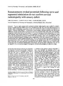
To match our control subjects for age with patients with cervical spondylotic myelopathy we selected from the laboratory normative data'6 those collected in normal subjects over 40 years of age (20 subjects (age range 40-82, mean 52-7, seven men) for median nerve SEPs, 16 subjects (age range 40-82, mean 53-2, seven men) for radial and ulnar nerve SEPs), see table 1. For assessing the conduction time in somatosensory pathways we measured the peaking latencies of Erb's point N9, cervical N13, scalp far-field P9 and P14, and contralateral parietal N20 potentials. To eliminate interindividual variations related to arm length, P9-N13, P9-P14, P14-N20, and P9-N20 interpeak intervals were also calculated. The amplitude of the N1 3 potential was assessed by calculating the N1 3/P9 amplitude ratio using the Cv6-AC traces'3-'6 as shown in fig 1.
Evoked potentials after upper limb stimulation in diagnosis of cerical spondylotic mydopathy Figure 1 SEPs evoked by stimulation of (A) nght radial, (B) median, and (C) ulnar nerves in a 46-year-old control subject. Two traces obtained on two successive runs of 2024 (for the median and ulnar nerves) or 4096 (for the radid nerve) are superimposed. Erb-Fz = Erb's point referred to a forehead electrode (Fz); the P14 potential picked up from the frontal electrode is injected as an "N14" negatit*y; Cv6AC = spinous process of the sixth cervical vertebra, referred to an anterior cerical electrode (AC); Parc-Sh = partetal electrode contralateral to the stimulus, referred to the shoulder contralateral to the stimulus (noncephalic reference electrode); ParcPar, = parietal electrode contralateral to the stimulus, referred to the parietal electrode ipsilateral to the stimulus; using this derivation the subcortical far-f eld potentials preceding the N20 response are canceled out. (D) Method used for measuring the amplitude of P9 (1) and N13 (2) as well as the N13/P9 amplitude ratio in the Cv6-AC trace.
303
-11 gv
A
L;R
AI-
Erb-Fz
Erb-Fz
4
N9 >
Cv6-AC Cv6-AC N13 N9P4
Parc-Sh
Parc-Sh
Parc-Pari
Par,-Pari N20
0
C
Time (ms)
0
30
30
Time (ms)
.1 V
Erb-Fz N9 D
Cv6-AC
Cv6-AC
: N13
2 RV
N9 P1 4 I
N
Parc-Sh
Parc-Pari N20
0
Time (ms)
0
30
Results All patients but one showed abnormal SEPs on both sides, at least for one of the stimulated nerves. In only one patient SEP abnormalities were limited to the right side. The latency of the N9 and P9 responses were always within normal limits, as well as the P9-N13 interpeak interval and the N13 latency, when the N13 response was identifiable. Abnormalities in N20 potential were always associated with an abnormal P14 potential. N13 ABNORMALITS Reduced or absent
Ni 3 potential
was
found
Time (ms)
30
after stimulation of at least one nerve in all patients but two. N13 potential was absent or reduced in 71/76 of radial, 64/76 of median, and 49/76 of.ulnar nerve SEPs. Radial, median, and ulnar spinal SEP abnormalities were combined as follows. N13 was abnormal for the three tested nerves in 49 upper limbs (64-5%). These abnormalities were bilateral in 23 patients and unilateral in three. When N13 was abnormal for only two of the three tested nerves, the pattern consisted in all cases of abnormal N13 in radial and median nerve SEPs with normal ulnar nerve spinal SEPs. This was found in 15 upper limbs (19-7%). These abnormalities
Restuccia, Valeriani, Di Lazzaro, Tonali, Mauguiere
304
Table 2 Correlation between clinical signs and distribution ofSEP abnormalities Normal
Abnormal Clinical signs
In all three nerves
Radial, median, ulnar
Radial, median
Radial 3/7 upper limbs 3/7 upper limbs
0/5 upper limbs 0/5 upper limbs
Lower motor neuron signs Pain and temperature segmental hypesthesia Joint and touch hypesthesia
31/49 upper limbs 16/49 upper limbs
N13 response 8/15 upper limbs 4/15 upper limbs
22/49 upper limbs
4/15 upper limbs
1/7 upper limbs
4/5 upper limbs
Lower motor neuron signs Pain and temperature
19/30 upper limbs 8/30 upper limbs
P 14 response 3/6 upper limbs 6/6 upper limbs
5/11 upper limbs 4/11 upper limbs
15/29 upper limbs 5/29 upper limbs
segmental hypesthesia Joint and touch hypesthesia
23/30 upper limbs
1/6 upper limbs
7/11 upper limbs
0/29 upper limbs
18/29 upper limbs 7/29 upper limbs
N 20 response 3/4 upper limbs 4/4 upper limbs
3/8 upper limbs 4/8 upper limbs
18/35 upper limbs 7/35 upper limbs
22/29 upper limbs
1/4 upper limbs
6/8 upper limbs
2/35 upper limbs
Lower motor neuron signs Pain and temperature segmental hypesthesia Joint and touch hypesthesia
were bilateral in four patients and unilateral in seven. When N13 was abnormal for only one nerve, only radial nerve SEPs were affected. This was the situation in seven upper limbs (9.2%). These abnormalities were bilateral in two patients and unilateral in three. A normal N13 was found whatever the stimulated nerve in five upper limbs; these normal spinal responses were bilateral in two
patients and found after stimulation of the left side in one patient. Table 2 shows correlations betweeq N13 findings and clinical signs in the corresponding upper limb. P14 ABNORMALITIES
The P14 potential was absent or delayed after stimulation of at least one nerve in 24 patients (63-1%). Abnormal P14 was found in 30, 36, and 47 of the 76 radial, median,
N14~~I I% ,, i,. .: . . . ;. ......i.~~4~ t~~!r~~\ .
1ieY:]E' ''' _ a.fZi-
!Y~ I~ xi -
~ ~ ~
~
~
~
~
~
~
T rT txX .
.e
r~~~~~~~'T
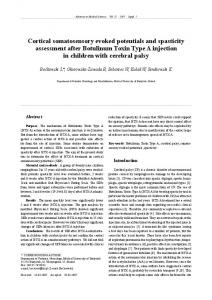
Figure 2 (A) Delayed or absent P14 with normal latency N20 after stimulation of the median nerve. Right and left median nerve SEPs are illustrated with the same abbreviations as in fig 1. The peak latencies of N9 and P9 responses were within normal limits. The N13 potential was absent after stimulation of the right median, and reduced after stimulation of the left median nerve. Whereas the N20 latencies, as weUl as the P9-N20 intervals, were stiU within normal limits, the P9-P14 interval was slighdy increased after stimulation of the right median nerve and the P14 was unrecognisable after stimulation of the left median nerve. (B, C) The cervical spinal cord MRI (Tl- and T2-weighted) showed a stenosis of the cervical cord with spondylotic cord compression at the C3-C4, C4-C5, and C5-C6 levels.
305
Evoked potentials after upper limb stimulation in diagnosis of cervical spondylotic myelopathy
B
A
+1 1
C
AV
-1J2 [tV
Erb-Fz
Erb-Fz
Erb-Fz
N9:
N13 ?
N 13 ?
Cv6-AC
N9 _fN13 ?
N9
Cv6-AC
2 RV +I2V
+
Cv6-AC P9
pg P14 A
Parc-Sh
P9
_
P14
P14
Parc-Sh
Parc-Sh
Parc-Pari
Parc-Pari
~N0:
Parc-Par
N20
30 lime
N20
N20 30
0
(ms)
30
0
Time (ms)
Time (ms)
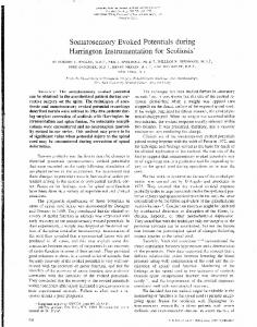
Figure 3 Association of normal scalp SEPs and abnormal spinal SEPs after stimulation of all three nerves. (A) Right radial, (B) median, and (C) ulnar nerve SEPs. Same presentation as infigs 1 and 2. The peaking latencies of N9, P9, P14, and N20 responses, as well as P9-P14 and P14-N20 interpeak intervals were within normal limits. N13 potential was absent or reduced after stimulation of all three nerves. E
D
for only two of the three tested nerves the consisted of abnormal P14 in median and ulnar nerve SEPs with normal radial nerve scalp SEPs. This was found in six upper limbs (7.9%). This type of SEP abnormality was bilateral in one patient and unilateral in four. When P14 was abnormal for only one nerve, only ulnar nerve SEPs were affected. This was found in 11 upper limbs (145%), bilaterally in five patients, and unilaterally in one. Normal radial, median, and ulnar nerve P14 potentials were found in 29 upper limbs (38- 1%). This finding was bilateral in 14 patients and unilateral in one. Table 2 gives the correlations between P14 findings and clinical signs in the corresponding upper limb.
pattern
N20 ABNORMALITIES .
Figure3contd compression
at
(DE)
Cervical
spinal
cordMR(lad2wihe)hwd
CS-C6 level
and ulnar nerve SEPs respectively; P14 was abnormal for the three tested nerves in 30 upper limbs (39 5%). These abnormalities were bilateral in 13 patients and unilateral in four. In all cases where the P14 was abnormal
N20 potential was absent or delayed after stimulation of at least one nerve in 22 patients (57-9%). The N20 potential was abnormal in 29/76 of radial, 33/76 of median, and 41/76 of ulnar nerve SEPs. Radial, median, and ulnar scalp N20 abnormalities were combined as follows. N20 was abnormal for the three tested nerves in 29 upper limbs (38'1%). These abnormalities were bilateral in 13 patients and unilateral in three. When N20 was abnormal for only two of the three tested nerves the pattern consisted in all cases of abnormal N20 in median and ulnar nerve SEPs with normal radial nerve N20. This was found in four upper limbs (5'3%), bilaterally in one patient, and unilaterally in two. When N20 was abnormal for only one nerve, only ulnar nerve SEPs were affected. This was the situation in eight upper limbs (10-5%). These abnormalities were bilateral in three patients and unilateral in two. The N20 potential was normal for all tested nerves in 35 upper limbs (46%). This finding was bilateral in 16 patients and uni-
Restuccia, Valeriani, Di Lazzaro, Tonali, Mauguiere
306
A
B
C
+ 12
Erb-Fz
,\> ? Erb-Fz
Erb-Fz
N9 N13 ?
Cv6-AC
+I 2 [V
AV
Cv6-AC
N9
N9