Viewing from: Google Indexer. BMJ Journals. Subscribe · Log In More .... RSS · Facebook · Twitter · Blog · SoundCloud · YouTube · Website Terms & Conditions ...
Journal of Neurology, Neurosurgery, and Psychiatry 1988;51:182-187
Somatosensory evoked potentials following nerve and segmental stimulation do not confirm cervical radiculopathy with sensory deficit URS D SCHMID, CHRISTIAN W HESS, HANS-PETER LUDIN From the Department ofNeurology and Neurosurgery, University Hospital, Berne, Switzerland
SUMMARY Twenty eight patients with unilateral cervical radiculopathy were studied by somatosensory evoked potentials (SEPs) from nerve stimulation at the wrist and from skin stimulation at the first, third or fifth finger depending on the root involved. In order to evaluate the reliability of various "radicular SEP patterns" as described in the literature, absolute latencies and side-to-side differences of the brachial plexus component from the supraclavicular fossa (N9), the medullary component (N1 3) from the cervical vertebra Cv7, and the primary cortical component (N20, P25) were assessed. Side-to-side differences of the amplitudes of N20/P25 and of the conduction times across the intervertebral fossa (interval N9-N13) were analysed. After nerve stimulation, 68% of the patients had false negative findings on the symptomatic, while 36% had positive findings on the asymptomatic side. After segmental stimulation, 72% of the patients had false negative findings on the symptomatic, while 22% had positive findings on the asymptomatic side. It is concluded that SEPs following nerve and segmental stimulation do not reliably confirm clear-cut already established diagnoses of unilateral radiculopathy with sensory and motor deficit. Therefore, they will not be helpful in the electrophysiological investigation of cervicobrachialgias of unknown origin. Several electrophysiological methods are employed in the diagnosis of cervical radiculopathy. Needle electromyography and electroneurography are helpful in that they may show a segmental pattern of denervation in muscles when nerve conduction studies are normal. However, the diagnostic yield of these methods remains limited because they cannot provide direct proof of impaired nerve conduction in the very proximal segment of peripheral nerves.' 3 With the technique of somatosensory evoked potentials (SEPs) the sensory pathways can be assessed along the whole of their course from the peripheral nerve to the contralateral hemisphere. Hence, SEPs have been used in patients with cervical radiculopathy by several authors.' 16 Increased conduction time between the plexus component and the first medullary component,89 diminished amplitude6 8-10 or deletion of the first medullary,2 3 as well as diminished amplitude Address and address for reprint requests: Dr Med. LJrs D Schmid, Department of Neurosurgery, University-Hospital, CH-3010, Berne, Switzerland Received 5 June 1987 and in revised form 23 July 1987. Accepted 27 July 1987
182
or deletion of the primary cortical component2 37 have been described as indicating cervical root lesions, whereas other authors failed to find any SEP alterations in cervical radiculopathies.'5 The sensitivity of the SEP was found to be higher when considering the intraindividual side-to-side differences.3 10 In single root lesions, SEP from segmental stimulation of the skin, which requires averaging of a greater number of samples, was reported to be superior to SEP from nerve stimulation at the wrist,7 1213 and correct positive results of 50%,12 57%,3 or 86-6%7 in segmental SEP have been reported. Only a few of these investigations are based on larger groups of patients,2 3 7 and reports giving detailed analysis of the rate of correct negative and false positive or negative SEP findings are rarely given.23 The question of diagnostic reliability of these SEPpatterns is of obvious importance. We therefore prospectively examined a series of 28 patients with clear-cut one-sided cervical radiculopathies and radicular sensory and motor deficits, using SEP from nerve stimulation at the wrist and from segmental stimulation at the fingers on both sides.
Somatosensory evoked potentials following nerve and segmental stimulation Table 1 Clinical signs indicating cervical root impairment
183
For recording, needle electrodes were placed over the ipsilateral supraclavicular fossa (SF), the 7th cervical verteSensory and motor deficit and reflex abolished 16 bra (Cv7), and over the contralateral parieto-occipital scalp Sensory and motor deficit I Sensory deficit and reflex abolished 5 (Cx), 2cm posterior to C3/C4 placement according to the Sensory deficit alone 3 10-20 system. The reference electrodes were placed at Fz Motor deficit and reflex abolished 2 (10-20 system) for the Cv7 recording and at the contralateral Motor deficit alone I ear lobe for the SF recording. Two different montages were Total 28 simultaneously used for the Cx recording, one with reference at Fz and one with reference at the contralateral ear lobe. The former was used for measurement of the cortical comMaterial and methods ponent, the latter helped identify the medullary components. Twenty eight patients ranging in age from 25 to 70 years The electrode impedance was kept below 3 kfl in all record(mean 45,8 years, 16 males) were studied. The history of ings by means of careful skin preparation. The signals were amplified and averaged by a conventional cervicobrachialgia lasted over periods of 14 days to 17 years, with typical symptoms of cervical radiculopathy. Only cases four channel recording machine (Medelec ER 94a) except for with strictly unilateral clinical signs and with symptoms of eight cases, where SEPs from nerve stimulation were unilateral functional impairment of one or two cervical roots recorded using a two channel machine (DISA System-15). Filtering bandpass was 5-2000 Hz for the Cx recording and were included: seven patients with C6 radioculopathy, nine with C7 radiculopathy, four with C8 radiculopathy, six with 20-1000 Hz for the Cv7 and SF recording. To assess remixed C7/C8 radiculopathy and one with mixed Q6/C7 radi- producibility, on each side 2-3 recordings of 256 to 512 culopathy. Patients with peripheral neuropathy or cervical stimuli were averaged for SEPs from nerve stimulation and myelopathy were excluded. The neurological deficits of var- of 1012 to 2024 stimuli were averaged for SEPs from segious degrees are summarised in table 1. With the exception mental stimulation. of three patients, all had radicular sensory deficit. In all but two patients, clinical diagnosis was complemented by radio- Evaluation logical and/or intraoperative findings. The subsequent treat- The SEP components from nerve stimulation at the wrist ment was not influenced by the SEP findings, and was were named according to the normal mean peak latencies of conservative in 17 patients, and surgical in 11 patients. the negative peaks as follows. N9 (supraclavicular electrode), On the 28 patients, 44 investigations were performed (88 N13 (spinal electrode), and N20 (cortical electrode).2 - 10 The sides). Twenty two patients had a total of 26 investigations same nomenclature was used for the analogous SEP com(52 sides) following nerve stimulation at the wrist; 18 median ponents from segmental stimulation regardless of their nerve stimulations (36 sides), and eight ulnar nerve stimulonger latencies. The amplitude of the cortical component lations (16 sides); four of these 22 patients had both SEPs was measured peak-to-peak from N20 to the adjacent posiafter median as well as ulnar stimulation. In 18 patients SEPs tive maximum P25. from segmental stimulation at the fingers were recorded (36 Comparing the two or three consecutive recordings of sides), 12 of which had both nerve and segmental stimu- identical stimulation site, components were defined relation. Twenty healthy subjects ranging in age from 16 to 52 producible if they showed similar wave forms and peak laten(mean 27 6 years) served as a control group for SEPs from cies which did not differ by more than 1 ms for the cervical stimulation of the nerve at the wrist.5 For the segmental and 3 ms for cortical components respectively, and a mean of stimulation, normal values of Synek16 served as controls. the two or three values was taken for the analysis. If the components were not reproducible by this definition, they Stimulation and recording procedures were rated as "not obtained". When the corresponding comThe patients and subjects were comfortably positioned on a ponents were reproducible, proximal sensory conduction bed in a quiet room with their eyes closed. No muscle relax- time across the intervertebral foramen from the latency interants, analgesic or sedative drugs were used. Room- val N9-N1 3 and side-to-side differences were calcuated. temperature was about 23°C. Latencies, conduction times, amplitudes and the side-toFor nerve stimulation at the wrist, electrical pulses of side differences (A) of SEPs from nerve stimulation were con0-2 ms duration were applied over the median or ulnar nerve sidered as prolonged when their value exceeded the mean with surface electrodes (cathode 2.3 cm proximal to the an- plus 2-5 standard deviations (SD) of the normal values of the ode) at a rate of 3/s with a stimulus intensity of 4 mA above control group.5 The upper limits of normal were as follows: the motor threshold of the thenar or hypothenar muscles N9: 114 ms; N13: 15-8 ms; N20: 22-3 ms; N9-N13: 5-2 ms; of respectively. the side-to-side differences: AN9: 056ms; AN13: 077ms; For segmental stimulation, a pair of ring electrodes was AN20: -1 Ims; AN9-N13: 095ms; for the side-to-side attached to one finger with the cathode at the proximal difference of the amplitude AN20/P25. values of 50% or interphalangeal and the anode at the distal interphalangeal more were taken as abnormal. joint. Depending on the clinical diagnosis, the electrodes For SEPs from segmental stimulation only side-to-side were attached to the thumb, to the third or the fifth finger for differences (A) were taken into consideration, since side-tostimulation of the sensory skin supply of the 6th, 7th, or 8th side comparison in clinically unilateral radiculopathy has cervical root respectively. Stimulus intensity was 3-4 times been considered to be most sensitive.2 3 A side-to-side the sensory threshold which was always below pain thresh- difference was taken as abnormal when it exceeded the mean old. The pulse duration was 0-2 ms and stimulation rate was plus 4 SD,16 that is 2 ms for AN 13 and AN20. For the ampli3/s. tude AN20/P25, 50% of the contralateral side,7 and for the
Schmid, Hess, Ludin
184
Table 2 Nerve stimulation at the wrist: Reproducibility of components, absolute latencies, intervals, amplitudes and side-to-side differences (N of reproducible or calculable parameters/N ofparameters evaluated) Incidence of abnormal side-to-side
Parameter
N9 N13 N20 N9-N13 N20/P25
differences of latencies, conduction times
Reproducibility of parameters Symptomatic Asymptomatic side side
Incidence of abnormal absolute latencies and conduction-times Symptomatic Asymptomatic side side
and amplitudes* Symptomatic side
21/21 25/26 26/26 21/21 22/22
0/21 0/25 2/26 2/21
0/19 1/24 0/26 1/19
1/19 1/24 2/26 1/19
-
-
2/22
19/21 24/26 26/26 19/21 22/22
Asymptomatic side 0/19 3/24 1/26 3/19 1/22
*Prolonged latencies and conduction times or diminished amplitudes compared with the contralateral side
Table 3 Segmental stimulation: Reproducibility of components, and side-to-side differences oflatencies, conduction times and amplitudes (of reproducible or calculable parameters/ofparameters evaluated)
Reproducibility of Parameter parameters symptomatic asymptomatic side side N9 N13 N20 N9-N13 N20/P25
7/18 13/18 18/18 7/18 17/17
9/18 16/18 18/18 8/18 17/17
Incidence of abnormal side-to-side differences* symptomatic asymptomatic side side 0/5 2/13 2/18 0/5 3/17
0/5 0/13 2/18 1/5 1/17
*Prolonged latencies and conduction times or diminished amplitudes compared with the contralateral side
proximal sensory conduction time AN9-N 13, 1 ms side-toside difference was defined as abnormal.
Results SEPs from nerve stimulation at the wrist The frequency ofreproducible components or evaluable parameters at the symptomatic as well as at the
asymptomatic side from nerve stimulation are listed in table 2. All but five patients had all components reproducible, so that proximal sensory conduction time, amplitude of the primary cortical responses and side-to-side differences in 21 and 25 of the 26 patients were evaluable. Table 2 gives also the incidence of abnormal latencies, conduction times and amplitudes at the symptomatic and asymptomatic side. Absolute latencies, conduction times and amplitudes were normal in all but five cases. Side-to-side comparison revealed prolonged N1 3 in one of 24 of the symptomatic, but also in three of 24 of the asymptomatic sides. Prolonged proximal conduction time N9-N13 was found in one of 19 of the symptomatic, and in three of 19 of the asymptomatic sides. N20/P25 was significantly reduced in two of 21 of the symptomatic, and in one of 21 of the asymptomatic sides. In the patients as a group, there was no statistically significant difference (paired t test) between the two sides for the proximal conduction time N9-N 13 (p < 0O375), N13 (p > 04), and N20 (p < 0375).
Table 4 Accuracy* of various radicular SEP-patterns2 36-10 12 13 in confirming unilateral cervical radiculopathies with neurological deficit Segmental stimulation (n = 18) found at the
found at the symptomatic side ("correct positive")
asymptomatic side ('false positive")
N13 abolished N13 delayedt AN13 delayedt
28%
11%
15%
0%
N20 abolished AN20/P25 reducedt N20 delayedt AN20 delayedt
0% 18%
0% 6%
Radicular SEP-pattern
N9-N 13 prolongedt AN9-N13 prolongedt
Total of patients with "radicular SEP signs"I
Nerve stimulation at the wrist (n = 26)
found at the symptomatic side ("correct positive")
found at the asymptomatic side ('false positive")
4% 0% 4%
8% 4% 12%
0%
5% 0% 4%
-
-
11%
11%
0% 9% 8% 8%
0%
25%
10% 5%
5% 16%
28%
22%
32%
36%
-
*percentage of patients after segmental and nerve stimulation respectively tpercentage of the number of patients where the respective parameters were reproducible lpercentage of patients where one or more "radicular SEP signs" were positive
Somatosensory evoked potentials following nerve and segmental stimulation 185 SEP from segmental stimulation: the greater temporal dispersion of afferent influx The frequency of reproducible components and evalu- would result in amplitude diminution of the comable parameters following segmental stimulation are ponents. Diminished amplitude or deletion of primary listed in table 3. Of the 18 patients, N9 was obtained medullary or cortical components have been dein seven of the symptomatic and in nine of the asymp- scribed,2 3 and the latter is considered by some2 3 7 to tomatic sides, and N13 was reproducible in 13 of the be the only reliable SEP parameter in cases of cervical symptomatic, and in 16 of the asymptomatic sides. radiculopathy. But in view of the broad range of the Therefore, a proximal sensory conduction time was amplitudes of evoked potentials in general, and of the calculable in seven and eight cases respectively, while cervical components in particular, the non-specific side-to-side comparison for N13 and for N9-N1 3 re- finding of an attenuated component is of limited mained possible in only 13 and five patients value. Even if the side-to-side-differences were taken respectively. into consideration, amplitude reductions of 50% and The incidence ofabnormal side-to-side-differences is more of the N20/P25 component when comparing listed in table 3. In the cases where side-to-side com- with the contralateral side, or abolition of component parison remained possible, the values failed to point N13, was a rare finding and occurred just as often on to the symptomatic side, and the amplitude N20/P25 the symptomatic as on the asymptomatic side. in 17 patients was reduced only in three of the It follows that SEPs after nerve stimulation are not symptomatic, and in one of the asymptomatic sides. a sensitive diagnostic method for radiculopathy. It In the patients as a group, no significantly pro- seems that a sufficient number of afferent nerve fibres longed -latencies of the medullary components N13 contributing to the generation of cervical and cortical (p > 0-4, paired t test) or cortical components N20 SEP components remain preserved, probably because (p < 0-375, paired t test), and no significantly pro- they are using an entry other than that of the symplonged proximal conduction time N9-N13 (p = 0-1, tomatic root. The fast-conducting la afferents, which Wilcoxon-Mann-Whitney U Test), could be demon- do not follow the segmental distribution of sensory strated on the clinically symptomatic side. skin supply, are preferentially stimulated at the wrist In 11 cases with a clinical and neuroradiological because of their low threshold. With median nerve diagnosis of cervical radiculopathy, nerve root com- stimulation in case of C7 radiculopathy or with ulnar pression was verified during surgery. Taking only nerve stimulation in case of C8 radiculopathy, these these surgically verified compressive root lesions, all afferents will bypass a lesion affecting one or two roots with sensory deficit, the incidence of abnormal values at the cervical foramina. obtained from the clinically symptomatic sides On theoretical ground, this shortcoming should be remained unchanged. ruled out by using segmental stimulation of the skin at a finger, as has been suggested by several auDiscussion thors.2371316 Because of their smaller amplitude, segmental SEPs require more averages. Our results The aim of this study was to examine the usefulness of from segmental SEPs do show absence of cervical SEPs in the diagnosis of compressive cervical root components in some cases, and this occurred more lesions. SEPs following nerve stimulation at the wrist frequently on the symptomatic than on the asympand/or segmental stimulation of a finger were used in tomatic side. But, because in an important number of 28 patients. All presented with clear-cut unilateral patients either the plexus or the medullary component cervical radiculopathies with neurological deficit of or both could not be obtained on either side, a side-tovarious degrees, and all but three had a sensory deficit. side comparison of the proximal sensory conduction Either the median or ulnar nerve and the first, third, time was impossible in 73% of the patients. or fifth finger were stimulated according to the The frequent absence of the plexus component cervical root involved. needs to be explained. It could be caused by a longSEPs from nerve stimulation were mostly normal on standing root compression due to retrograde degenerthe clinically symptomatic side of these patients, and ation when the damage is situated distal to the sensory this was particularly so for the proximal conduction ganglia. However, the fact that this component was time N9-N 13. This finding is in contrast to previously also missing on the asymptomatic side in many inreported investigations,6 89 but confirms the negative stances points to a more technical reason. Although findings of others.15 Although not a specific sign, a plexus and cervical components following segmental prolonged conduction time across the cervical stimulation can safely be obtained in healthy subjects foramen due to compression-induced focal demy- when a sufficient number of sweeps are averaged,16 elination would have been the most convincing good relaxation is crucial to minimise muscle interalteration indicating a cervical root lesion. ference, and this is difficult to achieve in patients who On the other hand, drop-out of damaged fibres and are in pain.
186 Any attempt to compensate for muscle artefacts by assembling more averages will fail in these patients, because the longer duration of the recording process makes muscle relaxation more difficult, so that after some time of averaging, the base-line noise gets worse instead of better. The cortical component N20/P25 proved to be very stable. N20 was obtained in all cases from segmental stimulation. Abnormal side-to-side differences of the amplitudes were observed in 4/17 cases. But, while in three cases the amplitude was diminished on the clinically symptomatic, in one case the amplitude was diminished on the asymptomatic side. This finding contradicts the statements of some authors who consider diminished amplitude of the cortical component as most reliable sign in cervical radiculopathies.7 Of the 18 patients with segmental stimulation, five (28%) had one or more abnormal SEP results on the symptomatic side, but this was also found in four (22%) patients on the asymptomatic side. Even when taking the 11 surgical patients only, where the root compression was verified by neuroradiological investigation and during surgery, the results were only slightly better. A possible explanation for the bilaterally affected plexus or medullary components would be the assumption that subclinical and radiologically silent involvement of the nerve root or of the spinal cord of the asymptomatic side cannot be completely ruled out. If
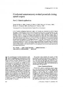
Schmid, Hess, Ludin true, this would mean, that SEPs after segmental stimulation may detect even subclinical changes, yet such high sensitivity makes the method useless for clinical purposes. Conclusions In a group of patients with clear-cut cervical radiculopathy with unilateral neurological deficit including sensory impairment (table 1), a standard SEP technique was employed, and a "correct positive" SEP pattern as described in the literature was rarely found (table 4). Even when the segmental stimulation technique was used, 72% of the patients had false negative findings on the clinically symptomatic side, while 22% of the patients had positive "radicular SEP signs" on the asymptomatic side. The figure gives an example of such a false negative segmental SEP recording in a left sided cervical radiculopathy with sensory deficit. As SEP following nerve and segmental stimulation have only rarely confirmed a clear-cut cervical radiculopathy with sensory deficit, we do not expect them to be helpful in the electrophysiological investigation of doubtful cases of cervicobrachialgias.
The authors express their gratitude to Andrew Wade, for reviewing the English manuscript and to HansJurgen Reuter for his support.
23.2 C'3 -Fz
C'4- Fz
Ii 1.5pV
1--
7t'.,
C'4-A2
C'3-A1
-.1 .....
2 8
-'Il 1.0'Aiv ez
17.6
Cv7- Fz
D.I OjuV
I .
Cv7-Fz
ERB-A1
,
el..
1:1
Ii.
Y./-
I 5ms
..
5 ms
Fig Normal segmental SEP after stimulation of the left and right thumb of a 60 year oldfemale with history of repeated left sided severe cervico-brachialgia for 1 year and signs of strictly left-sided cervical radiculopathy C6 with sensory and reflex deficit. Myelography and surgery revealed cervical disc herniation at the level C5/6. (a) symptomatic left side, (b) asymptomatic right side.
Somatosensory evoked potentials following nerve and segmental stimulation 187 References eral, cervical and cortical evoked potentials in patients with cervical spondylosis. J Neurol, Neurosurg, 1 Stohr M, Buettner UW, Wietholter H, Riffel B. ComPsychiatry 1980;43:683-9. bined recording of compound action potentials and 10 Siivola J, Sulg I, Heiskari M. Somatosensory evoked po2 3
4 5 6 7
8
9
spinal cord evoked potentials in differential diagnosis of spinal root lesions. Arch Psychiat Nervenkr 1983;233: 103-10. Pilade JP, Pelissier J, Georgescu M, Simon L, et al. Spinal somesthetic evoked potentials and cervicobrachial neuralgia. Revue du Rhumatisme 1984;51:7-13. Eisen A, Hoirch M, Moll A. Evaluation of radiculopathies by segmental stimulation and somatosensory evoked potentials. Can J Neurol Sci 1983;10: 178-82. Iragui VJ. The cervical somatosensory evoked potential in man. Electroencephalogr Clin Neurophysiol 1984;57:228-35. Vlach L, Ludin HP. Spinale evozierte Potentiale beim Gesunden. Thesis 1982, University of Berne. Stohr J, Dichgans J, Diener HC, Buettner UW. Evozierte Potentiale. Heidelberg, Springer Verlag 1982. Chapter 1-2. Jorg J. Praktische SEP-Diagnostik. Stuttgart Enke Verlag (1983). El Negamy E, Sedgwick EM. Delayed cervical somatosensory evoked potentials in cervical spondylosis. J Neurol, Neurosurg, Psychiatry 1979;42:238-41. Ganes T. Somatosensory conduction times and periph-
tentials in diagnostics of cervical spondylosis and herniated discs. Electroencephalogr Clin Neurophysiol
1981;52:276-82. 11 Hacke W, St6hr M, Diener HCW, Buettner U. Recommendations for recording techniques regarding evoked potentials as a routine diagnostic procedure. Z EEGEMG 1985;16:162-4. 12 Schramm J. Somatosensorisch evozierte Potentiale in der Differentialdiagnose spinaler Erkrankungen. In: Schramm J. Evozierte Potentiale in der Praxis.
Heidelberg Springer-Verlag 1985;98-127. 13 Jorg J. SEP-Diagnostik in der Neurologie. Z EEG-EMG
1985;16:34-89. 14 Dal Bianco P, Mamoli B, Dorda W. The identification and variation of NSEP waveforms. Z EEG-EMG 1985;16:206-1 1. 15 Yu YL, Jones SJ. Somatosensory evoked potentials in cervical spondylosis. Correlation of median, ulnar and posterior tibial nerve components with clinical and radiological findings. Brain 1985;108:273-300. 16 Synek VM. Somatosensory evoked potentials after stimulation of digital nerves in upper limbs. Normative data Electroencephalogr Clin Neurophysiol
1986;65:460-3.