nutrients Article
Anti-Inflammatory Mechanism Involved in Pomegranate-Mediated Prevention of Breast Cancer: the Role of NF-κB and Nrf2 Signaling Pathways Animesh Mandal 1 , Deepak Bhatia 2 and Anupam Bishayee 3, * 1
2 3
*
Cancer Therapeutics and Chemoprevention Group, Department of Pharmaceutical Sciences, College of Pharmacy, Northeast Ohio Medical University, Rootstown, OH 44272, USA;
[email protected] Department of Pharmacogenomics, Bernard J. Dunn School of Pharmacy, Shenandoah University, Ashburn, VA 20147, USA;
[email protected] Department of Pharmaceutical Sciences, College of Pharmacy, Larkin University, Miami, FL 33169, USA Correspondence:
[email protected] or
[email protected]; Tel.: +1-305-760-75211
Received: 1 February 2017; Accepted: 5 April 2017; Published: 28 April 2017
Abstract: Pomegranate (Punica granatum L.), a nutrient-rich unique fruit, has been used for centuries for the prevention and treatment of various inflammation-driven diseases. Based on our previous study, a characterized pomegranate emulsion (PE) exhibited a striking inhibition of dimethylbenz(a)anthracene (DMBA)-initiated rat mammary tumorigenesis via antiproliferative and apoptosis-inducing mechanisms. The objective of the present work is to investigate the anti-inflammatory mechanism of action of PE during DMBA rat mammary carcinogenesis by evaluating the expression of cyclooxygenase-2 (COX-2), heat shock protein 90 (HSP90), nuclear factor-κB (NF-κB) and nuclear factor erythroid 2p45 (NF-E2)-related factor 2 (Nrf2). Mammary tumor samples were harvested from our previous chemopreventive study in which PE (0.2–5.0 g/kg) was found to reduce mammary tumorigenesis in a dose-dependent manner. The expressions of COX-2, HSP90, NF-κB, inhibitory κBα (IκBα) and Nrf2 were detected by immunohistochemical techniques. PE decreased the expression of COX-2 and HSP90, prevented the degradation of IκBα, hindered the translocation of NF-κB from cytosol to nucleus and increased the expression and nuclear translocation of Nrf2 during DMBA-induced mammary tumorigenesis. These findings, together with our previous results, indicate that PE-mediated prevention of DMBA-evoked mammary carcinogenesis may involve anti-inflammatory mechanisms through concurrent but differential regulation of two interrelated molecular pathways, namely NF-κB and Nrf2 signaling. Keywords: anti-inflammatory effects; breast tumor; COX-2; DMBA; HSP90; NF-κB; Nrf2; Punica granatum
1. Introduction Pomegranate (Punica granatum L.) is a nutrient-rich fruit which represents a reservoir of bioactive phytochemicals with exceptional medicinal values. The pomegranate is a plant native from the Himalayas to Iran and has been cultivated and naturalized throughout the world and in the United States, including Arizona, California and Texas. Pomegranate, known as “a pharmacy unto itself ” has been used for centuries in various traditional and folk medicine for the treatment of a large number of ailments [1–3]. During the last decade, pomegranate fruit has been gaining a widespread reputation as a dietary supplement as well as a functional food due to emerging scientific evidence on potential health benefits, including prevention and/or treatment of cardiovascular ailments, neurological disorders, oncologic diseases, dental problems, inflammation, ulcer, arthritis, microbial
Nutrients 2017, 9, 436; doi:10.3390/nu9050436
www.mdpi.com/journal/nutrients
Nutrients 2017, 9, 436
2 of 13
infection, obesity, diabetes, acquired immune deficiency syndrome and erectile dysfunction [4–9]. Pomegranate fruit contains phytochemicals, including flavonoids (e.g., anthocyanins and catechins), flavonols (e.g., kaempferol and quercetin), flavones (e.g., apigenin and luteolin), conjugated fatty acids, hydrolyzable tannins and related compounds which are thought to be responsible for various biological and pharmacological activities [4,10–14]. Based on preclinical and clinical studies conducted by various laboratories worldwide, pomegranate-derived substances, such as juice, extracts and phytoconstituents exhibited cancer preventive and therapeutic effects against colon, liver, lung, prostate and skin cancer [4,15–18]. Various extracts, fractions and phytochemicals from pomegranate fruit, peel, seed and flower demonstrated cytotoxic, antiproliferative, proapoptotic, antiangiogenic, anti-invasive, and antimetastatic properties against estrogen receptor-positive and -negative breast cancer cells [19–32]. Pomegranate seed oil and fermented juice concentrate were found to suppress 7,12-dimethyl benz(a)anthracene (DMBA)-induced preneoplastic mammary gland lesions ex vivo in a mouse mammary organ culture model [33]. Oral administration of pomegranate juice concentrate reduced the volume and weight of xenografted BT-474 tumors in female athymic nude mice [26]. Recently, we have documented the novel finding that oral feeding of a pomegranate emulsion (PE), containing most bioactive phytochemicals present in the whole fruit, exerted a significant chemopreventive activity against DMBA-initiated mammary tumorigenesis in female rats [34]. PE reduced the incidence, total burden and average weight of mammary tumors with a concomitant inhibition of intratumor cell proliferation, induction of apoptosis, and it altered the expression of Bax, Bcl2, Bad, caspase-3, caspase-7, caspase-9, poly (ADP ribose) polymerase and cytochrome c [34]. We have also observed that PE diminished the expression of estrogen receptor-α (ER-α), ER-β and cyclin D1 and abrogated the expression, cytoplasmic accumulation and nuclear translocation of β-catenin, an essential transcriptional cofactor for Wnt/β-catenin signaling, during DMBA mammary carcinogenesis [35]. Emerging studies indicate that chronic inflammation is involved in the development and progression of mammary carcinoma [36–40] and pomegranate phytochemicals are endowed with anti-inflammatory properties [4,8,11,12]. Accordingly, this study was conducted to investigate the anti-inflammatory mechanisms of PE administration by analyzing various proinflammatory and stress markers, such as cyclooxygenase-2 (COX-2) and heat shock protein 90 (HSP90) as well as several inflammation-regulatory pathways, namely nuclear factor-κB (NF-κB) and nuclear factor erythroid 2p45 (NF-E2)-related factor 2 (Nrf2) signaling, during DMBA-inflicted mammary gland tumorigenesis in rats. 2. Materials and Methods 2.1. Test Materials, Chemicals and Antibodies PE, a proprietary formulation containing pomegranate aqueous extract and seed oil, was purchased from Rimonest Ltd. (Haifa, Israel). A detailed description of the preparation of this emulsion has been published previously [15]. The chemical analyses of this formulation revealed the presence of caffeic acid, corilagin, ellagic acid, ferulic acid, gallic acid, 5-hydroxymethylfurfural, protocatechuic acid, punicalagins (A and B) and trans-p-coumaric acid in the aqueous phase and mixed octadecatrienoic acids, sterols and steroids (e.g., 17-α-estradiol), tocol and γ-tocopherol in the lipid phase [15]. The mammary carcinogen DMBA was procured from Sigma-Aldrich (St. Louis, MO, USA). Paraformaldehyde was obtained from Ted Pella (Redding, CA, USA). Primary antibodies, such as COX-2, inhibitory κBα (IκBα), NF-κB-p65, Nrf2 as well as the ABC staining kit were purchased from Santa Cruz Biotechnology (Santa Cruz, CA, USA). HSP90 was a product of Enzo Life Sciences (Farmingale, NY, USA). 2.2. Experimental Protocol and Tumor Tissue Harvesting Breast tumor sections for this work were collected from our previously completed chemopreventive study [34] based on an animal protocol approved by the Institutional Animal
Nutrients 2017, 9, 436
3 of 13
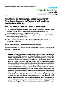
Care and Use Committee of Northeast Ohio Medical University (Rootstown, OH, USA). In short, female Sprague-Dawley rats (Harlan Laboratories, Indianapolis, IN, USA) were divided into six separate groups. All animals were fed a basal diet (LabDiet, St. Louis, MO, USA) ad libitum. Group A (n = 12) and group B (n = 11) were kept untreated. The remaining rats were orally administered (gavaged) with PE three times per week as follow: 0.2 g/kg (group C, n = 8), 1.0 g/kg (group D, n = 8) and 5.0 g/kg (group E, n = 7 and group F, n = 5) for a total of 18 weeks. Two weeks following the commencement of the study, animals from groups B, C, D and E were orally administered with a single dose of DMBA (50 mg/kg body weight) to induce mammary tumorigenesis. Eighteen weeks after initiation of the study, all animals were sacrificed and mammary glands were harvested and fixed in 4% paraformaldehyde for further analysis. 2.3. Immunohistochemical Analysis The intratumor protein expressions of COX-2, HSP90, IκBα, NF-κB-p65 and Nrf2 were analyzed by methods described previously [41]. In brief, tumor tissue sections were first hydrated using phosphate-buffered saline followed by incubation in sodium citrate buffer for antigen retrieval. Endogenous peroxidases were blocked by treating the sections with 1% H2 O2 . Tissue sections were then treated with blocking solution followed by washing with phosphate-buffered saline and incubating overnight at 4 ◦ C with primary antibodies (1:100 dilution). After several washes, tissue sections were treated with horseradish peroxidase-conjugated secondary antibody (1:200 dilution) for 30 min at room temperature, and then with 3,30 -diaminobenzidine tetrahydrocholoride solution to visualize brown antigen-antibody complexes. Finally, the sections were counterstained with Gill’s hematoxylin solution. The immunohistochemical slides were viewed under a light microscope (BX43, Olympus, Center Valley, PA, USA) and 1000 tumor cells/rat were analyzed. 2.4. Statistical Analyses All data are expressed as mean ± standard error of mean (SEM). Statistical analyses were performed by using a commercial software (SigmaPlot 11.0, Systat Software, Inc., San Jose, CA, USA). One-way analysis of variance with least significant difference post hoc analysis was employed to compare various parameters among different treatment and control groups. A p-value less than 0.05 was considered statistically significant. 3. Results 3.1. PE Abrogated Elevated COX-2 Expression during DMBA-Induced Mammary Tumorigenesis A substantial expression of COX-2 was observed predominantly in the cytoplasm of tumor cells of DMBA control animals (Figure 1(Aa)). PE at a dose of 0.2 g/kg slightly reduced intratumor COX-2 immunopositivity compared to the DMBA control (Figure 1(Ab)). On the other hand, a moderate and drastic inhibition of COX-2 expression was noticed following PE treatment at a dose of 1 and 5 g/kg, respectively (Figure 1(Ac,Ad)). Although PE at 0.2 g/kg slightly decreased the percentage of COX-2-positive cells, this result was statistically insignificant (Figure 1B). On the other hand, there was a significant (p < 0.01 or 0.001) inhibition of the percentage of intratumor COX-2-positive cells in animals administered with 1 or 5 g/kg PE compared to the DMBA control, respectively.
Nutrients 2017, 9, 436
4 of 13
Nutrients 2017, 9, 436 Nutrients 2017, 9, 436
4 of 13 4 of 13
Figure 1. COX‐2 expression during 7,12‐dimethyl benz(a)anthracene (DMBA)‐induced breast
Figure COX‐2 expression expression during 7,12-dimethyl 7,12‐dimethyl benz(a)anthracene (DMBA)‐induced breast Figure 1. 1. COX-2 (DMBA)-induced tumorigenesis in rats in the during presence or absence of benz(a)anthracene pomegranate emulsion (PE) treatment. breast (A) tumorigenesis in rats rats in in the the presence or absence of pomegranate emulsion (PE) treatment. (A) tumorigenesis in presence or absence of pomegranate emulsion (PE) treatment. Immunohistochemical localization of COX‐2‐positive cells (arrows) in tumor sections (magnification: (A)Immunohistochemical localization of COX‐2‐positive cells (arrows) in tumor sections (magnification: Immunohistochemical localization of COX-2-positive cells (arrows) in tumor sections ×200). The various treatment groups are: (a) DMBA control; (b) PE (0.2 g/kg) plus DMBA; (c) PE (1 ×200). The various treatment groups are: (a) DMBA control; (b) PE (0.2 g/kg) plus DMBA; (c) PE (1 (magnification: ×200). The are: (a)(B) DMBA control; (b) PE (0.2 plus g/kg) plus DMBA; and various (d) PE treatment (5 g/kg) groups plus DMBA. Quantitative analysis of g/kg) COX‐2‐ g/kg) plus DMBA; and (d) PE (5 g/kg) plus DMBA. (B) Quantitative analysis of COX‐2‐ DMBA; (c) PE (1 g/kg) plus DMBA; and (d) PE (5 g/kg) plus DMBA. (B) Quantitative analysis of immunopositive cells from representative images. Results (mean ± SEM) are based on 1000 cells per immunopositive cells from representative images. Results (mean ± SEM) are based on 1000 cells per COX-2-immunopositive cells from representative images. Results (mean ± SEM) are based on 1000 animal and four animals per group. * p