and flutamide. The platelet count on admission was within normal limits âTable 1â. The results of chest and abdominal radiographs were unremarkable.
Hematopathology / SPURIOUS AUTOMATED PLATELET COUNT
Spurious Automated Platelet Count Enumeration of Yeast Forms as Platelets by the Cell-DYN 4000 Shahnila Latif, MD, Diana M. Veillon, MD, Donald Brown, MT(ASCP), Jenny Kaltenbach, MT(ASCP), Sherry Curry, MT(ASCP)SH, Andrea J. Linscott, PhD, Arnold Oberle, MS, and James D. Cotelingam, MD Key Words: Optical and impedance platelet count; Hematology analyzer; Spurious platelet count DOI: 10.1309/H2K6E59LYFE63GPB
Abstract We recently encountered a patient with thrombocytopenia secondary to multiple drug therapy, disseminated prostatic adenocarcinoma, and sepsis who had a sudden decrease in his platelet count as enumerated by the Cell-DYN 4000 hematology analyzer (Abbott Diagnostics, Santa Clara, CA). A manual platelet count performed thereafter was even lower. The etiology of the spurious platelet count was clarified when numerous yeast forms were observed on routine microscopy of the peripheral blood smear. Subsequently, these organisms were identified as Candida glabrata from a positive blood culture (BACTEC 9240, Becton Dickinson, Cockeysville, MD). To our knowledge, this is the first report of spurious enumeration of yeast forms as platelets in an automated hematology system. The principle underlying platelet enumeration by the Cell-DYN 4000 system and other hematology analyzers and the value of microscopy on peripheral smears with unexpected CBC count results are discussed.
882 882
Am J Clin Pathol 2003;120:882-885 DOI: 10.1309/H2K6E59LYFE63GPB
The timely reporting of accurate platelet counts is part of standard operating procedure in a hematology laboratory. The accuracy of platelet counts is of particular clinical relevance when thrombocytopenic patients near the transfusion threshold. The former “gold standard” of manual or hand platelet counting has a long turnaround time, is costly, and has questionable accuracy. Automated methods for platelet counting have been in existence since the 1950s when the impedance principle was introduced by Wallace Coulter. Despite substantial technical advances in hematology analyzers, a number of factors continue to interfere with automated platelet counts.
Case Report A 75-year-old man who was a nursing home resident was admitted to the Louisiana State University Health Sciences Center, Shreveport, with a diagnosis of urosepsis and hypothermia. His relevant medical history included hypertension and prostate cancer with bone metastases. Admission vital signs included a blood pressure of 150/82 mm Hg, a pulse rate of 62 beats per minute, a respiratory rate of 16 breaths per minute, and a temperature of 35.7°C. The remainder of the physical examination was noncontributory. At admission, the patient was taking quinapril, hydrochlorothiazide, leuprolide, and flutamide. The platelet count on admission was within normal limits ❚Table 1❚. The results of chest and abdominal radiographs were unremarkable. Urine cultures grew coagulase-negative staphylococci, and treatment with ciprofloxacin and piperacillin was begun. The antibiotic regimen was changed to vancomycin, gentamicin, © American Society for Clinical Pathology
Hematopathology / CASE REPORT
❚Table 1❚ Relevant Hematology Data Laboratory Test WBC count, /µL (× 109/L) RBC count, × 106/µL (× 1012/L) Hemoglobin, g/dL (g/L) Hematocrit, % (proportion of 1.0) Mean corpuscular volume, µm3 (fL) Mean corpuscular hemoglobin, pg Mean corpuscular hemoglobin concentration, % (proportion of 1.0) RBC distribution width, % Platelet count, × 103/µL (× 109/L) Mean platelet volume, fL
Admission 14,400 (14.4) 3.55 (3.55) 10.4 (104) 31.9 (0.319) 89.9 (89.9) 29.3 32.6 (0.326) 13.9 190.0 (190) 9.8
Day 19 12,400 (12.4) 3.30 (3.30) 10.2 (102) 29.9 (0.299) 90.5 (90.5) 30.8 34.0 (0.340) 15.6 Automated, 42 (42); manual 20 (20) 12.7
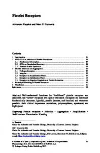
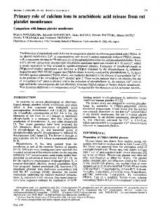
and ceftriaxone following completion of susceptibility studies. The patient’s clinical condition and laboratory parameters improved. On the 19th hospital day, thrombocytopenia was noticed and considered secondary to sepsis. The platelet count as enumerated by the Cell-DYN 4000 (Abbott Diagnostics, Santa Clara, CA) was 42 × 103/µL (42 × 109/L). No platelet flags were generated, and the platelet histograms were unremarkable. In keeping with laboratory review criteria, microscopic examination of the peripheral blood smear was performed. Herein ❚Image 1A❚, the platelet count seemed to be lower than that enumerated by the analyzer. In addition, numerous extracellular ❚Image 1B❚ and intracellular yeast forms were observed. A manual platelet count was performed subsequently by light microscopy using a hemacytometer chamber (Unopette method, Becton Dickinson, Franklin Lakes, NJ) and enumerated at 20 × 103/µL (20 ×
A
Discharge
Reference Range
24,100 (24.1) 2.84 (2.84) 8.7 (87) 27.1 (0.271) 95.1 (95.1) 30.4
3,900-9,200 (3.9-9.2) 4.30-5.60 (4.30-5.60) 12.6-16.6 (126-166) 39.0-49.0 (0.390-0.490) 81.0-97.0 (81.0-97.0) 27.0-33.0
32.0 (0.320) 16.9 179.0 (179)
32.0-35.0 (0.320-0.350) 10.2-13.4 151-368 (151-368)
10.3
6.5-10.9
10 9 /L). Microbial culture of this blood specimen grew Candida glabrata (BACTEC 9240, Becton Dickinson, Cockeysville, MD). Intravenous line colonization was considered the source of fungemia and was managed by line replacement. Microbial blood culture also revealed persistent bacteremia with Providencia stuartii. Gentamicin therapy was begun; therapy was switched to ceftriaxone after completion of susceptibility studies. The patient subsequently was discharged on the 25th hospital day and returned to the nursing home in stable condition, where he completed his ceftriaxone therapy.
Discussion The methods commonly used for enumerating platelets on high-volume hematology analyzers include electrical
B
❚Image 1❚ A, Peripheral blood smear demonstrating variation in platelet size (Wright-Giemsa, ×1,000). B, Peripheral blood smear demonstrating extracellular yeast forms (Wright-Giemsa, ×1,000).
© American Society for Clinical Pathology 883
Am J Clin Pathol 2003;120:882-885 883 DOI: 10.1309/H2K6E59LYFE63GPB 883
Latif et al / SPURIOUS AUTOMATED PLATELET COUNT
impedance, optical scatter, or a combination of these methods.1 Spurious elevation of the platelet count has been reported when nonplatelet material such as cytoplasmic fragments of RBCs and WBCs are enumerated erroneously as platelets.2-7 Other reports of interference with the platelet count on some of the newer large-volume hematology analyzers include autoagglutinins, cryoglobulins, cold agglutinins, platelet satellitosis, platelet clumps, giant platelets, microcytic RBCs, malaria-infected RBCs, bacteria, and leukemic cell fragments in the tumor lysis syndrome.8-12 The Cell-DYN 4000 uses optical scatter and electrical impedance to count and size platelets.13 By optical scatter, platelets are counted by measurement and analysis of scattered light from an argon-ion laser as it intersects and disperses off the platelet surface.1 Electrical impedance is used to evaluate platelet size and verify the platelet count by measuring the platelet’s ability to impede the movement of the electrical charge.1 By combining optical scatter and electrical impedance, the requirement for smear verification, hand counting, and other external validation has been reduced.13 The Cell-DYN 4000 also has the capability of performing an immunophenotypic platelet count. This assay uses the monoclonal antibody to CD61 and enumerates platelets by fluorescence.14 The immunoplatelet count by flow cytometric analysis, also known as the RBC/platelet ratio, is now considered the gold standard for platelet counting.15 This assay is not performed in our laboratory, and the immunophenotypic platelet count on the Cell-DYN 4000 became commercially available only after the present investigation. Flow cytometric studies performed on cultured C glabrata revealed no evidence of CD61 expression, and no nonspecific fluorescence was detected. Since the yeast form in this patient’s blood ranged from 2 to 4 µm, the inability of the analyzer to distinguish yeast forms from platelets is readily explained. No other potential interfering substances were identified on the peripheral blood smear. Subsequent analysis of aliquots of different yeast forms suspended in saline and platelet-poor plasma revealed that C glabrata demonstrated patterns of electrical impedance and optical scatter similar to those of platelets. The volume of the organism by impedance averaged 12.3 fL. Since our patient was thrombocytopenic with increased numbers of large platelets and increased mean platelet volume and platelet distribution width values (Image 1A), these small yeast forms (Image 1B) were not distinguished easily from large platelets. Subsequent analysis of larger yeast forms, including Candida albicans and Candida parapsilosis, did not demonstrate electrical impedance or optical scatter patterns similar to those of platelets. Although many laboratories use stringent criteria for confirmation of thrombocytopenia, spurious increases in platelet counts are verified less frequently. Also, as the 884 884
Am J Clin Pathol 2003;120:882-885 DOI: 10.1309/H2K6E59LYFE63GPB
technical sophistication of hematology analyzers continues to increase, results frequently are reported without microscopic verification. While the controversy regarding which technology (optical scatter or electrical impedance) is superior for platelet counting likely will continue,13,16 in this patient, both methods incorrectly identified the yeast forms as platelets. With the increasing sophistication of automated hematology analyzers, the microscopic examination of peripheral blood smears has been limited by criteria defined by each laboratory. However, as exemplified in the present case, it is unlikely that light microscopy will be eliminated completely. Interfering substances continue to be reported for most of the routinely measured analytes. Substances most commonly reported to interfere with platelet counts are listed in the preceding text. One would expect that with the incorporation of immunophenotyping into routine hematology analyzers, the number of reported interfering substances will decrease. Many smaller laboratories, however, will not be able to afford the cost of immunoplatelet counting. Despite technical advances, laboratory professionals will need to retain a knowledge base concerning basic principles of instrumentation and the substances or conditions that interfere with clinical laboratory assays such as the platelet count. Unusual or unexpected results constitute a red flag that requires confirmation of results by ancillary methods and encourages laboratory professionals to continue to modify and refine review criteria as instrument technology and clinical practice evolve. From the Department of Pathology, Louisiana State University Health Sciences Center, Shreveport. Address reprint requests to Dr Veillon: Dept of Pathology, LSUHSC, 1501 Kings Highway, Shreveport, LA 71130.
References 1. Morris MW, Davey FR. Basic examination of blood. In: Henry JB, ed. Clinical Diagnosis and Management by Laboratory Methods. Philadelphia, PA: Saunders; 2001:479-519. 2. Cornbleet J. Spurious results from automated hematology cell counters. Lab Med. 1983;14:509-514. 3. Patrick CH, Lazarchick J. The effect of bacteremia on automated platelet measurements in neonates. Am J Clin Pathol. 1990;93:391-394. 4. Akwari AM, Ross DW, Stass SA. Spuriously elevated platelet counts due to microspherocytosis. Am J Clin Pathol. 1982;77:220-221. 5. Di Giovanni S, De Matteis MA, Ciocca D, et al. Pseudothrombocytosis and pseudoleukocytosis in a case of essential mixed cryoglobulinemia (type II). Clin Exp Rheumatol. 1986;4:143-145. 6. Maitra A, Ward PC, Kroft SH, et al. Cytoplasmic inclusions in leukocytes: an unusual manifestation of cryoglobulinemia. Am J Clin Pathol. 2000;113:107-112.
© American Society for Clinical Pathology
Hematopathology / CASE REPORT
7. Bonifazi F, Stanzani M, Bandini G. A case of pseudothrombocytosis. Haematologica. 1999;84:275. 8. Survey of instruments: high-volume hematology analyzers. CAP Today. December 2001;15:39-41, 44-46, 48. 9. Von Ahsen N, Ehrlich B, Scott CS, et al. Cryoglobulins interfere with platelet counts by optical and impedance methods but not with the CD61 immunoplatelet count. Clin Chem. 2001;47:1858-1860. 10. Gloster ES, Strauss RA, Jimenez JF, et al. Spurious elevated platelet counts associated with bacteremia. Am J Hematol. 1985;18:329-332. 11. Crabbe G, Van Poucke M, Cantinieaux B. Artefactuallynormal automated platelet counts due to malaria-infected RBC. Clin Lab Haematol. 2002;24:179-182. 12. Li S, Salhany KE. Spurious elevation of automated platelet counts in secondary acute monocytic leukemia associated with tumor lysis syndrome. Arch Pathol Lab Med. 1999;123:1111-1114.
13. Kunicka JE, Fischer G, Murphy J, et al. Improved platelet counting using two-dimensional laser light scatter. Am J Clin Pathol. 2000;114:283-289. 14. Gill JE, Davis KA, Cowart WJ, et al. A rapid and accurate closed-tube immunoassay for platelets on an automated hematology analyzer. Am J Clin Pathol. 2000;114:47-56. 15. International Council for Standardization in Haematology Expert Panel on Cytometry and International Society of Laboratory Hematology Task Force on Platelet Counting. Platelet counting by the RBC/platelet ratio method: a reference method. Am J Clin Pathol. 2001;115:460-464. 16. Sandhaus LM, Osei ES, Agrawal NN, et al. Platelet counting by the Coulter LH 750, Sysmex XE 2100, and Advia 120. Am J Clin Pathol. 2002;118:235-241.
© American Society for Clinical Pathology 885
Am J Clin Pathol 2003;120:882-885 885 DOI: 10.1309/H2K6E59LYFE63GPB 885