mitogen phytohaemagglutinin stimulated respiration to the same extent as ConA, but did not increase the mitochondrial calcium pool. In addition, respiration was ...
Biochem. J. (1988) 256, 167-173 (Printed in Great Britain)
167
Stimulation of respiration by mitogens in rat thymocytes is independent of mitochondrial calcium Patricia L. LAKIN-THOMAS and Martin D. BRAND Department of Biochemistry, University of Cambridge, Tennis Court Road, Cambridge CB2 IQW, U.K.
The role of calcium in the control of respiration by the mitogen concanavalin A (ConA) was investigated in rat thymocytes. ConA induced an increase in both mitochondrial respiration and the mitochondrial calcium pool. The stimulation of respiration was shown to be independent of the increase in mitochondrial calcium: the calcium pool declined after 3 min, whereas the respiration increase was persistent, and was not affected by depletion of the calcium pool or by buffering intracellular Ca2" transients with quin2. The mitogen phytohaemagglutinin stimulated respiration to the same extent as ConA, but did not increase the mitochondrial calcium pool. In addition, respiration was unaffected by changes in the mitochondrial calcium pool induced by increasing or decreasing extracellular calcium. These results indicate that control of respiration is not located in the Ca2"-sensitive mitochondrial dehydrogenases. The ConA-induced increase in respiration could be blocked by oligomycin, suggesting control by cytoplasmic ATP turnover, and was not associated with detectable changes in NAD(P)H fluorescence, indicating a balance between increased electron transfer and increased supply of reduced substrates. INTRODUCTION The factors which control respiration rate, and the mechanisms by which changes in respiration rate are brought about, have not been thoroughly described for any intact cell system. Comparatively more is known about the control of respiration in isolated mitochondria; when this information is extrapolated to intact cells, it can be argued that respiration is probably controlled by relatively few factors: the kinetic properties of the components of oxidative phosphorylation, the supply of substrates (NADH, reduced ubiquinone and 02) and the phosphorylation state of the adenine nucleotides (ATP/ ADP ratio) (for review, see [11). One mechanism for controlling the supply of reduced substrates is by controlHing the activity of intramitochondrial dehydrogenases. Denton & McCormack [2,3] have proposed that increases in cytoplasmic free Ca2" can lead to parallel increases in mitochondrial Ca2" and activation of Ca2"-sensitive dehydrogenases. Extracellular stimuli which activate ATP-utilizing processes often increase cytoplasmic Ca2" as well, and the Ca2+activation of dehydrogenases may be a mechanism for enhancing the supply of ATP without decreases in the ATP/ADP and NADH/NAD+ ratios [2]. There is now convincing evidence that Ca2"-mobilizing stimuli do lead to a Ca2"-dependent activation of dehydrogenases in rat liver [4], rat heart [5] and pig lymphocytes [6]. We have been studying the mitogenic stimulation of rat thymocytes as a model system for the control of respiration. Mitogens have previously been shown to stimulate respiration in lymphocytes in general and in rat thymocytes in particular (reviewed in [7]). A number of growth factors and mitogens rapidly mobilize calcium and increase cytoplasmic free Ca2" in many cell types, and this has also been demonstrated in mouse and rat thymocytes [8,9]. It has been proposed [10,11] that the
increase in cytoplasmic Ca2" in lymphocytes may increase mitochondrial Ca2+ and activate dehydrogenases, accounting for the observed stimulation of respiration. In a previous paper [12], we described a method for estimating the size of the mitochondrial calcium pool in rat thymocytes, and demonstrated an increase in the size of this pool after mitogenic stimulation by concanavalin A (ConA). In the present paper we investigate the relationship between the mitochondrial calcium pool and respiration rate, and demonstrate that the mitogeninduced increase in respiration does not require an increase in the mitochondrial calcium pool; moreover, the mitochondrial calcium pool does not play a significant role in the control of respiration in these cells under these conditions.
EXPERIMENTAL Preparation of thymocytes Thymocytes were prepared from 4-6-week-old female rats as previously described [12]. Incubation medium was RPMI 1640 as previously described [12], containing 0.42 mM-calcium, 2 mM-glutamine and buffered with 10 mM-Hepes and 24 mM-NaHCO3. For fluorimetry experiments [intracellular free Ca2" in quin2-loaded cells or NAD(P)H fluorescence], RPMI was prepared by the formula supplied by Flow Laboratories, but without adding vitamins or Phenol Red (which increase the fluorescence of the medium). Viability of freshly isolated cells as determined by Trypan Blue exclusion was greater For all experiments, cells were incubated at than 95 5 x 10⁷ cells/ml at 37 °C unless otherwise indicated. Mitochondrial calcium pool 45Ca labelling and calculation of intracellular calcium were as previously described [12]. Briefly, cells were 00.
Abbreviations used: Ca2 , free calcium; calcium, total or exchangeable calcium; ConA. concanavalin A:FCCP, carbonyl cyanide ptrifluoromethoxyphenylhydrazone; TPA, 12-O-tetradecanoylphor-bol 13-acetate.
Vol. 256
P. L. Lakin-Thomas and M. D. Brand
168
labelled with 45Ca and 3H20 and rapidly centrifuged through a wash medium containing 2 mM-EGTA (final concn.) and an oil layer. Total pellet radioactivity was corrected for extracellular volume, and the ratio of 45Ca/3H20 in the cell pellet was converted into intracellular [calcium] in 4umol/l of cell water. The technique measures exchangeable calcium, which is assumed to be proportional to total calcium and free Ca2+. To measure the mitochondrial calcium pool, cells were incubated with 10 ,g of oligomycin/ml with or without 5 ,uM-rotenone, for 5 min before centrifugation. The difference in intracellular calcium between these two treatments is described as the rotenone-releasable calcium pool, and is taken to represent the exchangeable mitochondrial calcium pool, as discussed previously [12]. For experiments involving 4 mm and 5 mm external calcium, the EGTA concentration in the wash medium was increased from 2 mm to 20 mm, resulting in a final free Ca2+ concentration of about 80 nm (calculated by the method of Fabiato & Fabiato [13]); the time of exposure of the cells to the wash medium was increased from less than 10 s to about 20 s. Respiration Oxygen consumption was measured in a Clark-type oxygen electrode at 37 °C, 1 ml of cell suspension being used. Additions were made after the cells had established a basal rate of respiration (for 3-5 min). ToQ correct for any small non-linearity of the electrode response and changes in basal rates of respiration, experimental runs were alternated with control runs, and all additions (including solvent controls) were made when the medium had reached the same percentage saturation of oxygen. The rate of respiration was calculated immediatedly before and 3 min after addition, and the percentage change in the rate after addition was determined. Several replicate runs were performed on each cell preparation, and the effect of a treatment was calculated as the difference between the mean percentage changes of the experimental and control runs. Basal rates of respiration were 4.2 + 0.2 nmol of 0/min per 107 cells (mean + S.E.M. for 13 cell preparations). Quin2 loading and intracellular free Ca2` Thymocytes were incubated at 37 °C for 30 min with 10 /IM-[3H]quin2 acetoxymethyl ester (5.4 Ci/mol) in dimethyl sulphoxide, and washed twice by centrifugation and resuspension in fresh medium at room temperature; viability was unaffected by the loading and washing procedure. Considerable leakage of quin2 out of the cells was found when cells were maintained at 37 °C after labelling and washing, and therefore labelled cells were kept at room temperature and warmed to 37 °C for several minutes before measurement of fluorescence. The intracellular quin2 concentration was determined from the radioactivity in a sample of the washed cell suspension and the average cell volume (0.1 1 pl/cell) obtained in the 45Ca-labelling experiments; intracellular quin2 ranged from 0.62 to 0.84 mM. Quin2 fluorescence was measured in a fluorescence spectrophotometer with excitation and emission wavelengths of 339 and 495 nm, by using a I ml quartz cuvette thermostatically maintained at 37 'C. Intracellular free Ca2. was calibrated by the method of Tsien et al. [14]: F.. was obtained by adding 0.012% (v/v) Triton X- 100+50,M-EGTA, and Fm1n was determined by a subsequent addition of 20 mM-EGTA. The
observed fluorescence was corrected for extracellular quin2 (estimated by adding 20 mM-EGTA in the absence of Triton) and for the small change in autofluorescence on adding Triton (determined in cells without quin2). NAD(P)H fluorescence Autofluorescence was measured with a fluorescence spectrophotometer by using excitation and emission wavelengths of 355 and 450 nm. These wavelengths were chosen after determining reduced-versus-oxidized spectra, scanning emission at a fixed excitation wavelength and vice versa. Cells were maintained at 37 °C in a thermostatically controlled 1 ml quartz cuvette. Baseline fluorescence was established for 3-5 min before additions. The observed changes in fluorescence after additions were corrected for the autofluorescence of the reagents themselves; these addition artifacts were estimated from sequential additions to cells. Statistics Results are reported as means+ S.E.M. (n). Data were evaluated by Student's t test for paired or unpaired data as appropriate; P values of less than 0.05 were considered significant. RESULTS The- dose-response relationship between ConA and stimulation of respiration is presented in Fig. 1. Respiration in these cells was inhibited to approx. 20 of the basal rate by 1 /tM-myxothiazol, indicating that nearly all respiration was mitochondrial; the increase in respiration with ConA was completely abolished by myxothiazol. The response of the mitochondrial calcium pool to ConA is presented in Fig. 1 for comparison. The curves are roughly similar, although the optimal ConA -
4
25
60
'&, 0
50
20
0 w-
0
4)
40
U,
a)
15
T 0
30
10
E E ._5
20
C-
.a U,
m
'70
QC 4,
s°0
10
0
0 0
10
20
30
40
50
60
7(
ConA (pg/mi) Fig. 1. Concentration-dependence of ConA effects on respiration and mitochondrial calcium
Thymocytes were treated with various concentrations of ConA for 3 min before measurement of respiration increase (A) or mitochondrial calcium (A) as described in the Experimental section. Results shown are means + S.E.M. for 3-16 cell preparations for respiration, and 4-12 preparations for calcium determinations.
1988
Respiration and calcium in thymocytes
169
.--
0)
:3 C.) 0
-3
E E .ID 0 0 c 0 0
8 Time (min)
3
18
13
20
01) U)
15
01) E.)
10 0
.a U) 0)
5
0
2
6 4 Time (min)
8
10
Fig. 2. Time course of ConA effects on mitochondrial calcium and respiration Thymocytes were treated with ConA for various times before measurement of mitochondrial calcium or respiration increase as described in the Experimental section. ConA concentrations: El, 2.5 ,g/ml; *, 12.5 ,ug/ml. Results shown are means + S.E.M. for n cell preparations as indicated below. (a) Mitochondrial calcium; n = 6 for 2.5,g of ConA/mi, n = 7 for 12.5 ,g/ml; * significantly different from control (zero time). (b) Respiration increase: n = 4; ** not significantly greater than zero. concentration for the increase in respiration may be higher than the optimum for the increase in mitochondrial calcium. The optimal mitogenic concentration of ConA was previously determined [12] to be 5-7.5 ,g/ 108 cells, or approx. 2.5-3.8,ig/ml for the cell densities used in these experiments. ConA at 12.5 ,ug/ml was used for the following experiments (unless otherwise indicated), as this concentration gave reproducible and readily detectable responses. The time course for the ConA effect on the mitochondrial calcium pool was previously reported for 2.5 ,ug of ConA/ml [121. Fig. 2 shows the effects of 2.5 and 12.5 ,ug of ConA/ml on the mitochondrial calcium pool and on respiration. Although the calcium pools had reached a peak and subsequently declined by 8 min after ConA addition (Fig. 2a), the stimulation of respiration was persistent (Fig. 2b) and had not declined even after 60 min (results not shown). These results indicated that
Vol. 256
the persistent stimulation of respiration was not correlated with changes in the mitochondrial calcium pool. To determine whether the initial increase in respiration (within 3 min) required an initial increase in the mitochondrial calcium pool, we pre-treated cells with EGTA before ConA addition. A 3 min pre-treatment with EGTA completely blocked the increase in the calcium pool and depleted the pool found in resting cells as well, but did not block the ConA-induced increase in respiration (Table 1, line 3). The mitogenic lectin phytohaemagglutinin, which has been shown to be mitogenic for these cells [15], also induced an increase in respiration, but did not increase the mitochondrial calcium pool (Table 1, line 4). From these results it appeared that the stimulation of respiration did not require an increase in the mitochondrial calcium pool at 3 min. It is possible, however, that a rapid Ca2" 'spike' could be necessary to trigger some process required for the increase in respiration, and such a spike would not be detected by the method used to measure the mitochondrial calcium pool. Rapid Ca"+ transients have been seen in several cell types loaded with the sensitive Ca2" indicators fura2 or indol: for example, in mouse B lymphocytes stimulated by anti-Ig, a rapid Ca2" spike is seen in individual cells loaded with fura2 [16] and in cell populations loaded with indol [17]. Such rapid Ca2" transients can be prevented by loading cells with a high concentration of a calcium buffer [17], and we have therefore measured respiration in cells loaded with the fluorescent Ca2" indicator quin2. Fig. 3 presents typical fluorimeter traces of the ConA effects on cytoplasmic free Ca2" in quin2-loaded cells. The mean Ca2" concentrations measured for three cell preparations were 76 + 6 nm in resting cells and 301 + 56 nm 3 min after ConA addition. Pre-treatment with EGTA for 3 min largely blocked the increase in Ca2+, and no rapid transients were seen; the increase in cytoplasmic free Ca2` after ConA was less than 25 nM. The effects of quin2 loading on respiration are presented in Table 1, lines 5 and 6. Quin2 did not significantly affect the respiration rate of resting cells (results not shown), and did not block the ConA-induced increase in respiration. Pre-treatment with EGTA partially blocked this increase. This experiment does not rule out the possibility that the small increase in Ca2" seen with EGTA-treated quin2loaded cells was necessary for the increase in respiration, but it is clear that no large Ca2` transients were required. The lack of effect of calcium depletion on the ConAinduced respiration increase led us to consider whether changes in mitochondrial calcium by other means could affect respiration. In quin2-loaded cells, increasing the extracellular calcium concentration from 0.42 mm to 4 or 5 mm produced large and variable increases in cytoplasmic free Ca2` (roughly 10-fold; results not shown). From studies with isolated mitochondria [18], it can be predicted that such an increase in cytoplasmic Ca2` should lead to an increase in mitochondrial Ca2", and we assayed the expected increase in the exchangeable mitochondrial calcium pool in 45Ca-labelled cells. With high external calcium, it was necessary to modify the technique for removing extracellular bound 45Ca (see the Experimental section). With this wash' procedure, the ConA-induced increase in mitochondrial calcium appeared to be only 9 Ismol/l (rather than 46 umol/I; see Table 1, line 2), and therefore the mitochondrial calcium pools reported for high external "calcium may be
170
P. L. Lakin-Thomas and M. D. Brand
Table 1. Effects of various treatments on mitochondrial calcium and respiration
Thymocytes were assayed for mitochondrial calcium and respiration increase (as described in the Experimental section) 3 min after additions. Respiration increase was measured relative to untreated controls (line 1). ConA was 12.5 /tg/ml in all cases. For rows 3 and 6, 5 mM-EGTA was added 3 minutes before ConA. For rows 7 and 8, 'calcium' is total extracellular calcium. Results are shown as means +S.E.M. for (n) cell preparations; N.D., not determined. *not significantly greater than zero; **not significantly different from ConA alone; *** significantly different from ConA alone. Mitochondrial calcium pool (4umol/l of cell water)
Addition
1 2 3 4
None ConA EGTA + ConA Phytohaemagglutinin
5 6 7 8 9
Quin2-loaded cells: ConA EGTA + ConA Calcium (4 mM) Calcium (5 mM) EGTA
11.1±4.6(6)
Respiration
(0O increase) (0.0)
46.3+ 8.3 (10) 0.5 ± 3.0 (3)* -5.1+0.8 (3)*
11.3+ 1.5 (16) 17.6+ 3.9 (3)** 15.6+ 1.9 (4)
N.D. N.D. 220 ± 34 (4) 251 ±42 (4) -0.3± 3.6 (8)*
16.5+0.9 (4) 12.4+ 1.0 (4)*** 3.0+0.5 (4) 3.0+ 3.2 (4)* -5.5+2.1 (4)*
(5 ,ug/ml)
2
c
270
(a)
CllonA
._
X0
77 _ 2 min
(b)
i
EGTA
C.onA
Fig. 3. Effect of ConA on intracellular free Ca2` Thymocytes were loaded with quin2 before assay of intracellular free Ca2+ by fluorimetry. Ca2+ was calibrated as described in the Experimental section. (a) 12.5 ,g of ConA/ml; (b) S mM-EGTA was added 3 min before ConA. Traces shown are typical of three cell preparations.
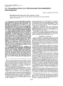
underestimated. Increasing the extracellular total calcium concentration from 0.42 mm to 4 or 5 mm resulted in very large increases in the mitochondrial calcium pools without large increases in respiration (Table 1, lines 7 and 8). Extracellular calcium at 10 mm or higher resulted in a large increase in respiration (results not shown); however, this increase was abolished by 0.5 mM-ouabain, indicating that it resulted from increased Na+/K+ATPase activity, which in turn may have been driven by Na+ or other ion movements across the plasma membrane. Depletion of the mitochondrial calcium pool in resting cells by addition of EGTA did not significantly inhibit respiration (Table 1, line 9). Large changes in the mitochondrial calcium pool therefore had little or no effect on respiration. Having demonstrated that the ConA-induced increase in respiration was independent of changes in the mitochondrial calcium pool, we attempted to identify
other factors which might control respiration in these cells. One possibility would be an increased supply of reduced substrates from glycolysis. When cells were treated for 20 min with the glycolytic inhibitor 2-deoxyD-glucose at -50 mm, ConA could still induce an increase in respiration (results not shown), indicating that glycolytic flux may not be required for the response. We next explored the possibility that the respiration response may result from a decrease in the cytoplasmic ATP/ADP ratio. Oligomycin, an inhibitor of the mitochondrial ATPase, completely reversed the ConA-induced stimulation of respiration (Fig. 4), indicating that the response requires mitochondrial ATP synthesis. Several inhibitors of ATP-utilizing processes were screened for their effects on the basal respiration rate and on the ConA response, but we were unable to identify a likely source for the stimulation. Inhibition of the Na+/K+-ATPase with 0.5 mM-ouabain inhibited basal respiration by less than 1 %, and had no effect on ConA stimulation. The cationophore gramicidin at 0.1 tM stimulated respiration by 3700, presumably by collapsing the Na+/K+ gradient and stimulating the Na+/K+-ATPase, and this stimulation was completely blocked by ouabain; this experiment demonstrated that respiration could be stimulated by ATP turnover in these cells, and that ouabain was effective as an inhibitor at this concentration. The protein-synthesis inhibitor cycloheximide at 100,ug/ml inhibited basal respiration by 10 , but had no effect on ConA stimulation. Actinomycin D, an inhibitor of RNA synthesis, and colchicine, an inhibitor of microtubule treadmilling, had no effect on either the basal or the stimulated respiration rate, at concentrations of 10 jug/ ml and 300 ng/ml respectively. Our results indicated that increases in calcium did not stimulate respiration in these cells, and that increased glycolytic flux was not required for the ConA response. The stimulation of respiration also appeared to be independent of protein kinase C activation: addition of 500 nM-TPA had no effect on respiration after 3 min (°0 increase= - 1.3+0.7 for three cell preparations), and pre-treatment with TPA for 3 minibefore addition of
1988
Respiration and calcium in thymocytes
120
*
100i ,
171
DISCUSSION *
\
The results presented here indicate that in rat
thymocytes changes in respiration rate and changes in
related. The ConA-induced in the absence of any increase in mitochondrial calcium (Table 1, line 3) and in No 60 ~ the absence of any large cytoplasmic Ca2l transients ae 60 \ (Table 1, lines 5 and 6). Not only does the ConA-induced respiration increase appear to be independent of calcium X 40 i ifluxes, but respiration in these cells is insensitive to .L 20 changes in calcium pools by other methods. A large increase in the mitochondrial calcium pool has only a small effect on respiration (Table 1, lines 7 and 8). We . . . , . , , 0 2 4 6 8 10 conclude that calcium-activated processes, either cytoplasmic or mitochondrial, are not sufficient to stimulate Time (min) respiration. This implies that calcium cannot control Fig. 4. Effect of oligomycin on ConA-induced increase in respiration by activation of Ca2"-sensitive ATPases in respiration the cytoplasm. It also implies that Ca 2 cycling across the mitochondrial inner membrane does not make a Thymocytes were treated with 12.5,ug of ConA/ml ) or significant contribution to respiration, even under conmedium (A) at zero time, followed by 50 ng of oligomycin/ ditions where the calcium load is increased by ConA or ml at 3 min (indicated by the arrow). Respiration was high extracellular calcium. A third implication is that assayed as described in the Experimental section, and Ca2' activation of intramitochondrial dehydrogenases respiration rate is reported as percentage of the initial rate does not have a significant effect on respiration rate in (at zero time). Results shown are means+S.E.M. for four these cells. cell preparations; *significantly greater than medium control. As reviewed by Denton & McCormack [2], an increase in mitochondrial calcium is expected to activate Ca2 sensitive dehydrogenases. Hume et al. [10] reported an ConA did not block the ConA-induced increase in activation of respiration by low concentrations of Ca2l in rat thymocyte mitochondria respiring on either pyruvate respiration. Thus none of the likely intracellular signals or oxoglutarate, suggesting that these two dehydro(Ca2+ mobilization, protein kinase C activation or genases are Ca2l-sensitive in these cells as they are in all stimulation of glucose uptake) was sufficient to account other vertebrate tissues surveyed [2]. Although we have for the stimulation of respiration. In addition to attempting to identify specific factors not directly measured the activities of these dehydrowhich may be controlling respiration, we also tried a genases in our system, it seems likely that their activities more general approach to determining whether the would have been affected by the changes in mitochondrial calcium. The purified dehydrogenases are reported to ConA-induced respiration increase was driven by substrate supply or ATP demand. Changes in the redox state respond to Ca2 , with half-maximal effects at around of the NAD(P)H pool should indicate changes in the I /tM-Ca2+[2]. Our values for exchangeable mitochondrial balance between the supply of reduced substrates and the calcium (in /tmol/l of cell water) can be used to make rate of electron transfer. To detect any changes in the rough estimates of intramitochondrial free Ca2+, by reduced nicotinamide nucteotide pool, the endogenous using the following assumptions: (1) mitochondria NAD(P)H fluorescence was measured. As discussed by occupy 5 0 of the total cell volume, as in pig lymphocytes Sies [19], the fluorescence signal will include contributions [20]; (2) about 200 of total mitochondrial calcium is from both mitochondrial and cytoplasmic NADH and exchangeable in 60 min, as in rat hepatocytes [21]; (3) intramitochondrial free Ca2` is about 0.0600 of total, as NADPH. To calibrate the response, cells were treated in rat heart [18]; (4) treatments do not affect these with either I ,aM-myxothiazol [an inhibitor of the mitochondrial cytochrome b-c, complex which will percentages; (5) all cells in the population give approxiblock mitochondrial electron transfer and reduce the mately the same response to treatments. By using these NAD(P)H pool] or 1 ,/M-FCCP (an uncoupler which will assumptions, free intramitochondrial Ca2+ in resting cells increase electron transport and oxidize the pool). (Table 1, line 1) is approx. 0.7 /,M; ConA (line 2) increases this to 2.8 /,M, and 5 mM-calcium (line 8) Gramicidin was added at 25 nm, a concentration which increases it to 15 ,tM; EGTA + ConA (line 3) decrease it stimulated respiration to about the same extent as to 0.03,tM, and EGTA alone (line 9) depletes the pool 12.5 ,g of ConA/ml and should therefore mimic any below the level of detection. Thus the changes that we ConA-induced cytoplasmic ATP turnover. A net reobserve in mitochondrial calcium may be in the right duction by myxothiazol could be detected (an increase of 21.6 + 3.7 arbitrary fluorescence units), and an oxidation range for modulating dehydrogenase activity. If the Ca2+sensitive dehydrogenases carried most of the control of by either FCCP or gramicidin (a decrease of 6.2 + 0.7 units for gramicidin), but no effect of ConA could be respiration in these cells, then changes in the mitochondrial calcium pool should produce corresponding detected (0.0 + 0.5 unit). After a 3 min pre-treatment with EGTA, the ConA effect was still not significant changes in respiration rate. With isolated mitochondria, for example from rat liver [22], increases in extra(- 1.3+1.3 units). (The data represent four replicate mitochondrial Ca2" within the physiological range can determinations from one thymocyte preparation, stimulate respiration. We can therefore conclude that in means+ S.E.M.; similar resufts were obtained with four these cells most of the control of respiration must be different preparations.) X 80
\\ \\
_:
-
Vol. 256
calcium fluxes
are not
respiration increase
can occur
172
located elsewhere. This does not rule out the possibility that changes in dehydrogenase activity may play a role in the control of other processes; for example, activation of pyruvate dehydrogenase may be significant in regulating substrate choice by controlling pyruvate use. Calcium effects on respiration have been reported for other cell types. Binet & Claret [23] reported a stimulation of respiration in rat hepatocytes by aadrenergic agonists or vasopressin; chelating extracellular calcium with EGTA inhibited the response. Similarly, the stimulation of respiration in perfused rat liver by phenylephrine [24] or glucagon [25] was reported to be blocked by depleting intracellular calcium pools. McCormack's work [4,5] has demonstrated that hormones which are known to mobilize Ca2" and stimulate respiration in intact cells also increase mitochondrial calcium and thereby activate intramitochondrial dehydrogenases. In all these cases, however, the stimulation of respiration in intact cells has not been shown to depend on Ca2" stimulation of dehydrogenases; the stimulation could be mediated by other effects of hormones, such as increased ATP turnover. Better indirect evidence for a link between respiration and activation of dehydrogenases comes from the analysis of changes in NAD(P)H reduction. In rat hearts, reduction of the NAD(P)H pool corresponding to a stimulation of respiration was observed during increased work load [26]. In rat liver and hepatocytes [27,28], hormones which stimulate respiration and activate pyruvate dehydrogenase also increase NAD(P)H reduction in a calciumdependent manner, and NAD(P)H changes in parallel with cytoplasmic free Ca2". In spite of these studies, there is not yet direct evidence in any intact cell system for control of respiration by Ca2"-sensitive intramitochondrial dehydrogenases. There are, however, several reports of a lack of correlation between respiration and calcium fluxes: calcium depletion was found to have no effect on pyruvate-stimulated respiration in guinea-pig synaptosomes [29], and stimulation of respiration by glucagon or isoprenaline did not correlate with changes in cytoplasmic free Ca2l in rat hepatocytes [30]. If the stimulation of respiration by ConA is not at the level of Ca2"-sensitive dehydrogenases, where is it? We have been unable in these experiments to identify any single process which could account for the change in respiration rate. The inhibition of the response by oligomycin (Fig. 4) initially suggested cytoplasmic ATP turnover. However, we were unable to identify any of the most likely ATP-utilizing processes in the cell as the source of a ConA-stimulated ATP demand, and, surprisingly, none of these processes appears to contribute significantly to the basal respiration rate, although they have been shown to do so in other cell types [31,32]. It should be noted that oligomycin inhibits basal respiration to about 29 0 of control, and the failure to see a ConA stimulation under these conditions may mean simply that control of respiration has shifted to a different, ConA-insensitive, step. Increased substrate supply from glycolysis also does not appear to be necessary for the ConA-induced stimulation of respiration: 2-deoxy-Dglucose had no effect on the ConA-induced increase in respiration. Glycolysis may play only an insignificant role in energy metabolism in lymphocytes [7]; glutamine and fatty acid oxidation may be the major fuels in these cells [I 1]. The measurement of NAD(P)H fluorescence did not
P. L. Lakin-Thomas and M. D. Brand
provide any clues as to the factors controlling respiration in these cells. Neither the reduction expected with increased substrate supply nor the oxidation expected from increased ATP demand (or uncoupling) could be detected after ConA treatment. This is consistent with a previous report from this laboratory that ConA had no effect on the mitochondrial membrane potential in pig and mouse lymphocytes [20]. The lack of effect on NAD(P)H fluorescence indicates that ConA may activate processes both upstream and downstream of the NAD(P)H pool. Ca2" activation of dehydrogenases is apparently not responsible for the upstream activation: if this were the case, then in the presence of EGTA the downstream activation by ConA would produce an oxidation of NAD(P)H, but this was not observed. Halestrap [33] has proposed a direct stimulation of the respiratory chain by glucagon in hepatocytes, but such a downstream activation alone cannot account for. our observations in thymocytes. One candidate for a regulatory signal might be ADP: an increase in the intramitochondrial ADP pool might activate the same dehydrogenases proposed to be Ca2"-regulated, as well as stimulating ATP synthesis. However, our results with gramicidin indicate that a stimulation of ATP turnover alone results in an oxidation of NAD(P)H, and therefore must have a greater effect on downstream that on upstream processes. Many factors may be involved in the ConA-induced increase in respiration in thymocytes, including the provision of reduced substrates from glutamine and fatty acid oxidation and ATP utilization by plasma-membrane transport systems and macromolecular synthesis. Thus a balance between 'push' and 'pull' on oxidative phosphorylation may maintain the NADH/NAD+ ratio and the mitochondrial membrane potential in these cells. We thank Dr T. R. Hesketh for the gift of [3H]quin2 acetoxymethyl ester and for advice on the quin2 experiments, and R. P. Hafner and Dr J. D. Johnston for helpful comments on the manuscript. This work was supported by a Damon Runyon-Walter Winchell Cancer Fund Fellowship (DRG-882) to P.L.L.-T.
REFERENCES 1. Brand, M. D. & Murphy, M. P. (1987) Biol. Rev. 62, 141-193 2. Denton, R. M. & McCormack, J. G. (1985) Am. J. Physiol. 249, E543-E554 3. McCormack, J. G. & Denton, R. M. (1986) Trends Biochem. Sci. 11, 258-262 4. Assimacopoulos-Jeannet, F., McCormack, J. G. & Jeanrenaud, B. (1986) J. Biol. Chem. 261, 8799-8804 5. McCormack, J. G. & Denton, R. M. (1987) Biomed. Biochim. Acta 46, S487-S492 6. Baumgarten, E., Brand, M. D. & Pozzan, T. (1983) Biochem. J. 216, 359-367 7. Hume, D. A. & Weidemann, M. J. (1980) Mitogenic Lymphocyte Transformation, pp. 141-181, Elsevier, Amsterdam 8. Hesketh, T. R., Moore, J. P., Morris, J. D. H., Taylor, M. V., Rogers, J., Smith, G. A. & Metcalfe, J. C. (1985) Nature (London) 313, 481-484 9. Guko,vskaya, A. S. Zinchenko, V. P., Petrunyaka, V. V., Khodorov, B. I. & Evtodienko, Y. V. (1986) Eur. J. Biochem. 161, 249-256
1988
Respiration and calcium in thymocytes 10. Hume, D. A., Vijaykumar, E. K., Schweinberger, F., Russel, L. M. & Weidemann, M. J. (1978) Biochem. J. 174, 711-716 11. Ardawi, M. S. M. & Newsholme, E. A. (1985) Essays Biochem. 21, 1-44 12. Lakin-Thomas, P. L. & Brand, M. D. (1987) Biochem. J. 246, 173-177 13. Fabiato, A. & Fabiato, F. (1979) J. Physiol. (Paris) 75, 463-505 14. Tsien, R. Y., Pozzan, T. & Rink, T. J. (1982) J. Cell Biol. 94, 325-334 15. Culvenor, J. G. & Weidemann, M. J. (1976) Biochim. Biophys. Acta 437, 354-363 16. Wilson, H. A., Greenblatt, D., Poenie, M., Finkelman, F. D. & Tsien, R. Y. (1987) J. Exp. Med. 166, 601-606 17. Bijsterbosch, M. K., Rigley, K. P. & Klaus, G. G. B. (1986) Biochem. Biophys. Res. Commun. 137, 500-506 18. Hansford, R. G. (1985) Rev. Physiol. Biochem. Pharmacol. 102, 1-72 19. Sies, H. (1982) in Metabolic Compartmentation (Sies, H., ed.), pp. 205-231, Academic Press, New York 20. Brand, M. D. & Felber, S. M. (1984) Biochem. J. 217, 453-459 Received 4 May 1988/28 June 1988; accepted 6 July 1988
Vol. 256
173 21. Foden, S. & Randle, P. J. (1978) Biochem. J. 170, 615-625 22. Johnston, J. D. & Brand, M. D. (1987) Biochem. J. 245, 217-222 23. Binet, A. & Claret, M. (1983) Biochem. J. 210, 867-873 24. Taylor, W. M., Reinhart, P. H. & Bygrave, F. L. (1983) Biochem. J. 212, 555-565 25. Kraus-Friedmann, N. (1986) FEBS Lett. 201, 133-136 26. Katz, L. A., Koretsky, A. P. & Balaban, R. S. (1987) FEBS Lett. 221, 270-276 27. Sugano, T., Shiota, M., Khono, H., Shimada, M. & Oshino, N. (1980) J. Biochem. (Tokyo) 87, 465-472 28. Staddon, J. M. & Hansford, R. G. (1987) Biochem. J. 241, 729-735 29. Kauppinen, R. A. & Nicholls, D. G. (1986) FEBS Lett. 199, 222-226 30. Crompton, M. & Goldstone, T. P. (1986) FEBS Lett. 204, 198-202 31. Siems, W., Dubiel, W., Dumdey, R., Muller, M. & Rapoport, S. M. (1984) Eur. J. Biochem. 139, 101-107 32. Muller, M., Siems, W., Buttgereit, F., Dumdey, R. & Rapoport, S. M. (1986) Eur. J. Biochem. 161, 701-705 33. Halestrap, A. P., Quinlan, P. T., Armston, A. E. & Whipps, D. E. (1985) Biochem. Soc. Trans. 13, 659-663