Plant Cell Monogr (000) : DOI 10.1007/7089_2007_135/Published online: 28 July 2007 © Springer-Verlag Berlin Heidelberg 2007
Stomatal Patterning and Guard Cell Differentiation Keiko U. Torii Department of Biology, University of Washington, Seattle, WA 98195, USA
[email protected]
Abstract Gas exchange between plants and the atmosphere takes place through stomata (singular, stoma), which are microscopic valves on the plant epidermis composed of paired guard cells. Stomatal differentiation involves a series of asymmetric divisions of precursor cells followed by a single symmetric cell division that produces terminally differentiated guard cell pairs. Stomatal development emerged as a model system to study how environmental- and cell-cell signals translate into site/orientation of asymmetric cell division and cell-type differentiation. This chapter focuses on cell-state transition events leading to guard cell differentiation in the model plant Arabidopsis, and cell-cell signaling mechanisms controlling stomatal patterning. Understanding how cell-cycle regulators influence stomatal patterning and differentiation will advance our knowledge of cell division control in plant development.
1 Introduction The evolution of land plants relied on the acquisition of mechanisms that protected themselves from the dry atmosphere and harmful UV rays, while allowing gas exchange for photosynthesis; and transpiration for stimulating water movement from the soil to aboveground tissues. The innovation of two distinct cell types on the plant epidermis was critical for solving this challenge. Epidermal pavement cells are tightly-sealed interlocking cells with thick cuticle layers. Stomata act as turgor-driven valves that allow gas exchange and transpiration. It is therefore not surprising that evolutionary biologists believe that the emergence of stomata predates the evolution of leaves, flowers, or even vasculature (Edwards et al. 1998). A stoma consists of a microscopic pore surrounded by a pair of guard cells, which open and close upon sensing environmental signals, such as drought, light, and CO2 concentrations. Given the importance of stomatal function for plant growth and survival, significant research has been done on physiological and molecular bases of stomatal opening/closure as well as their eco-physiological and environmental consequences (Assmann and Shimazaki 1999; Schroeder et al. 2001, 2001; Hetherington and Woodward 2003). For developmental biology, stomata serve as a superb system to understand cell-cell signaling, cell division, stem cell differentiation, cell polarity, and cellular morphogenesis in plants. The steps leading to the differentiation
344
K.U. Torii
of guard cells are uniquely coupled with specific types of cell divisions, the reiterative asymmetric division of precursor stem cells and a single symmetric division that generates a pair of guard cells. The simplicity and tractable nature of the leaf epidermis makes the study of stomatal development technically amenable (Nadeau and Sack 2002; Bergmann et al. 2004). Recent advances in model plant molecular genetics have begun to unravel how genetic and environmental signals act in controlling stomatal patterning. In this chapter, I will introduce the cellular processes of stomatal development with emphasis on the model plant Arabidopsis, and provide the latest updates on emerging cell-cell signaling mechanisms specifying the correct spacing and differentiation of stomata. Potential interactions of cell-cell signaling with intrinsic developmental regulators as well as environmental cues will be explored. Finally, future prospects on integrating cell cycle regulators in the context of stomatal patterning will be presented.
2 Stomatal Development in Arabidopsis Arabidopsis stomata are typically found in complexes with three subsidiary cells, one being distinctly smaller than the others, surrounding a pair of guard cells (Esau 1977; Zhao and Sack 1999; Serna and Fenoll 2000; Nadeau and Sack 2002). These are characteristic “anisocytic” stomatal complexes and are generated through stereotypical cell division patterns (Esau 1977). Stomatal development initiates post-embryonically when populations of protodermal cells, termed meristemoid mother cells (MMC), enter into asymmetric division (Nadeau and Sack 2002). This initial asymmetric division generates two daughter cells with distinct fates. The larger daughter cell differentiates into an epidermal pavement cell. In contrast, the smaller daughter cell, termed a meristemoid, possesses stem-cell like characteristics, as it continues to divide asymmetrically to renew itself over several rounds of divisions (Nadeau and Sack 2002). Typically, meristemoids reiterate 3 rounds of asymmetric division. The repeated asymmetric division of meristemoids will be hereafter referred to as amplifying asymmetric division, as each division increases the number of cells, which we termed stomatallineage ground cells (SLGC), larger daughter cells that function as subsidiary cells (Shpak et al. 2005). SLGCs are also referred to as “subsidiary cells” or “pavement cells” in literature. The meristemoid then differentiates into a round guard mother cell (GMC), which divides symmetrically once to generate a pair of guard cells (Nadeau and Sack 2002). This results in an anisocytic complex with three clonally-related subsidiary cells (Berger and Altmann 2000; Serna et al. 2002). However, the number of asymmetric divisions as well as the clonal relationship among cells constituting the stomatal complex is plastic and variable. For example, a detailed clonal analysis
Stomatal Patterning and Guard Cell Differentiation
345
of stomatal complexes in the adaxial epidermis of Arabidopsis Ler accession by Serna et al. (2002) revealed that, while the vast majority (87%) of anisocytic stomatal complexes derived from single precursor cells, the rest were of polyclonal origins. Geisler et al. (2000) reported that the number of asymmetric divisions in the Columbia accession varies from zero to three. This plastic nature of stomatal ontogeny reflects a dynamic intrinsic developmental program that integrates external cues for adaptation and survival. In fact, environmental factors, such as humidity and CO2 concentrations, are known to affect stomatal density and patterning (Gray et al. 2000; Lake et al. 2002).
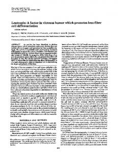
Fig. 1 Stomatal development in Arabidopsis. A Cartoon showing the key steps of stomatal differentiation. Undifferentiated cells in the protoderm can undergo either proliferative division to form pavement cells or asymmetric division to initiate stomatal development. Stage I: a subset of protodermal cells, a meristemoid mother cell (MMC) divides asymmetrically and forms a self-renewing meristemoid that reiterate a few rounds of asymmetric division. Stage II: the meristemoid then differentiates into a round, guard mother cell (GMC). Stage III: the GMC undergoes a single symmetric division. Stage IV: a pair of immature guard cells achieves final morphogenesis to form a functional stoma. The amplifying asymmetric division of meristemoids generates surrounding stomatallineage ground cells (SLGCs) that provide water and ions for stomatal opening and closure. B A polarity of asymmetric division during satellite meristemoid formation. The secondary asymmetric division occurs away from the existing stoma, thereby assuring that two stomata are separated by at least one cell apart (1-cell spacing rule). Modified from Torii (2006)
346
K.U. Torii
The entire process of stomatal patterning and differentiation in Arabidopsis can be divided into the following four critical stages (Fig. 1A). Stage I initiates the entry into the stomatal-lineage via emergence of MMCs (Stage I-a) and commitment to reiterative asymmetric division (Stage I-b). Stage II represents the differentiation of meristemoids into GMCs associated with the loss of potential for asymmetric division. Stage III includes the acquisition of symmetric division potential in GMCs, and finally Stage IV, or guard cell morphogenesis, concludes the process of stomatal development (Fig. 1A). Occasionally, SLGCs initiate asymmetric division and produce satellite meristemoids (Fig. 1B). This secondary asymmetric division occurs in a nonrandom fashon away from the existing stoma (Yang and Sack 1995; Geisler et al. 2000, 2003; Nadeau and Sack 2002). As a consequence, stomata are sep-
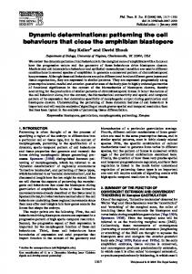
Fig. 2 Stomatal patterning mutants. Shown are the DIC (differential interference contrast) microscopy images of the abaxial rosette leaf epidermis of: A wild type; B tmm; C sdd1; D yoda; E erecta erl1 erl2; F flp-1; G flp-7; and H fama. Images are taken under the same magnification. A scale bar = 20 µm
Stomatal Patterning and Guard Cell Differentiation
347
arated by at least one cell (known as the “one-cell spacing rule”) (Fig. 1B, Fig. 2) (Nadeau and Sack 2002). Proper spacing is critical for physiological functions of stomata, because guard cells must exchange water and ions (e.g. K+ and Cl– ) with surrounding subsidiary cells in order to open and close (Assmann and Shimazaki 1999; Schroeder et al. 2001, 2001; Hetherington and Woodward 2003). The observed, “one-cell spacing rule” indicates that the newly forming meristemoid “knows” the location of pre-existing stoma and avoids stomatal cluster formation by orienting the site of secondary asymmetric division. This suggests the presence of cell–cell communication. In the following sections, I will describe the cytological events during stomatal differentiation and emerging roles of key regulatory genes of stomatal patterning.
3 Stage I-a: Entry Into Asymmetric Division 3.1 Regulation of Orientation and Frequency of Asymmetric Division Thus far, no molecular markers have been reported for MMC identity. The earliest cytological event that clearly distinguishes the MMC is the polarization of the cytoskeleton, which predicts the site of asymmetric division (Lucas et al. 2005). The orientation and frequency of the initial asymmetric divisions and cell-cell interaction among the daughter cells determine the proper density and spacing of stomata. Genes implicated in signal transduction play important roles in stomatal patterning. They include TOO MANY MOUTHS (TMM), STOMATAL DENSITY AND DISTRIBUTION1 (SDD1), YODA (YDA), and three ERECTA-family genes, ERECTA (ER), ERECTA-LIKE1 (ERL1), and ERL2 (Table 1) (Berger and Altmann 2000; Nadeau and Sack 2002; Bergmann et al. 2004; Shpak et al. 2005). Loss-of-function mutations in these genes confer clustered stomata, thus violating the “1-cell spacing rule” (Fig. 2). However, phenotypes of these mutants are not identical, suggesting that their relationships are not simply linear. TMM encodes a receptor-like protein with an extracellular leucine-rich repeat (LRR-RLP), which likely acts as a receptor for a positional cue that specifies the site of asymmetric division (Nadeau and Sack 2002). The phenotypes of tmm mutant plants are organ-dependent and complex: the cotyledons and leaves produce clustered stomata (Fig. 2B); the stems produce no stomata; and pedicels exhibit a gradient of no stomata to stomatal clusters (Yang and Sack 1995; Geisler et al. 1998). This complex phenotype implies that TMM triggers contrasting developmental events in a dosage-dependent manner and that each organ requires a different dosage of TMM. Perhaps, TMM potentiates
348
K.U. Torii
Table 1 Arabidopsis genes regulating stomatal development Gene name
AGI number
Gene Product (putative) Refs.
1: Genes regulating asymmetric division and stomatal patterning ERECTA (ER) a,b At2g26330 ERECTA-LIKE1 (ERL1) a,b At5g62230 ERECTA-LIKE2 (ERL2) a At5g07180 STOMATAL DENSITY At1g04110 AND DISTRIBUTION1 (SDD1) a TOO MANY MOUTHS At1g80080 (TMM) a YODA (YDA) a At1g63700
LRR-receptor-like kinase (Shpak et al. 2005) LRR-receptor-like kinase (Shpak et al. 2005) LRR-receptor-like kinase (Shpak et al. 2005) subtilicin-like (Berger & Altmann 2000; proteinase von Groll et al. 2002) LRR-receptor protein MAPkinase kinase kinase
2: Genes regulating guard cell differentiation FAMA a A3g24140 bHLH protein At1g14350 R2R3 Myb protein FOUR LIPS (FLP) a At2g02820 R2R3 Myb protein MYB88 b
(Yang and Sack 1995; Nadeau and Sack 2002) (Bergmann et al. 2004)
(Bergmann et al. 2004) (Lei et al. 2005) (Lei et al. 2005)
3: Genes regulating guard cell cytokinesis/morphogenesis CYCLIN-DEPENDENT KINASE B1;1 (CDKB1;1) CYTOKINESIS DEFECTIVE1 (CYD1) KEULE c STOMATAL CYTOKINESIS DEFECTIVE1 (SCD1) c
At3g54180 cyclin-dependent kinase (Boudolf et al. 2004) NA
NA (not cloned)
At1g12360 Sec1 protein At1g49040 DENN-WD40 protein
(Yang et al. 1999) (Sollner et al. 2002) (Fabel et al. 2003)
4: Genes mediating environmental control of stomatal density HIGH CARBON At2g46720 Long-chain fatty acid (Gray et al. 2000) DIOXIDE (HIC) biosynthesis Notes: Genes are in alphabetical orders a Photographic images provided in Fig. 2 b Phenotypes largely redundant. Combination of double (or triple) mutations among closely related paralogs revealed synergistic interactions c Weak- or temperature sensitive alleles show guard cell cytokinesis defects. The defects in severe alleles are pleiotropic
the entry into the stomatal pathway at lower concentrations (e.g. in MMC), but at high concentration (e.g. in meristemoids) TMM ensures guard cell differentiation, while inhibiting its neighboring cells from further asymmetric division. SDD1 encodes a subtilicin-like putative extracytoplasmic protease (Berger and Altmann 2000). In animals, this protease family is known to process pep-
Stomatal Patterning and Guard Cell Differentiation
349
tidic ligands to a mature form (Cui et al. 1998). The sdd1 mutant exhibits high stomatal density but with few stomatal clusters (Fig. 2C). This implies that SDD1 may primarily regulate the frequency of initial asymmetric division with lesser effects on specifying the orientation in relation to existing stoma. YDA encodes a putative mitogen activated protein kinase kinase kinase (MAPKKK), a cytoplasmic protein kinase acting at the entry point of MAPK cascades (Bergmann et al. 2004). In plants, animal, and fungi, MAPK cascades integrate and amplify signals transmitted from the upstream cell-surface receptors and activate downstream gene expression in the nucleus to regulate cellular processes. (Serger and Krebs 1995). Unlike tmm and sdd1, the yda mutation is highly pleiotropic. yda was first reported as a mutation defective in the initial asymmetric division of zygote with disrupted embryo patterning (Lukowitz et al. 2004). In addition, yda plants show severe dwarfism, disrupted floral patterning, and male and female sterility (Lukowitz et al. 2004). The epidermis of yda leaves produces high-density stomatal clusters (Bergmann et al. 2004). This phenotype is much more severe than in tmm and sdd1 (Fig. 2D). YDA may act as an on-off switch to repress initial entry into stomatal development when a cell receives a signal from its neighbors, while TMM and SDD1 translate the signal gradient. Consistently, the overlyactive form of YDA (YDA∆NB) severely inhibits the onset of initial asymmetric divisions, resulting in the epidermis consisting solely of pavement cells (Bergmann et al. 2004). Unexpectedly, the ERECTA-family genes were recently shown to play a role in stomatal patterning. ERECTA is a well-known gene regulating plant architecture, in which loss-of-function mutation confers a characteristic compact inflorescence with short pedicels and blunt fruits (Torii et al. 1996). ERECTA encodes an LRR receptor-like kinase (LRR-RLK), a prevalent family of RLKs that play important roles in developmental, steroid-hormone signal transduction, and defense against pathogens (Torii et al. 1996, 2004; Becraft 2002). ERECTA and its two paralogous genes, ERL1 and ERL2, interact in a synergistic manner in regulating stomatal patterning (Shpak et al. 2005). The erecta erl1 erl2 triple loss-of-function mutations confer severe dwarfism, disrupted floral patterning, male- and female sterility, and a high-density stomatal clustering phenotype (Fig. 2E) (Shpak et al. 2004, 2005). Overall, these phenotypes highly resemble those of the yda single mutant, suggesting that the YDA MAPK cascade may function downstream of ERECTA-family RLKs in multiple developmental processes. 3.2 Genetic Interactions and Hierarchy of Signal Transduction Studies of genetic interactions are now illuminating possible cell-cell signal transduction mechanisms regulating stomatal patterning. The overexpres-
350
K.U. Torii
sion of SDD1 inhibited the initial asymmetric division and reduced stomatal density (von Groll et al. 2002). However, the effect was reversed by the tmm mutation, thus placing TMM downstream of SDD1 (von Groll et al. 2002). The simplest interpretation of these results is that the excessive production of mature ligands by overexpression of SDD1 overly inhibited entry into stomatal development via the TMM receptor. YDA most likely acts downstream of the SDD1-TMM pathway. A single copy of YDA∆NB, which by itself does not completely inhibit the entry into stomatal development, was able to suppress the stomatal cluster phenotype of sdd1 and tmm (Bergmann et al. 2004). This indicates that a slight increase in YDA activity was able to resume a normal level of signal transduction in the absence of SDD1 or TMM. Perhaps, signals mediated via TMM are transmitted to the YDA-MAPK cascades to suppress neighboring cells to enter the stomatal-lineage. TMM does not possess any cytoplasmic stretch (Nadeau and Sack 2002). In fact, the TMM protein molecule ends with a 23 amino-acid-long membranespanning region, suggesting that the C-terminal end of TMM does not extend from the plasma membrane to the cytoplasm. In addition, the C-terminus of TMM possesses a GPI (glycosylphosphatidylinositol)-anchor motif, suggesting that TMM may be anchored to the membrane surface (Nadeau and Sack 2002). Such structural features make TMM unlikely to transmit signals by itself, and it is reasonable to speculate that TMM forms a receptor complex with a partner molecule that possesses a cytoplasmic effector domain. Scientists predicted that the partner of TMM would be an LRR-RLK, in light of other systems such as CLAVATA (CLV) in shoot apical meristem development, whereby CLV1 LRR-RLK is thought to form a receptor dimer with CLV2 LRR-RLP (Clark et al. 1997; Jeong et al. 1999). The three ERECTA-family LRR-RLKs are attractive candidates of the TMM partner. The genetic interaction of TMM and ERECTA-family by Shpak et al. (2005) suggest that TMM may restrict the inhibitory action of ERECTAfamily RLKs on the initial entry into asymmetric division. This was evident from their interactions in the stems. The tmm stems give rise to no stomata, while the erecta erl1 erl2 stems form high-density stomatal clusters (Nadeau and Sack 2002; Shpak et al. 2005). The epistatic relationship of TMM and ERECTA-family genes in the stem epidermis exhibited stoiochiometric dynamics: While TMM is epistatic to each one of the ERECTA-family genes, three ERECTA-family genes together are epistatic to TMM. Removing TMM and two out of three ERECTA-family genes resumed nearly wild-type stomatal phenotype (Shpak et al. 2005). It is exciting to speculate that TMM modulates the activity of ERECTA-family RLKs via direct association. However, in cotyledons and leaves, both tmm and erecta-family triple mutants display stomatal clusters. Do they act cooperatively in these organs while acting antagonistically in the stems? Establishing the molecular bases of receptor interactions and identifying their ligands is critical for elucidating the
Stomatal Patterning and Guard Cell Differentiation
351
complex action of TMM and ERECTA-family receptors. By analogy to known LRR-RLPs and LRR-RLKs, the ligands for TMM/ERECTA-family receptors are most likely small peptides.
4 Stage I-b and II: Amplifying Asymmetric Division and Differentiation of Meristemoids The amplifying asymmetric division of a meristemoid occurs in an inward spiral. Serna et al. (2002) documented that the division angle of meristemoids in the Ler adaxial leaf epidermis is exactly 60 degrees with little deviation. Therefore, a newly formed, triangular-shaped meristemoid likely re-establishes its polarity away from the polar end of the previous asymmetric division. This implies the presence of a chemical gradient and a cellular system to translate gradient to determine the site of cytokinesis. Lucas et al. (2006) reported that it is common to have two adjacent meristemoids in wild-type epidermis, given that the initial entry asymmetric division occurs randomly. However, two adjacent meristemoids always divide away from each other to avoid further contact. Such polarity is disrupted in the tmm mutant, indicating that TMM functions in perceiving the positional cues during amplifying asymmetric division. Flexible numbers of amplifying asymmetric division allows developmental plasticity to adjust stomatal density in response to environmental changes. Furthermore, it provides a way to “correct” erroneous division to avoid clustered stomata. The number of amplifying asymmetric divisions may be regulated by ERECTA-LIKE (ERL) genes. The pedicel epidermis of erl1 erl2 double mutants gave rise to stomatal complexes with reduced number of SLGC (stomatal lineage ground cells), implying that the meristemoids completed reduced rounds of asymmetric division and precociously differentiated into GMCs. ERECTA-family regulates the binary decision of daughter cells of an asymmetric division to adopt stomata vs. SLGC fates. Consistently, the epidermis of erecta single mutant produced occasional patches of 2–3 cells that appear to have undergone asymmetric division but failed to differentiate into guard cells. Expression of stomatal-lineage markers, TMM::GUS and ERL1::GUS in these groups of cells supports the hypothesis that both daughter cells became SLGCs. The tmm mutation greatly enhanced this “patches of cells with no stomata” phenotype. (Shpak et al. 2005). Indeed, the tmm erecta double mutations completely eliminated stomata from cauline-leaf and carpel epidermis, leaving numerous small cells that likely underwent asymmetric division and then became SLGC without accompanying guard cells. (Shpak et al. 2005). In both cases, termination of stomatal fate is likely due to misregulation of ERL1, which becomes overly inhibitory in repressing GMC
352
K.U. Torii
differentiation. Consistent with this hypothesis, the additional erl1 mutation reversed the no-stomata phenotype of tmm erecta. Whether TMM regulates ERL1 via direct association awaits further biochemical analysis. A positive regulator of differentiation of a meristemoid has not yet been identified. If such a gene exists, then the loss-of-function mutation may extend the lifespan of meristemoids’ stem cell-like activity, and consequently mutant plants should exhibit excessive rounds of amplifying asymmetric division. If a default pathway of a meristemoid is to adopt pavement cell fate, then the mutant plants lacking the positive regulator may form two pavement cells after entry into asymmetric division, just like those observed in the tmm erecta double mutant background (Shpak et al. 2005). Further isolation of such mutants and molecular cloning of causal genes will help us elucidate the molecular mechanisms of stem cell differentiation in the plant epidermis.
5 Stage VI: GMC Division – Intrinsic Regulation by Transcriptional Factors Once the meristemoid commits to becoming a GMC, it loses its potential for asymmetric division. GMC differentiation is evident from its changes in cellular morphology. The triangular meristemoid cell expands and become oval in shape with characteristic, end wall thickening (Lucas et al. 2006). The GMC achieves, precisely, a single symmetric division. This reflects the fundamental importance of having a pair of guard cells for proper stomatal function. Therefore, mechanisms must be present to ensure exactly one symmetric division occurs. Two genes, FOUR LIPS (FLP) and FAMA, prevent excessive (more than one) symmetric division of the GMC (Bergmann et al. 2004; Lai et al. 2005) The flp-1 mutant was initially isolated from the phenotype of paired stomata, which gives four aligned guard cells (Fig. 2F) (Lai et al. 2005). While adjacent stomata are arranged randomly in stomatal patterning mutants (tmm, sdd1, erecta-family, and yoda), paired stomata in flp are in parallel due to their origin from a single GMC. In the severe allele flp-7, the GMC undergoes reiterative symmetric divisions, which result in formation of a row of “caterpillar-like” guard cell clusters (Fig. 2G). FLP encodes an atypical R2R3type Myb protein, which most likely functions as DNA-binding transcription factor (Lai et al. 2005). Therefore, an attractive hypothesis is that FLP suppresses expression of positive regulators of cell cycle progression promoting GMC division. Interestingly, both weak- (flp-1, flp-2) and severe alleles (flp-7) are predicted to produce truncated proteins with incomplete Myb domains, with weaker alleles producing shorter fragments than the severe one (Lai et al. 2005). This apparent discrepancy between the severity of phenotypes and impacts on protein structure implies a dominant-negative activity of flp-7 gene products, which may interfere with redundant components. Consistent with
Stomatal Patterning and Guard Cell Differentiation
353
this idea, the T-DNA inserted knockout alleles of flp display weak phenotypes (Lai et al. 2005). The paralogous Myb gene, MYB88, is most likely the redundant factor. MYB88 shares high sequence identity (91% amino-acid identity in the MYB domain, 71% overall) and exhibits similar expression pattern with FLP (Lai et al. 2005). Interestingly, while complete loss-of-function mutations in MYB88 failed to confer any visible phenotype, they dramatically enhanced the size of “caterpillar-like” guard cell stacks in the flp mutant (Lai et al. 2005). Moreover, introduction of an extra genomic copy of MYB88 with its own promoter rescued the flp phenotype. The combined dosage of FLP/MYB88 may be critical for guard cell differentiation. FAMA was identified from transcriptional profiling as a gene upregulated in yoda (in which the epidermis is predominantly stomata) compared to YDA∆NB (in which the epidermis is predominantly pavement cells) (Bergmann et al. 2004). The loss-of-function fama phenotype highly resembles that of flp, suggesting that FAMA and FLP function in the same step of GMC differentiation. However, unlike flp, the abnormal “caterpillar-like” clusters in fama never form mature guard cells, suggesting that FAMA suppresses GMC division but in addition promotes the guard cell differentiation program (Fig. 2H). FAMA encodes a bHLH (basic-Helix Loop Helix) protein and most likely acts as a DNA- binding transcription factor. The molecular identity of FLP and FAMA as Myb and bHLH transcription factors highlights an intriguing link to underlying mechanisms of epidermal cell-type differentiation in leaves and roots. Differentiation of trichomes and root hairs requires orchestrated actions of Myb transcriptional activators, which associate with bHLH proteins (Schiefelbein 2000, 2003). It would be therefore of special interest to address whether FLP and FAMA physically associate with each other and constitute a transcriptional regulatory complex.
6 Stage IV: Guard Cell Morphogenesis The final stage of stomatal differentiation involves guard cell morphogenesis, a step leading to the formation of paired guard cells. After a symmetric division of the GMC, the new cell wall forms along the side of division, which develops a pore. The guard cells adopt a characteristic microtubule and microfibril organization (Hepler and Palevitz 1974). Defective cytokinesis leads to abnormal guard cell morphology. The cytokinesis defective1 (cyd1) mutant forms abnormal guard cells with various degrees of cytokinesis defects: ∼ 20% form a single, large round cell lacking any ventral wall or pore, ∼ 10% have incomplete wall with pore, and ∼ 5% form a single round guard cell with partial cell wall protrusions (Yang et al. 1999). These abnormal cells are either single- or bi-nucleated, each correlating with the extent of cytoki-
354
K.U. Torii
netic defects (Yang et al. 1999). The molecular identity of CYD1 is not known. The temperature-sensitive stomatal cytokinesis-defective1-1 (scd1-1) mutant exhibits abnormal guard cells similar to the cyd1 mutant (Falbel et al. 2003). SCD1 encodes a protein with two domains (DENN domain and WD-40 repeats), and these structural features imply a possible role for SCD1 in vesicle trafficking during cell-plate formation. Consistently, the null alleles of scd1 exhibit pleiotropic defects in cytokinesis and polar cell expansion. Therefore, abnormal guard cell morphology may be a sensitive indicator of general cytokinesis defects, which can be exploited to recover weak alleles of key regulatory genes for cytokinesis. Consistently, the weak alleles of KNOLLE and KEULE, two genes initially isolated as regulators of embryogenesis, display abnormal stomatal morphology (Sollner et al. 2002). KNOLLE and KEULE encode syntaxin and Sec1, respectively, two physically-interacting proteins required for vesicle fusions at the nascent cell plate (Lukowitz et al. 1996; Waizenegger et al. 2000). In addition to cytokinesis, the cell cycle defects may confer abnormal guard cells (see below).
7 Cell Cycle Regulation in Stomatal Patterning Stomatal patterning and differentiation is tightly coupled with specific types of cell division: the initial asymmetric division of MMCs, the amplifying asymmetric division of meristemoids, and a single symmetric division of GMCs. In addition, genome replication is strictly controlled during stomatal development, as guard cells remain at 2C (diploid) unlike the rest of epidermal cells that undergo endoreduplication (Melaragno et al. 1993). The obvious questions are whether specific cell cycle regulators control distinct cell division types during stomatal development and, if so, whether forcing cell cycle switches can invoke/suppress stomatal development. Studies suggest that cell cycle regulators may influence stomatal patterning, but they do not impinge on stomatal differentiation. The promoter of the Arabidopsis CTD1 gene, which encodes a subunit of the DNA-replication licensing complex together with AtCDC6, is highly active in stomatal-lineage cells (Castellano Mdel et al. 2004). The AtCTD1::GUS promoter activity resembles that of TMM and ERL1, with highest activity in meristemoids and GMCs, and moderate activity in SLGCs (Castellano Mdel et al. 2004). Overexpression of AtCDT1 and AtCDC6 slightly increased the numbers of stomata, but it did not lead to formation of adjacent stomata. These results suggest that the DNA-replication licensing complex may promote stomatal asymmetric division and that forcing G1-to-S phase transition may slightly increase the MMC specification. However, this is not sufficient to overcome negative regulation by cell–cell signaling components encoded by SDD1, TMM, YODA, and ERECTA-family genes.
Stomatal Patterning and Guard Cell Differentiation
355
Arabidopsis B-type cyclin-dependent kinase gene CDKB1,1 is also expressed in stomatal-lineage cells with high expression in meristemoids, GMCs, and in guard cells (Boudolf et al. 2004). Overexpression of a dominantnegative form of CDKB1,1 led to a significant reduction in SLGCs due to reduced amplifying asymmetric division. Intriguingly, the mature stomata in the dominant-negative transgenic plants exhibited aberrant morphology, with unicellular round or kidney-shaped single guard cells without a pore (Boudolf et al. 2004). These unicellular stomata have a nuclear content of 4C, indicating that they are arrested in the G2 phase. Therefore, inhibition of CDKB1,1 prevents division of both meristemoids and GMCs without interfering with the guard cell differentiation program. How stomatal developmental regulatory genes influence cell cycle machinery is an open question. At least four members of stomatal cell-cell signal transduction, YDA and three ERECTA-family RLKs, are required for cell proliferation during normal plant growth, as both yda and erecta erl1 erl2 triple mutant plants are severely dwarfed with reduced cell numbers (Lukowitz et al. 2004; Shpak et al. 2004). Conversely, the overly-active YDA∆NB plants show excessive stem elongation due to increased cell numbers (McAbee and Torii, unpublished). How do YDA and three ERECTA-family RLKs promote cell proliferation while suppressing entry into the stomatal lineage? RT-PCR analysis of erecta erl1 erl2 triple mutant plants by Shpak et al. (2004) did not reveal any increase in mRNA levels of G1-cyclins that are known to promote auxinmediated organ growth (Mizukami and Fischer 2000; Hu et al. 2003). It is possible that cell proliferation is modulated by a mechanism other than G1cyclin expression. Better understanding of the exact cell cycle defects in these mutants may link cell cycle regulation and stomatal patterning.
8 Environmental Control of Stomatal Patterning Plants sense environmental changes and adjust stomatal density accordingly. Numerous environmental factors, including light, humidity, drought, ozone, and atmospheric CO2 concentrations affect stomatal density and/or stomatal index (Holroyd et al. 2002). Among these factors, CO2 concentrations and stomatal density show an inverse correlation in a wide variety of plant species (Holroyd et al. 2002). How do plants integrate environmental signals to modulate intrinsic stomatal developmental programs? Identification of the HIGH CARBON DIOXIDE (HIC) gene by Gray et al. (2000) brought new insight into this important question. The Arabidopsis hic mutant has no apparent phenotype in ambient conditions. However, the hic mutant is greatly increased in stomatal density (approx. 40% increase) under the elevated CO2 concentration (Gray et al. 2000). HIC encodes a putative 3-keto acyl Co-A synthase, an enzyme regulating synthesis of very-long-chain fatty acids (VLCFA), which
356
K.U. Torii
constitute epicuticular wax. HIC is expressed specifically in developing guard cells (but not in the meristemoid or GMC). Consistent with the role of HIC in epicuticular wax biosynthesis, mutations in two additional epicuticular wax biosynthesis genes, CER1 and CER6, confer significant increases in stomatal density even in the ambient CO2 levels (Gray et al. 2000; Holroyd et al. 2002). Unlike HIC, CER1 and CER6 affect wax composition in the entire epidermis, including pavement cells (Aarts et al. 1995; Fiebig et al. 2000). One scenario is that the altered composition in the guard cell extracellular matrix changes the concentration gradient of a diffusible inhibitor of stomatal development to neighboring cells. Under high CO2 concentrations, altering wax composition only in the guard cells (but not the entire epidermis) is sufficient to trigger excess stomatal formation. Obviously, identifying the elusive diffusible signal is the key for understanding environmental control of stomatal patterning.
9 Future Perspectives Recent years have seen a dramatic advancement in our understanding of molecular mechanisms of stomatal patterning and differentiation. The identification of SDD1, TMM, YODA, and ERECTA-family genes now allows us to investigate the biochemical basis of stomatal cell-cell signaling. Establishing molecular interactions among these signaling molecules is the obvious next step. It would be particularly interesting to see whether TMM and ERECTAfamily RLKs form receptor heterodimers. However, several key regulatory molecules are still missing. For example, we do not know the identity of ligands or downstream MAPK components for stomatal patterning. Likewise, nothing is known about the positive regulators, which specify meristemoid identity as well as differentiation of meristemoids to GMCs. In animals, control of asymmetric division and cell-type differentiation is controlled by the orchestrated actions of cell-cell signaling, cell division programs, and transcription factors that drive the fate decision. Some key transcription factors for stomatal differentiation may have yet to be discovered. An integrated approach, taking advantage of modern “omics” as well as classical forward- and reverse genetics, may lead to a breakthrough in filling the gap in our knowledge of the molecular bases of stomatal development. Updates: Since the original book chapter was submitted, four significant publications have appeared. Wang et al. (2007) identified two MAPKs (AtMPK3 and AtMPK6) and two upstream MAPKKs (AtMKK4/AtMKK5) as redundant negative regulators of stomatal differentiation. Both biochemical and genetic data indicate that these kinases act downstream of YODA. Interestingly, AtMPK3/6 and AtMKK4/5 are known to regulate environmental-
Stomatal Patterning and Guard Cell Differentiation
357
and biotic (pathogen) stress. Findings by Wang et al. (2007) provide tantalizing evidence that both developmental and stress-induced signaling pathways converge at the downstream MAPK cascades. Second, a trio of genes directing three key steps of stomatal differentiation was identified (MacAlister et al. 2007; Ohashi-Ito and Bergmann, 2006; Pillitteri et al. 2007). Loss-of-function mutations in the gene SPEECHLESS (SPCH) confer an epidermis solely made of pavement cells, thus lacking any stomatal lineage cells. Loss-of-function mutations in MUTE lead to excessive asymmetric division of the meristemoids that fail to differentiate into GMC. Both SPCH and MUTE encode basic helix-loop-helix (bHLH) proteins closely-related with each other as well as with FAMA. Therefore, stomatal differentiation is directed by sequential actions of the three “key switch” bHLH genes: SPCH at initiation (from MMCs to meristemoids), MUTE at precursor differentiation (from meristemoids to GMCs), and FAMA at terminal differentiation of guard cells (from GMCs to guard cells). The findings highlight an intriguing parallel between stomatal cell-type differentiation and muscle- and neuron cell-type differentiation in animals. Acknowledgements I thank members of my laboratory, especially Lynn Pillitteri, Jessica McAbee, and Naomi Bogenschutz for providing photographic images and helpful comments. My research programs are supported by the US-Department of Energy (DEFG02-03ER15448), Japan Science & Technology Agency (CREST award), and by the US-National Science Foundation (IOB-0520548).
References Aarts M, Keijzer C, Stiekema W, Pereira A (1995) Molecular characterization of the CER1 gene of Arabidopsis involved in epicuticular wax biosynthesis and pollen fertility. Plant Cell 7:2115–2127 Assmann S, Shimazaki K (1999) The multisensory guard cell. Stomatal responses to blue light and abscisic acid. Plant Physiol 119:809–816 Becraft PW (2002) Receptor kinase signaling in plant development. Annu Rev Cell Develop Biol 18:163–192 Berger D, Altmann T (2000) A subtilisin-like serine protease involved in the regulation of stomatal density and distribution in Arabidopsis thaliana. Genes Dev 14:1119–1131 Bergmann DC, Lukowitz W, Somerville CR (2004) Stomatal development and pattern controlled by a MAPKK kinase. Science 304:1494–1497 Castellano Mdel M, Boniotti MB, Caro E, Schnittger A, Gutierrez C (2004) DNA replication licensing affects cell proliferation or endoreplication in a cell type-specific manner. Plant Cell 16:2380–2393 Clark SE, Williams RW, Meyerowitz EM (1997) The CLAVATA1 gene encodes a putative receptor kinase that controls shoot and floral meristem size in Arabidopsis. Cell 89:575–585 Cui Y, Jean F, Thomas G, Christian JL (1998) BMP-4 is proteolytically activated by furin and /or PC6 during vertebrate embryonic development. EMBO J 17:4735–4743
358
K.U. Torii
Edwards D, Kerp H, Haas H (1998) J Exp Bot 49:255–278 Esau K (1977) Stomata. In: Anatomy of seed plants. Wiley, New York, pp 88–99 Falbel TG, Koch LM, Nadeau JA, Segui-Simarro JM, Sack FD, Bednarek SY (2003) SCD1 is required for cytokinesis and polarized cell expansion in Arabidopsis thaliana [corrected]. Development 130:4011–4024 Fiebig A, Mayfield J, Miley N, Chau S, Fischer R, Preuss D (2000) Alteration in CER6, a gene identical to CUT1, differentially affect long-chain lipid contects on the surface of pollen and stems. Plant Cell 12:2001–2008 Geisler M, Nadeau J, Sack FD (2000) Oriented asymmetric divisions that generate the stomatal spacing pattern in Arabidopsis are disrupted by the too many mouths mutation. Plant Cell 12:2075–2086 Geisler M, Yang M, Sack FD (1998) Divergent regulation of stomatal initiation and patterning in organ and suborgan regions of the Arabidopsis mutants too many mouths and four lips. Planta 205:522–530 Geisler MJ, Deppong DO, Nadeau JA, Sack FD (2003) Stomatal neighbor cell polarity and division in Arabidopsis. Planta 216:571–579 Gray JE, Holroyd GH, van der Lee FM, Bahrami AR, Sijmons PC, Woodward FI, Schuch W, Hetherington AM (2000) The HIC signalling pathway links CO2 perception to stomatal development. Nature 408:713–716 Hepler P, Palevitz B (1974) Microtibules and microfilaments. Annu Rev Plant Physiol 25:309–362 Hetherington AM, Woodward FI (2003) The role of stomata in sensing and driving environmental change. Nature 424:901–908 Holroyd GH, Hetherington AM, Gray JE (2002) A role for the cuticular waxes in the environmental control of stomatal development. New Phytol 153:433–439 Hu Y, Xie Q, Chua NH (2003) The Arabidopsis auxin-inducible gene ARGOS controls lateral organ size. Plant Cell 15:1951–1961 Jeong S, Trotochaud AE, Clark SE (1999) The Arabidopsis CLAVATA2 gene encodes a receptor-like protein required for the stability of the CLAVATA1 receptor-like kinase. Plant Cell 11:1925–1933 Lai LB, Nadeau JA, Lucas J, Lee EK, Nakagawa T, Zhao L, Geisler M, Sack FD (2005) The Arabidopsis R2R3 MYB proteins FOUR LIPS and MYB88 restrict divisions late in the stomatal cell lineage. Plant Cell 17:2754–2767 Lake JA, Woodward FI, Quick WP (2002) Long-distance CO2 signalling in plants. J Exp Bot 53:183–193 Lucas JR, Nadeau JA, Sack FD (2006) Microtubule arrays and Arabidopsis stomatal development. J Exp Bot 57:71–79 Lukowitz W, Mayer U, Jurgens G (1996) Cytokinesis in the Arabidopsis embryo involves the syntaxin-related KNOLLE gene product. Cell 84:61–71 Lukowitz W, Roeder A, Parmenter D, Somerville C (2004) A MAPKK kinase gene regulates extra-embryonic cell fate in Arabidopsis. Cell 116:109–119 MacAlister CA, Ohashi-Ito K, Bergmann DC (2007) Transcription factor control of asymmetric cell divisions that establish the stomatal lineage. Nature 445:537–540 Mizukami Y, Fischer RL (2000) Plant organ size control: AINTEGUMENTA regulates growth and cell numbers during organogenesis. Proc Natl Acad Sci USA 97:942–947 Nadeau JA, Sack FD (2002) Control of stomatal distribution on the Arabidopsis leaf surface. Science 296:1697–1700 Nadeau JA, Sack FD (2002) Stomatal Development in Arabidopsis. In: Somervile CR, Meyerowitz EM (eds) Arabidopsis Book. ASPB, Rockville, MD
Stomatal Patterning and Guard Cell Differentiation
359
Ohashi-Ito K, Bergmann DC (2006) Arabidopsis FAMA controls the final proliferation/differentiation switch during stomatal development. Plant Cell 18:2493–2505 Pillitteri LJ, Sloan DB, Bogenschutz NL, Torii KU (2007) Termination of asymmetric cell division and differentiation of stomata. Nature 445:501–505 Schiefelbein J (2003) Cell-fate specification in the epidermis: a common patterning mechanism in the root and shoot. Curr Opin Plant Biol 6:74–78 Schiefelbein JW (2000) Constructing a plant cell. The genetic control of root hair development. Plant Physiol 124:1525–1531 Schroeder JI, Allen G, Hugouvieux V, Kwak JM, Wagner D (2001) Guard cell signal transduction. Annu Rev Plant Physiol Plant Mol Biol 52:627–658 Schroeder JI, Kwak JM, Allen GJ (2001) Guard cell abscisic acid signalling and engineering drought hardiness in plants. Nature 410:327–330 Serger R, Krebs E (1995) The MAPK signaling cascade. FASEB J 9:726–735 Serna L, Fenoll C (2000) Stomatal development and patterning in Arabidopsis leaves. Physiol Plant 109:351–358 Serna L, Torres-Contreras J, Fenoll C (2002) Clonal analysis of stomatal development and patterning in Arabidopsis leaves. Dev Biol 241:24–33 Shpak ED, Berthiaume CT, Hill EJ, Torii KU (2004) Synergistic interaction of three ERECTA-family receptor-like kinases controls Arabidopsis organ growth and flower development by promoting cell proliferation. Development 131:1491–1501 Shpak ED, McAbee JM, Pillitteri LJ, Torii KU (2005) Stmatal patterning and differentiation by synergistic interactions of receptor kinases. Science 309:290–293 Sollner R, Glasser G, Wanner G, Somerville CR, Jurgens G, Assaad FF (2002) Cytokinesisdefective mutants of Arabidopsis. Plant Physiol 129:678–690 Torii KU (2004) Leucine-rich repeat receptor kinases: structure, function, and signal transduction pathways. Int Rev Cytol 234:1–46 Torii KU (2006) Stomatal patterning and differentiation: emerging role of cell-cell signaling (in Japanese). Tanpakushitsu Kakusan Koso 51:145–154 Torii KU, Mitsukawa N, Oosumi T, Matsuura Y, Yokoyama R, Whittier RF, Komeda Y (1996) The Arabidopsis ERECTA gene encodes a putative receptor protein kinase with extracellular leucine-rich repeats. Plant Cell 8:735–746 von Groll U, Berger D, Altmann T (2002) The subtilisin-like serine protease SDD1 mediates cell-to-cell signaling during Arabidopsis stomatal development. Plant Cell 14:1527–1539 Wang H, Nugwenyama N, Liu Y, Walker JC, Zhang S (2007) Stomatal development and patterning are regulated by environmentally responseive mitogen activated protein kinases in Arabidopsis. Plant Cell doi10.1105/tpc106.048298 Waizenegger I, Lukowitz W, Assaad F, Schwarz H, Jurgens G, Mayer U (2000) The Arabidopsis KNOLLE and KEULE genes interact to promote vesicle fusion during cytokinesis. Curr Biol 10:1371–1374 Yang M, Nadeau JA, Zhao L, Sack FD (1999) Characterization of a cytokinesis defective (cyd1) mutant of Arabidopsis. J Exp Bot 50:1437–1446 Yang M, Sack FD (1995) The too many mouths and four lips mutations affect stomatal production in Arabidopsis. Plant Cell 7:2227–2239 Zhao L, Sack FD (1999) Ultrastructure of stomatal development in Arabidopsis (Brassicaceae) leaves. Am J Bot 86:929–939