Phil. Trans. R. Soc. B (2008) 363, 1317–1332 doi:10.1098/rstb.2007.2250 Published online 11 January 2008
Dynamic determinations: patterning the cell behaviours that close the amphibian blastopore Ray Keller* and David Shook Department of Biology, University of Virginia, Charlottesville, VA 22904, USA We review the dynamic patterns of cell behaviours in the marginal zone of amphibians with a focus on how the progressive nature and the geometry of these behaviours drive blastopore closure. Mediolateral cell intercalation behaviour and epithelial–mesenchymal transition are used in different combinations in several species of amphibian to generate a conserved pattern of circumblastoporal hoop stresses. Although these cell behaviours are quite different and involve different germ layers and tissue organization, they are expressed in similar patterns. They are expressed progressively along presumptive lateral–medial and anterior–posterior axes of the body plan in highly ordered geometries of functional significance in the context of the biomechanics of blastopore closure, thereby accounting for the production of similar patterns of circumblastoporal forces. It is not the nature of the cell behaviour alone, but the context, the biomechanical connectivity and spatial and temporal pattern of its expression that determine specificity of morphogenic output during gastrulation and blastopore closure. Understanding the patterning of these dynamic features of cell behaviour is important and will require analysis of signalling at much greater spatial and temporal resolution than that has been typical in the analysis of patterning tissue differentiation. Keywords: blastopore; gastrulation; morphogenesis; patterning; Xenopus
1. INTRODUCTION Patterning is often thought of as the process of specifying a region of the embryo to differentiate into a particular cell or tissue type, but in the context of morphogenesis, patterning is the specification of a dynamic, spatio-temporal pattern of cell behaviours and tissue properties that generate and transmit the forces driving morphogenic (shape generating) processes. Spemann (1938) distinguished between patterning of morphogenesis, which he referred to as ‘dynamic determination’ of regionally autonomous ‘formative tendencies’, and patterning of tissue types, which he referred to as ‘material determination’, and he discussed at length how the two might be related. Here we will discuss the aspects of patterning the morphogenic cell behaviours that close the blastopore during gastrulation of amphibians. Some of these formative movements were already described at the tissue level in Spemann’s day, but what we have learned about the cell behaviours underlying these regional movements since then may offer new insights into how genes encode the forces that shape the embryo. The major points are the following. First, the cell behaviours driving blastopore closure are expressed in a progressive pattern of high spatio-temporal resolution. Second, the progressive aspect of their expression is important for the morphogenic function of these cell behaviours. Third, these cell behaviours have boundaries of expression and axial properties that impart function in the context of the geometry and * Author for correspondence (
[email protected]). One contribution of 11 to a Discussion Meeting Issue ‘Calcium signals and developmental patterning’.
biomechanics of a particular gastrulation strategy. Fourth, different cellular mechanisms of force generation are used in the same presumptive tissues, and in similar spatial patterns, even in fairly closely related species. Fifth, the specific combination of cellular mechanisms used to generate force varies in different species, but the pattern of forces is conserved within eggs of the particular architecture examined here. Finally, the question of how these very precise spatial and temporal progressions are regulated, and how their regulation is linked to the much better understood regulation of patterning tissue types is an important one and will require analysis of signalling events with high spatio-temporal resolution in living tissues.
2. SUMMARY OF THE FUNCTION OF CONVERGENT EXTENSION AND CONVERGENT THICKENING IN Xenopus GASTRULATION One of the regional, ‘formative tendencies’ observed by Walter Vogt and Hans Spemann was the ‘constriction’ and ‘stretching’ of the marginal zone, the collar of involuting tissue that surrounds the blastopore and turns inside during amphibian gastrulation (Vogt 1929; Spemann 1938). Here the marginal zone will be referred to as the involuting marginal zone (IMZ). Collectively, these movements of convergence (narrowing or constriction) and extension (lengthening or stretching) of the IMZ are thought to squeeze the blastopore shut and elongate the anterior–posterior body axis (Vogt 1929; Keller et al. 1992a). In the early amphibian embryos, tissue volume is conserved, and convergence produces predominantly extension, in which case it is often called ‘convergent extension’, or produces predominantly thickening, in which case it
1317
This journal is q 2008 The Royal Society
1318
R. Keller & D. Shook
Dynamic patterns of cell behaviours
is called ‘convergent thickening’ (Keller & Danilchik 1988; Keller & Shook 2004). In Xenopus, the African clawed frog, convergent extension is driven by active mediolateral intercalation of cells in the presumptive notochordal and somitic mesoderm, which generates hoop stress, an arc of tension, around the inside of the blastoporal lip, thus aiding involution of the IMZ and the squeezing of the blastopore shut, while simultaneously elongating the body axis (figure 1a). The active wedging of cells between one another along one axis (the presumptive mediolateral axis of the tissue and the embryo) results in a very efficient elongation of the tissue in the transverse direction (the presumptive anterior–posterior axis; Keller et al. 2000; Keller 2002; figure 1b). This process of intercalation is driven by a complex of cell behaviours called mediolateral intercalation behaviour (MIB), which consists of expression of mediolaterally polarized protrusive activity, and presumably traction of cells on one another (Shih & Keller 1992a), or on tethers of extracellular matrix lying across the cells (Davidson et al. 2004b). This traction generates tension in the mediolateral axis of the tissue, which first elongates the cells parallel to the mediolateral axis, making them fusiform in shape, and then pulls them between one another to form a narrower, longer array (figure 1b,c). The expression of convergent extension in the presumptive notochordal and somitic mesoderm (see fusiform shapes, dark and light grey areas in figure 1a) on the dorsal side of the gastrula is complemented by the expression of convergent thickening on the ventral side (rectangles, medium grey in figure 1a). In convergent thickening, convergence appears to generate a thicker tissue rather than a longer one, but the cellular basis is not known (Keller & Danilchik 1988). Because convergence on the dorsolateral aspect of the gastrula produces much extension, whereas convergence on the ventral aspect produces little extension and more thickening, the blastopore closes asymmetrically towards the ventral side (arrows, figure 1a,d ). Convergent thickening is expressed in the ventral IMZ by a tissue that ultimately forms both dorsal (somitic) and ventral mesoderms. The cells in the early involuting, vegetal edge of the ventral IMZ crawl out of the IMZ and migrate across the inner blastocoel roof, and thus escape dorsalizing signals that arrive later in this region, whereas the tissue in the animal, late involuting IMZ of this region converges and thickens to form the thick ring of presumptive mesoderm around the ventral side of the blastopore, which is later swept into the posterior somitic mesoderm (Keller et al. 1989; Wilson et al. 1989; Keller & Shook 2004; figure 1d ).
3. THE SPATIAL AND TEMPORAL PATTERN OF EXPRESSION OF MIB IS AN IMPORTANT ASPECT OF ITS FUNCTION IN GASTRULATION The axial orientation of MIB with respect to the embryonic architecture, and the progressive nature of its expression are critical aspects of its function. The expression of MIB, the resulting cell intercalation and the convergence that results from intercalation can all be represented as occurring along a series of arcs, arcing across the dorsal blastoporal lip with the Phil. Trans. R. Soc. B (2008)
presumptive lateral ends next to the vegetal endoderm (curved lines, thick in the notochordal mesoderm, thinner in the somitic mesoderm). MIB occurs progressively, beginning in the presumptive anterior somitic and notochordal mesoderm, and progressing posteriorly in each tissue (from a to p in figure 1d ), on the inside of the blastoporal lip (figure 1d ). In addition to its anterior-to-posterior progression, MIB progresses from the presumptive lateral-to-medial aspect of each tissue (l to m in figure 1d ). Therefore, the progress of MIB generates a posteriorly travelling hoop stress lying just inside the blastoporal lip, which tends to roll the IMZ over the lip, and also squeezes the blastopore shut. This means that front of MIB normally does not pass beyond the blastoporal lip. However, because this is difficult to illustrate, MIB is shown somewhat outside the lip (see fusiform cells in figure 1a). Also, MIB and the resulting convergence occur in a continuous progression, but their function in driving involution, blastopore closure and body plan elongation is easier to illustrate if convergence is visualized as the shortening of a series of discrete hoops that are pulled over the vegetal endoderm during their shortening (figure 1d ). A key element in this process is the anchorage of the ends of the arcs at the edge of the vegetal endoderm or completion of the arcs ventrally by the sister process of convergent thickening (rectangles, figure 1a ).
4. WHAT IS THE EVIDENCE FOR PROGRESSIVE EXPRESSION OF MIB ALONG THE ANTERIOR– POSTERIOR AND LATERAL–MEDIAL AXES? Fate mapping evidence (Keller 1975, 1976, 1991) shows the convergence of the IMZ along the arc-like patterns described above, and more recent mapping confirms the mediolateral orientation of the somitic mesoderm described above (Shook et al. 2004). Mapping of tissue fates to tailbud stages shows the locations of presumptive ventral tissues at the vegetal edge of the IMZ and the orientation of presumptive anterior–posterior axis of the somitic mesoderm from dorsal to ventral around the sides of the blastopore of the gastrula (Lane & Smith 1999; Lane & Sheets 2000, 2002a,b, 2006). The evidence for the arc-like patterning and the spatio-temporal progresssion of MIB was first seen in the ‘open-faced’ explants of a large sector of the IMZ of the early Xenopus gastrula, which consisted of the deep mesodermal cells and the overlying epithelial layer confined between two coverslips (Shih & Keller 1992a,b ). Under these conditions, high-resolution video microscopy shows the progressive expression of the bipolar protrusive activity, the characteristic fusiform shape of the cells and the cell intercalation to form a narrower, longer array (Shih & Keller 1992b; figure 1e). If convergent extension of the explants is slowed or stopped by excess pressure and friction (Shih & Keller 1992b), or by culturing on a rigid fibronectin substrate (Davidson et al. 2004a), the cells display MIB in situ, without global movements (figure 1 f ). MIB begins bilaterally in the proximity of the organizer in the lateral regions of the presumptive anterior somitic mesoderm in the early gastrula, before
Dynamic patterns of cell behaviours
R. Keller & D. Shook
1319
(b)
(a) (i)
(ii)
B (c)
B
(e) (i) V
a a a
(d )
D
VAZ
V
a a a (ii) p
p
p
m
p m
1
p
1 p
p (f)
p
(i) V
(g)
D
VAZ
V
(ii) n
s
2
l
3 s
4
(iii) 5
(h)
(i)
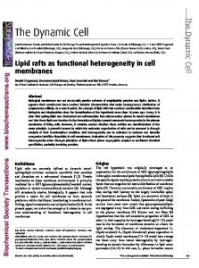
Figure 1. (a) The vegetal view of the IMZ of (i) the early Xenopus gastrula and (ii) the early neurula shows movements of involution (black curved arrows), convergence (short black, facing arrows) and cell intercalation (mediolateral intercalation behaviour, MIB; grey fusiform shapes) in the presumptive notochord (dark grey) and somitic mesoderm (light grey). Also, shown are the closure of the blastopore over the vegetal endoderm (straight arrows) and the site of convergent thickening (grey rectangles). The presumptive path of future shearing of the notochord with the somitic mesoderm is indicated (dotted grey arrow). Dorsal and anterior are at the top. (b) The magnified view (the circled areas denoted by B in (a)) of how cell intercalation produces convergent extension. (c) The MIB; the cells become polarized with large filo-lamelliform protrusions on their medial and lateral ends (dark grey), which extend (black arrows), attach to adjacent cells or extracellular matrix and shorten (light grey arrows), thereby pulling the cells between one another along the mediolateral axis to from a narrower, longer array. (d ) The vegetal view, comparable to (a) shows the presumptive anterior– posterior axes in the notochord (dark grey) and somitic mesoderm (light grey) (curved arrows, a, anterior; p, posterior). The presumptive mediolateral axis is indicated in the notochord and somitic mesoderm by curved lines (l, lateral; m, medial). (e) An explant of the IMZ shows progression of MIB expression (dark fusiform shapes) at the (i) early and (ii) late gastrula stages (bottom) of convergence and extension. D, dorsal; V, ventral; VAZ, vegetal alignment zone. The progression of MIB along the presumptive anterior–posterior axis (grey arrows) and presumptive lateromedial axis (black arrows) is shown. ( f ) The same events in explants prevented from undergoing convergent extension at the (i) early, (ii) mid and (iii) late gastrula stages (bottom). ( g) A photomicrograph shows an explant of the type illustrated in (e) at a late neurula stage, showing the elongated cells (MIB) in the notochord (n) and in the somitic mesoderm (s) and the segmentation of the anterior somites (numbered arrows). (h) The presumptive anterior–posterior (black dashed arrows) and presumptive lateromedial axes (grey dashed arrows) mapped on to the presumptive fates of the IMZ: notochord (dark grey); somitic mesoderm (stippled, medium grey). The dorsal midline (organizer region) is in the middle and the ventral midline is at the left and right ends. (i ) The features shown in (h) are also shown on the vegetal view of the gastrula. (e) Modified from (Keller & Danilchik 1988); (i) Adapted from Shih & Keller (1992b).
the notochordal–somitic boundary is formed, and it proceeds medially (black arrows, figure 1e, f ) to form an arc of fusiform cells, the vegetal alignment zone (VAZ, figure 1e, f ). It proceeds posteriorly from this Phil. Trans. R. Soc. B (2008)
origin along the lateral boundary of presumptive somitic mesoderm, from the anterior-to-posterior aspect of this tissue (grey arrows, figure 1e, f ). Within the VAZ, the notochordal–somitic mesodermal
1320
R. Keller & D. Shook
Dynamic patterns of cell behaviours
boundary forms and progresses from anterior to posterior (dashed arrows, figure 1e, f ). MIB is then expressed along the lateral boundary of the notochord in the same type of anterior-to-posterior (grey arrows, figure 1e, f ) and lateral-to-medial (black arrows, figure 1e, f ) progression seen in the somitic mesoderm. In the late neurula stages of these explants, the somites segment in the anterior-to-posterior order, the intersegmental boundaries forming arcs that originate laterally and proceed medially, parallel to the preexisting, mediolateral axis of cell polarization during MIB and the axis of cell intercalation (Shih & Keller 1992d; Davidson et al. 2004a; figure 1g). Therefore, three major events follow a near cell-by-cell progressive order along the anterior–posterior axis in these explants: the progress of MIB; the progress of the notochordal–somitic mesodermal boundary; and segmentation. The anterior-to-posterior (black arrows, figure 1h) and lateral-to-medial progressions (grey arrows, figure 1h) are shown diagrammatically on the IMZ explant and the IMZ of the vegetal view of the gastrula (figure 1i ). A fourth anterior-to-posterior progression, that of bottle cell formation and re-spreading, occurs in the epithelial endoderm just superficial to the deep IMZ (discussed in §13).
5. PATTERNING OF CELL BEHAVIOURS ANTICIPATES AND IS ESSENTIAL FOR THEIR FUNCTION Having the cell behaviours organized as hoops around the blastopore looks to be inane and inordinately complex when looked at with the embryologist’s eye for geometric simplicity, but viewed with function in mind, it is an elegantly simple and effective mechanical integration of local cell behaviour to drive large-scale tissue movements. By organizing MIB and convergence along the presumptive mediolateral axis, as hoops anchored in the vegetal region, and by progression of this behaviour along the transverse, presumptive anterior–posterior axis, convergent extension is aligned such that the progressive shortening of the arcs of converging cells anchored laterally at the vegetal endoderm accomplishes three things in one stroke: it rolls the IMZ inside and across the vegetal endoderm, thus covering it over; it closes the blastopore; and it elongates the body axis. Thus, the cell behaviours are patterned on the presumptive body plan such that it is pre-loaded to perform a specific biomechanical strategy of gastrulation. Because material or tissue-type determinations are coupled, perhaps variably, to these dynamic determinations, the shapes of presumptive tissue areas reflect the biomechanics of their future movements. The importance of this hoop stress strategy is supported by the fact that interrupting these arcs of cell intercalation (and convergence) results in catastrophic failure of blastopore closure and gastrulation (Schechtman 1942; Keller 1981). Preliminary biomechanical measurements show that these circumblastoporal, tensile forces are, in fact, developed (D. Shook et al. 2005, unpublished results), and they are comparable to the pushing, extension forces measured previously (Moore 1994; Moore et al. 1995). Phil. Trans. R. Soc. B (2008)
The progressive aspect of MIB is essential for its function. MIB is correlated with stiffening of the notochordal–somitic tissue by a factor of 3–4 in an anisotropic manner, the increase being along the anterior–posterior axis (Moore et al. 1995). If MIB were expressed simultaneously throughout the IMZ, regardless of the progress of involution, it is unlikely that the tissue could bend over the blastoporal lip and involute. When the extent of anterior–posterior axis is experimentally expanded into the pre-involution region of the dorsal IMZ, it fails to bend inwards and involute, but instead, it extends vegetally in a type of exogastrulation (Branford & Yost 2002). A similar disruption of progression, and also the anchor points of the hoop stress, may account for the tendency of lithiumdorsalized embryos, in which all the sectors of the IMZ behave as dorsal, organizer tissue (Kao & Elinson 1988, 1989), to extend an exogastrulated proboscis. Normally, the convergence hoop stress builds progressively from the dorsal side and rolls the dorsal and dorsolateral IMZ over the vegetal endoderm (yolk plug). In contrast, if all the sectors of the IMZ in lithium-dorsalized embryos behave as organizer (dorsal) tissue, convergence forces would develop simultaneously all around the blastopore, and the hoop stress would also encircle the blastopore rather than terminate at the lateral edges of the vegetal endoderm. As a result of this altered timing and anchorage, the constricting IMZ would probably squeeze the endoderm inwards and extend itself outwards in a symmetrical proboscis. Conversely, an uncoordinated or patchy expression of MIB should result in loss of continuity of hoop stress and failure to build up large-scale, tissue-level convergence forces, and as in the case of experimentally breaking hoop stress continuity, would result in locally independent extensions and failure of blastopore closure and also proper involution (Schechtman 1942; Keller 1981). Involution and internalization can occur without convergent extension (Schechtman 1942; Keller & Danilchik 1988; Ewald et al. 2004) but it is a short, shallow, symmetrical slit of an archenteron, very unlike the asymmetric, dorsally elongated one normally produced by involution with convergent extension.
6. THE SPECIAL PROBLEM OF THE NOTOCHORD The notochord presents an interesting problem in the simplified hoop stress mechanism, because its presumptive area does not bound the medial aspect of more than a few presumptive anterior somites (figure 1a,d ), whereas in the late neurula, the posterior end of the notochord has come to lie medial to the posterior somites (Keller & Shook 2004). The question then is how do all the ps in figure 1d come to lie posteriorly and next to one another? One mechanism is shearing of the notochord posteriorly with respect to the somitic mesoderm. The presumptive notochord and anterior somites are thought to involute, converge and extend together during gastrulation, and then, in the neurula stage, the notochord extends posteriorly faster than the somitic mesoderm, and thus the notochord shears posteriorly along its boundary with the somitic mesoderm. As it does so, the notochord
Dynamic patterns of cell behaviours pushes the blastopore into the thick ring of mesoderm encircling the lateral and ventral sides of the blastopore, and these cells move around both sides of the posterior notochord and join the posterior segmental plate (Keller & Shook 2004). This ventrally located, presumptive posterior somitic mesoderm previously expressed convergent thickening, and now belatedly displays the same sequence of cell behaviours previously showed by their more dorsal (presumptive anterior) counterparts, including radial intercalation followed by mediolateral intercalation (Keller et al. 1989; Wilson et al. 1989; Wilson 1990). This posterior movement of the notochord along the somitic mesoderm also can be seen in explants cultured on fibronectin (Davidson et al. 2004a). Mapped on to the gastrula, the path of this future shearing is along the upper (animal) edge of the IMZ, which is the future medial edge of the posterior somitic mesoderm (dotted arrows, figure 1d ). Although the arcs of convergence are shown diagrammatically as being continuous (figure 1d–f ), they are not. Despite the sliding of the tissues at the notochordal–somitic boundary, the tissues maintain contact, and the fibrillin matrix in the notochordal–somitic mesodermal boundary appears essential for this dynamic, sliding adhesion (Skoglund et al. 2006). Although this posterior shearing has been documented only during neurula stages, some of it may also occur during gastrulation, albeit where it is difficult to see, just as the presumptive somitic mesoderm rolls over the lateral blastoporal lip and turns inside out (see the rolling over of the arrows in presumptive somitic regions at the blastopore, figure 1d ). If so, the notochord would act as a posteriorly moving ‘zipper’ that would tend to roll the somitic mesoderm inwards and contribute to blastopore closure. Such a mechanism remains an intriguing possibility.
7. THE SIGNIFICANCE OF VEGETAL-TO-ANIMAL PROGRESSION OF CELL BEHAVIOUR IN THE IMZ The anterior-to-posterior progression of MIB is slower in the presumptive medial aspects of the notochordal and somitic territories than it is in the presumptive lateral regions (figure 1e,f ). This could reflect better conditions for the propagation of a single, anteriorto-posterior signal in the lateral regions, possibly set up by an intersecting signal acting in presumptive lateral– medial axis. Or it could represent a cascade of two signals, a primary anterior-to-posterior signal that passes along the lateral region where it generates a secondary signal that then progresses medially. The lateral–medial axes of the notochord and somitic territories have different orientations that together complete the arcs of convergence. How this is set up is not known. In the somitic mesoderm, the vegetal–animal axis is more nearly a presumptive lateral-to-medial transition, whereas in the notochord, a vegetal-to-animal transition is more nearly a presumptive anterior-to-posterior transition. In addition, in other groups of amphibians, such the urodeles discussed below, these axes are tilted to varying degrees. These double progressions are probably set up by some intersection of signalling pathways (see Kumano & Smith 2002; Heasman 2006), which is not yet understood. Phil. Trans. R. Soc. B (2008)
R. Keller & D. Shook
1321
A vegetal–animal axis of signalling is supported by numerous vegetal–animal differentiations in the IMZ (for an excellent review, see Kumano et al. 2001; Kumano & Smith 2002). First, the early involuting, vegetal region of the IMZ is presumptive leading edge mesoderm, which is migratory in nature, shows an animally directed migration and spreading on the roof of the blastocoel (see Nagel et al. 2004), whereas the late involuting animal part of the IMZ is presumptive notochord and somitic mesoderm, which displays MIB and undergoes convergent extension (Keller 1991; Keller et al. 1992b; see diagrams in Keller & Davidson 2004). Second, the vegetal and animal regions of the IMZ likewise express different molecular markers that reflect later development of fates of leading edge, ventral tissues in the former, and reflect development of dorsal, axial and paraxial mesoderm in the latter (Stewart & Gerhart 1990; Zoltewicz & Gerhart 1997; Lane & Smith 1999; Kumano et al. 2001; Kumano & Smith 2002; Lane & Sheets 2002a,b). Third, the variable amount of mesoderm in the superficial epithelial layer of the IMZ of different amphibian species always occupies the animal edge of the IMZ and the superficial endoderm the vegetal edge (Keller & Shook 2004). Finally, in contrast to the relatively minor differences in the MIB progression along the vegetalto-animal aspect of the IMZ (presumptive lateral to medial of the body plan) in Xenopus, there are very strong vegetal-to-animal progressions in the IMZ of urodeles (see §11). The complex mapping of the progressions and axial orientation of MIB, and additional cell behaviours such as the epithelial–mesenchymal transition (EMT) in other species (see §12 and figure 4), on to the presumptive tissue axes in the IMZ reflect the necessity of coupling of patterning of tissue type and cell behaviour on the one hand, and generating a pattern of forces that will accomplish gastrulation and body axis elongation on the other hand. Variations in these parameters between species reflect functional adaptation of the morphogenic machinery and the diversity of gastrulation strategies. The question is, how do the underlying signalling pathways regulate the type, the progressive aspect and the geometric aspect of the cell behaviours, and what is the geometry of these signals?
8. THE TIMING OF SIGNALS ORGANIZING MIB AND MESODERM FORMATION The pattern of MIB could be specified at some earlier stage and executed during gastrulation, and this may be true, but the evidence suggests that signals regulating MIB also persist during gastrulation. Dorsal mesodermal cells lose their characteristic polarized behaviour and morphology when isolated in small groups, or when dissociated throughout gastrulation and neurulation, suggesting that persistent contextual cues are needed (R. Keller 1992, unpublished work). Scattering labelled notochordal and somitic cells from one embryo into the same regions of the early gastrula explants, without regard to their original order, results in those cells adopting MIB in the anterior-to-posterior and lateral-to-medial order of the host tissue (Domingo & Keller 1995). Cells from other presumptive areas
1322
R. Keller & D. Shook
(a)
Dynamic patterns of cell behaviours
(b)
(c)
(d) 100 µm Figure 2. Processes that may aid establishment of MIB are illustrated. (a) Densely crowded, elongated cells self-align to form parallel arrays as a result of cell shape and distribution of protrusions. (b) Monolayered fibroblasts of this morphology show such self-alignment into parallel arrays in culture (Elsdale & Wasoff 1976). (c) Traction-mediated alignment of matrix fibrils by fibroblasts in culture feeds back on cell behaviour and mediates oriented migration (Harris et al. 1981; Stopak & Harris 1982). (d ) Developing tension along the sides of elongating cells inhibits protrusive activity perpendicular to the surface, whereas tension at the ends stimulates protrusive activity (Kolega 1986; Katsumi et al. 2002). (b) Adapted from Elsdale & Wasoff (1976) and (c) adapted from Stopak & Harris (1982).
scattered into the notochordal and somitic territories of explants adopt the fates of the host tissue, although with a delay (Domingo & Keller 1995). Thus, the signals inducing both the dynamic determination of MIB and the material determination of mesodermal cell fates persist during gastrulation.
9. TENSILE, POSITIVE FEEDBACK MECHANISMS MAY ENHANCE MIB EXPRESSION Once MIB is underway, the convergent, tensile force it develops may act as a positive feedback, based on what is known from the cultured cell lines. Some cultured cell lines have elongated, fibroblastic morphology, similar to the elongated fusiform mesodermal cells, have a tendency to self-align, form parallel arrays and actually reduce the frequency of collisions between them at high density as a result of local cell contact interactions (Elsdale & Bard 1972; Elsdale & Wasoff 1976; figure 2a,b). A degree of similar self-alignment of the fusiform, intercalating mesodermal cells is probable. Also, cell traction tends to align collagen fibrils along the lines of tension in culture and, in turn, the movement of cells along the aligned fibrils is enhanced (Harris et al. 1981; figure 2c). Similar traction on the fibronectin matrix associated with intercalating mesodermal cells ( Davidson et al. 2004b) may generate a similar fibril-mediated, positive feed back on motility. Aligned fibrils would probably offer a stiffer substrate, and stiffer substrates positively feed back on the cell to stimulate greater protrusive activity and traction (Lo et al. 2000). Increased tension along the elongated, anterior and posterior sides of the fusiform mesodermal cells may suppress the protrusive activity there, thus enhancing the polarized distribution of tractive protrusions at the medial and lateral ends. In the cultured cells, protrusive activity perpendicular to the axis of tension is suppressed (Kolega 1986), and experimentally applied tension inhibits protrusive and Rac activities on the sides parallel to the tension and enhances both at the ends (Katsumi et al. 2002). Phil. Trans. R. Soc. B (2008)
Finally, tensile forces transmitted to neighbouring cells by mediolateral traction may activate stretch-activated calcium channels ( Jaffe 2007), and result in the propagation of contraction, development of traction force (Doyle et al. 2004) and the further development of mediolaterally oriented tension. The waves of calcium transients and associated contraction waves are propagated across the IMZ explants (Wallingford et al. 2001), and although they do not appear to be temporally or spatially correlated with the directions of MIB progression, or the axes of intercalation under the culture conditions in which they were observed, they may reflect a phenomenon used locally, with different timing and spatial parameters, in vivo. These mechanical feedback mechanisms apply to the tensile, mediolateral aspect of the converging tissue. Compressive forces in the anterior–posterior axis of extension (Moore 1994; Moore et al. 1995) may also affect the cell behaviour. The function of compressive and tensile loads on cell behaviour and force production could be experimentally probed with appropriate biomechanical manipulation of explants.
10. EPITHELIAL–MESENCHYMAL TRANSITION IN THE IMZ GENERATES CONVERGENCE FORCE DURING BLASTOPORE CLOSURE IN THE URODELE AMPHIBIAN The commonly studied urodeles (tailed amphibians) generate a circumblastoporal hoop stress similar to that seen in Xenopus, but they use a different combination of cell behaviours. Although blastopore closure looks superficially similar in Xenopus and in the urodeles (Ambystoma mexicanum, Ambystoma maculatum and Taricha granulosum), a closer inspection shows that the urodeles use the EMT and ingression of superficial presumptive mesodermal cells as a major force generator during blastopore closure (Shook et al. 2002). The EMT of surface presumptive mesoderm is a conserved process in the early morphogenesis of amphibians (Purcell 1993; Minsuk & Keller 1996, 1997;
Dynamic patterns of cell behaviours (a)
(b)
(c)
(d)
(e)
(f)
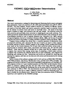
Figure 3. The presumptive somitic mesoderm (dark grey) and presumptive notochord (medium grey) in a lateral view of the anuran Xenopus at (a) the early gastrula and (b) mid-neurula stages, and in a lateral view of a typical urodele, Triturus (Triton) at the (c) early gastrula and (d ) neurula stages. (e) The vegetal view of the urodele (Triturus) early gastrula, with the dorsal side at the top, shows convergence, probably by cell intercalation, in the notochord (light grey area, grey arrows) and the removal of the somitic mesoderm from the surface by EMT (dark grey area, black arrows). The box and the inset in (e) show a magnification of the subduction zone (black line) in which EMT occurs. ( f ) The vegetal view of the same embryo shows the progression of EMT from dorsal to ventral in the gastrula, which is the presumptive anterior-to-posterior direction (black arrows), and in the vegetal-to-animal direction, which is the presumptive lateralto-medial direction (grey arrows).
Shook et al. 2002, 2004). However, the amount of mesoderm, the timing of its EMT and the functional significance of the force it generates during gastrulation vary greatly. Anurans have relatively little superficial mesoderm and thus relatively less EMT than urodeles, with Xenopus having the least of the species examined thus far (figure 3a). This superficial mesoderm consists of presumptive notochord and the medial edge of the presumptive somitic mesoderm, which is located at the animal edge of the IMZ; the cells of both tissues undergo EMT and ingression in an anterior-to-posterior progression, but it occurs from the gastrocoel roof during neurulation (Purcell 1993; Minsuk & Keller 1996, 1997; Phil. Trans. R. Soc. B (2008)
R. Keller & D. Shook
1323
Shook et al. 2004; black arrows, figure 3b), and thus is irrelevant to gastrulation and blastopore closure. In contrast, the urodeles examined thus far have a very large area of somitic mesoderm in the surface epithelium (figure 3c), and most of it is removed by a massive EMTat the lower edge of the mesodermal territory, next to the vegetal endoderm during gastrulation, leaving the notochord on the gastrocoel roof of the neurula (Shook et al. 2002; figure 3d ). The fact that such a massive EMT occurs during gastrulation suggests that it might have a functional role, and indeed, it appears to generate substantial convergence force. This EMT occurs in the ‘subduction zone’ along the vegetal edge of the IMZ at its boundary with the vegetal endoderm (black line, figure 3e), where the surface somitic mesoderm appears to move beneath the vegetal endoderm (Shook et al. 2002). The epithelial mesodermal cells in the subduction zone constrict their apices to point that they cannot be resolved, and then they ingress and re-appear in the deep region as mesenchymal cells (Shook et al. 2002; box, figure 3e). More mesodermal epithelial cells enter the subduction zone, as the epithelial sheet moves vegetally (black arrows, box, figure 3e). It is probably pulled vegetally by the apical constrictions, since the ingression occurs only after the apices are very small, and thus their leaving accounts for little change in area, and thus little force. Preliminary measurements show that the EMT and subduction generates a tensile force comparable to the force generated by MIB-mediated cell intercalation in Xenopus (D. Shook 2005, unpublished results). In the context of the embryo, towing of the sheet of superficial mesoderm vegetally, towards the endoderm (black arrows, figure 3e), occurs during involution of the notochord to the extent that in the post-involution region, the lateral edge of the notochord and the vegetal endoderm have met, and all or nearly all of the somitic mesoderm has ingressed (Vogt 1929; Shook et al. 2002); this convergence due to the removal of cells by EMT, together with convergent extension of the notochord, probably by cell intercalation (grey arrows, figure 3e), generates a continuous hoop stress similar to that seen in Xenopus. In this case, however, it is generated by two behaviours: convergent extension in the notochord and EMT in the somitic mesoderm (Shook et al. 2002).
11. EMT ALSO OCCURS IN A PROGRESSIVE PATTERN The EMT occurs in a progressive pattern similar to that displayed by MIB in Xenopus, beginning in the dorsolateral IMZ of the gastrula, and proceeding ventrally (black arrows, figure 3f ) such that its path forms the subduction zone (black line, figure 3f ). The rate of progress varies with the species. In Ambystoma, for example, the EMT appears to proceed quickly about half way around to the lateral lip where it often appears to slow until near the end of gastrulation when it crosses the remainder of the ventral blastoporal lip (figure 3f ). The progress of EMT around the blastopore correlates with the appearance of a blastoporal lip, or blastoporal cleft, in urodeles that have been studied thus far (Holtfreter 1943a,b; Shook et al. 2002). Some species,
1324
R. Keller & D. Shook
Dynamic patterns of cell behaviours
such as the gigantic salamander Megalobatrachus maximus (Ishikawa 1905) and Hemidactyium scutatum (D. Shook, unpublished work), do not form a ventral lip at all during gastrulation. Therefore, if the EMT of the superficial mesoderm occurs at all in these species (it has not been documented), it is apparently occurring solely through the dorsal and lateral subduction zones. There is also a vegetal-to-animal progression of the EMT in the urodeles, A. mexicanum and A. maculatum (Shook et al. 2002). Normally, the EMT is restricted to the subduction zone, and the vegetal-to-animal progression of EMT tows the IMZ vegetally, into the subduction zone. However, if an IMZ explant is pressed tightly between two coverslips, the epithelium cannot move, and the cells undergoing EMT have no place to ingress, so they undergo EMT in situ, and reveal a highly organized, nearly cell-by-cell front of the EMT, which sweeps from the vegetal-to-animal aspect of the explant, and leaves a mass of twisting and turning mesenchymal cells in its wake (Shook et al. 2002). This strong vegetal-to-animal progression of EMT in Ambystoma argues for a very strong vegetalto-animal axial patterning system for dynamic determinations in the IMZ of amphibians, as there is for tissue differentiation (Kumano & Smith 2002). When the paths of the EMT progression in urodeles are mapped on to the presumptive anterior–posterior and lateral–medial axes of their future body plan, similarities and differences with the progression of MIB in Xenopus emerge.
12. IS AXIAL PATTERNING IN URODELES THE SAME OR DIFFERENT FROM ANURANS? Vogt’s classic vital dye mapping of tissue fates and movements in the newt, Triton (now called Triturus), shows the superficial presumptive somitic mesoderm in the lateral IMZ with the presumptive medial edges against the notochord and the lateral edges bounding the lateral plate mesoderm, and the presumptive anterior somites located dorsally and the posterior ones sweeping laterally around the blastopore ( Vogt 1929; figure 4a,b), though not as far as in Xenopus. Pasteels’ fate map of Ambystoma (Pasteels 1942) is somewhat different from Vogt’s map of the newt. The somitic mesoderm bears a similar relationship to notochord and lateral plate, but the presumptive anterior–posterior axis of the somitic mesoderm (black arrows, figure 4c) is oriented more in the vegetal-to-animal direction, and the presumptive lateral–medial axis (grey arrows, figure 4c) is oriented more in ventral-to-dorsal direction than in the newt (black arrows in figure 4a,b). The vegetal placement of lateroventral mesoderm in Vogt’s map (figure 4a), in contrast to the ventral placement in Pasteels’ map (figure 4b), reinforces the differences between the maps. The progress of EMT has not been described in enough detail in both species to be definitive, but in explants it appears to be more from the anteriorto-posterior aspect of the somitic mesoderm in Ambystoma (Ambystomatidae; grey arrows, figure 4b) and more from the lateral-to-medial aspect in Taricha, which like Triturus, is a newt (Salamandridae; black Phil. Trans. R. Soc. B (2008)
arrows, figure 4a,b; Shook et al. 2002). Therefore, in different groups of amphibians, one or the other axis of pattern generation may dominate regulation of the EMT. The fate map of Cynops pyrrhogaster (Salamandridae; Nakamura 1938; figure 4e), which is closely related to Triturus, suggests that axial orientation is probably more similar to Triturus and Taricha than to Ambystoma. Despite these differences, it is clear from the explants of Taricha and Ambystoma that in both EMT makes a large contribution to convergence, and thus probably contributes to circumblastoporal hoop stress in the IMZ of the whole gastrula of both species. Whether these rather subtle differences are of functional significance is an interesting question.
13. BOTTLE CELL FORMATION AND RE-SPREADING IS ALSO PROGRESSIVE, AND PARALLELS ASPECTS OF THE EMT AND MIB PROGRESSIONS The blastoporal ‘bottle cells’ of Xenopus undergo the first step in the EMT, the apical constriction, and they do so in the place that EMT occurs in the urodeles, at the lower edge of the IMZ that bounds the vegetal endoderm (figure 5a). And as with the EMT, bottle cell apical constriction also pulls the IMZ epithelium vegetally (black arrows, figure 5a). But instead of completing the EMT by losing adhesion to the epithelial sheet and ingressing to form a mesodermal mesenchyme, the cells are carried inside by virtue of their attachment to the underlying deep mesoderm (figure 5b), and there they re-spread to form a large area of the endodermal lining of the archenteron (Keller 1981; Hardin & Keller 1988; figure 5c,d ). This behaviour is also found in the IMZ of other anurans and also in the small area of superficial pharyngeal endoderm found in the dorsal IMZ of the urodele amphibians (Vogt 1929; figure 3). In Xenopus, this apical constriction occurs progressively, though not in strict cell-by-cell order, from vegetal to animal in the IMZ, and also from the dorsal (presumptive anterior archenteron) to the ventral IMZ (presumptive posterior archenteron; Keller 1981; Hardin & Keller 1988; grey, dashed arrow in figure 5a,b). After being carried inside, the bottle cells then begin re-spreading progressively, in the same order of their formation (Keller 1978, 1981; Hardin & Keller 1988; grey, dashed arrows in figure 5c,d ). In Xenopus, this progressive apical constriction followed by re-spreading occurs in the same basic pattern as the progression of MIB, but it occurs in the surface epithelium and in a different germ layer, the presumptive endoderm. Bottle cell formation shares the overall pattern, the occurrence in the epithelium, the apical constriction as the first event, with the EMT in urodeles, but it occurs in the endoderm rather than the mesoderm. Therefore, the regulation bottle cell behaviour may be another manifestation of the same underlying pattern generator that regulates EMT and MIB, but with downstream differences associated with tissue organization and germ layer differentiation.
Dynamic patterns of cell behaviours (a)
6 7 8 9
32 1
5
K
J +
N 5 4
1325
dorsal
(b)
10 9 8 7
R. Keller & D. Shook
+ n
32 1
S
ventral
(c)
(d )
N P.P
S
(e)
T
P
N A S
Figure 4. The (a) right lateral side (b) and vegetal view of the Triturus (urodele) fate map, according to Vogt, (c) right lateral side (d ) and dorsal view of the Ambystoma (urodele) fate map, according to Pasteels, and the right lateral view of the fate map of Cynops (urodele), according to Nakamura. The presumptive notochordal (N) and presumptive somitic (S) mesoderm, the presumptive anterior–posterior axes ( black dashed arrows) and the presumptive lateral–medial axes (grey dashed arrows) are indicated. (a) Adapted from Vogt (1929), (b) adapted from Pasteels (1942) and (c) adapted from Nakamura (1938).
14. WHAT IS THE MOLECULAR MECHANISM OF PROGRESSIVE EXPRESSION OF CELL BEHAVIOURS IN THE IMZ? The patterning of tissue fates in the IMZ of amphibians is largely understood from the work on one model system, Xenopus, and it involves the interplay of a myriad of components, beginning with the localization of maternal factors in the oocyte, their redistribution on fertilization, which results in a complex interaction of positive and negative or antagonistic signals acting in the vegetal–animal and in the dorsoventral geometry of the gastrula (Kumano & Smith 2002; De Robertis & Kuroda 2004; Heasman 2006; Lane & Sheets 2006; for teleosts, also see Schier & Talbot 2005). But much less is known about the patterning of cell behaviours, particularly their axial and temporal components that define the dynamic progressions described above, and also how the behaviours are linked to tissue Phil. Trans. R. Soc. B (2008)
differentiation. The challenge is to understand how the embryo puts its cellular players on the field in an effective morphogenic plan. In regard to convergent extension, TGF-b signalling was implicated by the fact that activin-treated animal caps differentiate dorsal mesodermal tissues (notochord, somite) and show convergent extension in a dose-dependent fashion (Green et al. 1992). Although the cell behaviour driving these movements in animal caps could not be imaged, it has been assumed that they must be similar to MIB. Recent work showed that the non-canonical Wnt/PCP pathway, consisting of some components of the planar cell polarity (PCP) pathway of Drosophila (Adler & Lee 2001; Adler 2002), is essential for establishing the polarized protrusive activity displayed in the MIB underlying convergent extension in amphibians and teleost fish (Heisenberg et al. 2000; Sepich et al. 2000; Tada & Smith 2000;
1326
R. Keller & D. Shook
(a)
Dynamic patterns of cell behaviours (b)
An D
V veg
(c)
(d )
A veg
A veg
An P
P An
Figure 5. The behaviour of the presumptive bottle cells (dark grey) are shown in the surface epithelial layer of the Xenopus IMZ (a) just prior to gastrulation, (b) in the early gastrula, (c) in the mid-late gastrula and (d ) the neurula from the left side. The dorsal (D) side of the gastrula is at the left and the ventral (V) side is at the right. The animal (An) and vegetal (veg) ends of the IMZ and the anterior (A) and posterior (P) ends of the forming archenteron lining are shown. The progress of bottle cell formation from dorsal to ventral (presumptive anterior to posterior) (dashed arrows (a,b)), and the progress of re-spreading along the same axes (dashed arrows (c)) are shown.
Wallingford et al. 2000, 2002; Darken et al. 2002; Jessen et al. 2002; Marlow et al. 2002; Tada et al. 2002). But these findings did not reveal an obvious mechanism for orienting the axes of MIB and the resulting cell intercalation with respect to the axes of the body plan, nor do they account for its progressive nature. Ninomiya et al. (2004) found that graded differences in tissue properties along the anterior–posterior axis supply these large-scale, contextual cues for expression and orientation of MIB. Juxtaposing explants of tissue originating from different anterior–posterior axial levels, as assayed by the expression levels of chordin (high anteriorly) and brachyury (high posteriorly), resulted in cell intercalation at the interface, whereas juxtaposing the same anterior-to-posterior levels did not. This was also true for anterior-to-posterior differences induced in animal caps by different doses of activin. The resulting cell elongation and apparent cell intercalation was oriented perpendicular to the axis of the putative anterior-to-posterior differences, suggesting that cell recognition of some unknown antero-posteriorly graded positional property is essential for eliciting MIB in the correct perpendicular (mediolateral) orientation. These cues provide an essential, large-scale axial orientation to the downstream, PCPmediated polarization events (Ninomiya et al. 2004). It may be a general rule that differences along the presumptive axis of extension provide the primary cue for cell intercalation along the transverse axis, whether the PCP pathway is involved downstream or not. Other systems that converge and extend by cell intercalation, including hindgut convergent extension (Johansen et al. 2002; Lengyel & Iwaki 2002) and germ band convergent extension (Zallen & Wieschaus 2004) in Drosophila also depend on anterior-to-posterior differences in gene expression. In the latter case, the PCP pathway genes are not necessary (Zallen & Wieschaus 2004). Phil. Trans. R. Soc. B (2008)
The important, graded anterior-to-posterior property may involve differences in cell adhesion. Cells from different anterior-to-posterior levels recognize one another as different, and attempt to sort out when mixed (Ninomiya et al. 2004), suggesting that an antero-posteriorly graded cell affinity or cell adhesive property maintains anterior-to-posterior cell order and perhaps determines the axis of cell intercalation. Activin induction, which was used in a graded fashion to produce the regional differences in these experiments, also reduces C-cadherin activity in adhesion assays, and such a change appears essential for convergent extension, and presumably for the underlying cell intercalation in Xenopus (Brieher & Gumbiner 1994; Zhong et al. 1999). Taken together, these observations suggest the hypothesis that orientation of MIB may be cued by antero-posteriorly graded cell adhesion, perhaps mediated by graded C-cadherin activity, which in turn, may be induced by anteroposteriorly graded TGF-b signalling. The presumptive anterior–posterior axis turns out to be an axis of nodal/TGF-b-sensitive events in both the dorsal organizer region (the notochord) and in the lateroventral region (the somitic mesoderm). The expansion of the vegetal-to-animal (presumptive anterior–posterior) extent of dorsal tissues in the organizer by the manipulation of nodal and its antagonist, lefty (antivin; Branford & Yost 2002; also see Gritsman 2000), suggests that nodal signalling may play a role in the anterior-to-posterior position effect on MIB orientation in the dorsal midline. Similarly, in the dorsal-to-ventral (presumptive anterior–posterior) axis of the somitic mesoderm, which is much longer and arrayed around both sides of the blastopore, the progression of TGF-b signalling is seen by antibody staining of active SMAD2, which is initially high in the dorsal IMZ in the early gastrula, then rises laterally, and finally ventrally, followed by a decline first dorsally and then laterally, later in gastrulation (Lee et al. 2001; Hashimoto-Partyka et al. 2003). This progression is regulated by maternal Wnt/b-catenin signalling, one of the initial events in establishing dorsoventral asymmetry (Lee et al. 2001; Hashimoto-Partyka et al. 2003). Nodal signalling has been reported to be cell autonomous and non-cell autonomous, occurring over short and over long range and, indeed, it may differ with the specific nodal and the molecular context (Gritsman et al. 1999; Gritsman 2000; Chen & Schier 2001; Meno et al. 2001; Niederlander et al. 2001; Hashimoto-Partyka et al. 2003; Williams et al. 2004). The molecular context may be particularly important (Lander et al. 2002). Whatever the mechanism of its expression, both in the dorsal IMZ, where the presumptive anterior–posterior axis runs vegetalto-animal, and in the lateral-to-ventral IMZ, where this axis runs ventral to dorsal, a travelling wave of nodal signalling may be linked to the specification of local cellular differences that cue MIB in the progressive pattern seen in time lapse movies. But other signalling systems are also involved, and it is not commonly appreciated (Lane et al. 2004) that this dorsal–ventral axis of the IMZ in the gastrula is actually the presumptive anterior–posterior axis of the somitic mesoderm and is the axis of the ‘third signal’
Dynamic patterns of cell behaviours along which dorsalization, and thus specification of MIB, in more ventral tissues is thought to occur (Kumano & Smith 2002; Lane & Sheets 2006). Cell fate along this axis is regulated by ‘ventralizing’ signals from the ventral sector, countered by dorsal antagonists (De Robertis & Kuroda 2004; Lane et al. 2004; Marom et al. 2005; Heasman 2006). The progress of MIB, given its association with dorsal notochordal and somitic tissue, is a read-out of this process. Also, important in organizing MIB is fibroblast growth factor (FGF) signalling, which occurs in a positive feedback loop with Xenopus Brachyury (Xbra), which in turn, regulates Xwnt11, a key player in noncanonical Wnt/PCP regulation of MIB (Heisenberg et al. 2000; Tada et al. 2002). Most interesting in regard to regulation of cell behaviour, versus tissue differentiation is the discovery that cytoplasmic regulators of FGF signalling, Sprouty and Spred, toggle FGF signalling between differentiation and cell behaviour pathways. Sprouty blocks the protein kinase C (PKC) pathway that regulates convergent extension, and Spred blocks the mitogen-acivated protein kinase pathway that regulates tissue differentiation (Sivak et al. 2005), and thus is a downstream branch point in FGF signalling underlying dynamic and material determination. Finally, the tissue movements described above and progressive tissue interactions may come into play. A ‘time space regulator’ model proposes that patterning in the anterior-to-posterior dimension of the IMZ depends on stabilization of a transient expression of Hox genes in a specific temporal order in nonorganizer mesoderm by the organizer as convergent extension progressively brings these regions into proximity (see §6; Wacker et al. 2004a,b). Understanding how these signalling pathways converge to produce the polarized MIB in the highly specific progressions observed requires knowing much more about the downstream regulators of cell motility, polarity, adhesion and cell contact behaviour, and how regulation of these aspects is linked to tissue specification. The mechanisms of patterning tissue type along the relevant axial dimensions have, in general, been done with molecular and tissue recombination methods of relatively low temporal and spatial resolution, and thus to learn how the travelling front of MIB is regulated, and linked to the presumptive tissue axes, will require signalling reporters of high temporal and spatial sensitivity in living tissues (Williams et al. 2004), and also manipulations of cell–cell position and interactions at a finer level. Nearly nothing is known about regulation of the highly progressive EMTs in amphibians. More is known about the regulation of EMT in other embryos. In mice, the EMT of epiblast cells in the primitive streak is regulated by Brachyury and FGF signalling (Hashimoto et al. 1987; Wilson et al. 1993, 1995; Deng et al. 1994; Yamaguchi et al. 1994; Ciruna et al. 1997) and downstream regulation of the transcription factor snail, a negative regulator of E-cadherin expression (Batlle et al. 2000; Cano et al. 2000; Ciruna & Rossant 2001). FGF signalling must also regulate the EMT through other routes, since the EMT phenotype of cells lacking snail is less severe than those lacking FGFR1 (Deng et al. 1994; Yamaguchi et al. 1994; Ciruna et al. Phil. Trans. R. Soc. B (2008)
R. Keller & D. Shook
1327
1997). The first step in the EMT of Drosophila mesoderm is ventral furrow formation, which begins with an apical constriction and cell shape change similar to that seen in amphibian bottle cells. Coordination of apical constriction requires a secreted ligand, folded gastrulation (Fog; Costa et al. 1994) acting upstream of concertina (Cta), which encodes a Ga protein (Parks & Wieschaus 1991; Morize et al. 1998), through G-protein-coupled receptor that has not been identified. Downstream of concertina, the rho activator, DRhoGEF2, and Rho1 are essential for the accumulation of myosin II at the cell apices (Barrett et al. 1997; Hacker & Perrimon 1998; Nikolaidou & Barrett 2004; Rogers et al. 2004; Dawes-Hoang et al. 2005). But DRhoGEF2 also has a Fog/Cta-independent role in actin localization at the apices of the cells (Fox & Peifer 2007). This alternate route of regulating RhoGEF2 involves its apical localization by a transmembrane protein (Kolsch et al. 2006). Abelson (Abl) kinase also plays a role by suppressing Ena localization to the cell apices, and thus allows actin accumulation there (Fox & Peifer 2007). RhoGEF2-null mutants show no apical constriction and the mesoderm does not internalize at all, whereas fog-null and cta-null mutants show an uncoordinated constriction of the apices and the mesoderm eventually internalizes, though abnormally. However, it is still not known how the coordination works, and the possibility of similar mechanisms being used in coordinating the progressive phenomena in amphibians may be small, since the former does not involve the large area and highly progressive nature of the latter. Other questions that should be explored include differences regulating the timing of the gastrula stage EMT of the somitic mesoderm, versus the neurula stage EMT of notochord in urodeles, and the neurula stage EMT of both tissues in anurans (Shook et al. 2002, 2004). Also, how is the regulation of EMT of superficial mesoderm linked to its germ layer specification in the epithelial layer, and how does that compare with linkage of MIB and mesoderm specification in the deep region of Xenopus? Also, are there shared features between the EMT regulation in epithelial mesoderm in urodeles and the regulation of bottle cell formation in epithelial endoderm in urodeles and anurans, particularly since the first step in both appears to be an apical constriction? Apical constrictions in different regions of the same organism appear to be regulated differently. In Xenopus, the actinbinding protein Shroom is required for neural apical constriction but not blastoporal bottle cell apical constriction (Haigo et al. 2003). Although some molecular components are conserved between mice and Drosophila, others are not shared by all of the several early invaginations in Drosophila (Kolsch et al. 2006; Fox & Peifer 2007). Finally, do the differences in mapping of the axial patterns of the MIB and the EMT progression on to the gastrula IMZ reflect functional adaptations for different biomechanical strategies of gastrulation, and how are these axial differences regulated? Regulation of bottle cell formation in Xenopus is better understood that regulation of the EMT in any amphibian. Bottle cells can be experimentally induced
1328
R. Keller & D. Shook
Dynamic patterns of cell behaviours
in the animal cap by TGF-b family (VegT, Xnr1, BVg1, activin) growth factors, and as with mesoderm formation, FGF signalling is essential (Kurth & Hausen 2000). Cerberus, a secreted protein that binds and inhibits the activity of Xnr1 but not Vg1 or activin, blocks Xnr1-induced bottle cell formation and also endogenous bottle cell formation but not those induced by activin or Vg1, suggesting that Xnr1 is the endogenous inducer. The spatial pattern of bottle cell formation in relation to the expression of the mesodermal markers goosecoid (gsc) and Xbra in ectopic induction experiments suggests that epithelial IMZ cells respond to high levels of TGF-b signalling by forming bottle cells, whereas the corresponding deep IMZ cells respond by expressing gsc at high levels of signalling and Xbra at lower levels of signalling ( Kurth & Hausen 2000). These model fits are consistent with the spatial relationship of these tissues during gastrulation. The presumptive bottle cells overlie the gsc-expressing leading edge migratory mesendoderm cells at the vegetal end of the IMZ at the outset of gastrulation, lose contact with these leading edge cells and retract to the boundary of the gscexpressing leading edge cells and the axial Xbraexpressing notochord cells as they undergo apical constriction, and then they re-spread across the leading edge cells again in the late gastrula and neurula stages (Keller & Shook 2004). Kurth & Hausen (2000) propose that bottle cell formation spreads from dorsal to the lateral and ventral sectors of the IMZ due to a wave of Xnr1 signalling passing from dorsal to ventral (Agius et al. 2000), and they also propose a limiting feedback mechanism in which Xnr1 upregulates cerberus (Piccolo et al. 1999), which in turn blocks further Xnr1 activity. They could not uncouple bottle cell and mesoderm formation, suggesting that the two are strongly linked. They propose two models to explain this, one in which both are induced by a common, graded signal, and a second in which deep cells respond to a graded Nodal signal which then induce the bottle cells in the superficial layer. Downstream, not much is known about the mechanism of the molecular mechanics of the constriction events. Shroom, an actin-binding protein, is expressed in neural plate (hinge point) bottle cells, and it is essential for the localization of actin to their apices and for their constriction in Xenopus neural plates, but Shroom is not expressed in the blastoporal bottle cells (Haigo et al. 2003). Again, there appear to be differences in the regulation of this behaviour in the same embryo. These differences could be due to the differences in germ layer specification or tissue organization (epithelial versus mesenchymal). Bottle cell formation and EMT share a common epithelial origin and begin with apical constriction, but they occur or begin in different germ layers (endodermal and mesodermal, respectively). Both share aspects of their pattern of progression with MIB. Are these behaviours the product of one underlying patterning mechanism with different downstream effectors, and if so, at what level does this differentiation occur? The epithelial versus deep mesenchymal tissue organization and the endodermal versus mesodermal germ layer may provide a different context of signalling. Phil. Trans. R. Soc. B (2008)
15. CONCLUSIONS The axial and progressive aspects of the dynamic patterns of cell behaviours in the IMZ of amphibians supports the notion that there is an underlying pattern generator, or generators, of high precision regulating these features of several major cell behaviours (MIB, EMT and bottle cell formation/re-spreading). These behaviours are progressive along the presumptive lateral-to-medial (dorsoventral) and presumptive anterior–posterior axes of the participating tissues, and in the case of MIB, its mediolateral polarization is parallel to the presumptive mediolateral tissue axis. The progressive aspect of MIB and its anchorage in the vegetal endoderm is essential for function. It is not the nature of the cell behaviour alone, but the context, the biomechanical connectivity and spatial pattern of the exercise of cell behaviours that determine specificity of morphogenic output. In the case of EMT, details of its progressive aspect and its axial orientation vary somewhat between several species examined thus far, but in all cases the EMT, in collaboration with convergence of the notochord, generates a tensile force that appears to be involved in blastopore closure. Within these eggs of similar architecture (egg size, yolk content and distribution, rate of development), a circumblastoporal hoop stress is a conserved feature, but it is generated by convergent extension (MIB) and convergent thickening in Xenopus, and by notochord convergent extension and somitic EMT in the urodeles. The pattern of force is more conserved than the cell behaviours that generate the force. The details of dynamic patterning of cell behaviours are important for function, and this highlights the need for probing the regulation of these fast and precise progressions of cell behaviour, and how they are coupled to, or uncoupled from tissue-type regulation. Short time scale, live imaging methods of analysing cell–cell and intracellular signalling events are needed to analyse the regulation of the patterns seen in time lapse recordings of emerging cell behaviours. This work was supported by the NICHD Merit Award R37 HD25594.
REFERENCES Adler, P. N. 2002 Planar signaling and morphogenesis in Drosophila. Dev. Cell 2, 525–535. (doi:10.1016/S15345807(02)00176-4) Adler, P. N. & Lee, H. 2001 Frizzled signaling and cell–cell interactions in planar polarity. Curr. Opin. Cell Biol. 13, 635–640. (doi:10.1016/S0955-0674(00)00263-5) Agius, E., Oelgeschlager, M., Wessely, O., Kemp, C. & De Robertis, E. M. 2000 Endodermal Nodal-related signals and mesoderm induction in Xenopus. Development 127, 1173–1183. Barrett, K., Leptin, M. & Settleman, J. 1997 The RhoGTPase and a putative RhoGEF mediate a signaling pathway for the cell shape changes in Drosophila gastrulation. Cell 91, 905–915. (doi:10.1016/S00928674(00)80482-1) Batlle, E., Sancho, E., Franci, C., Dominquez, D., Monfar, M., Baulida, J. & De Herreros, G. 2000 The transcription factor snail is a repressor of E-cadherin gene expression in epithelial tumor cells. Nat. Cell Biol. 2, 84–89. (doi:10. 1038/35000034)
Dynamic patterns of cell behaviours Branford, W. & Yost, H. J. 2002 Lefty-dependent inhibition of Nodal- and Wnt-responsive organizer gene expression is essential for normal gastrulation. Curr. Biol. 12, 2136–2141. (doi:10.1016/S0960-9822(02)01360-X) Brieher, W. M. & Gumbiner, B. M. 1994 Regulation of C-cadherin function during activin induced morphogenesis of Xenopus animal caps. J. Cell Biol. 126, 519–527. (doi:10.1083/jcb.126.2.519) Cano, A., Pe´rez-Moreno, M. A., Rodrigo, I., Locascio, A., Blanco, M. J., del Barrio, M. G., Portillo, F. & Nieto, M. A. 2000 The transcription factor snail controls epithelial– mesenchymal transitions by repressing E-cadherin expression. Nat. Cell Biol. 2, 76–83. (doi:10.1038/ 35000025) Chen, Y. & Schier, A. F. 2001 The zebrafish Nodal signal Squint functions as a morphogen. Nature 411, 607–610. (doi:10.1038/35079121) Ciruna, B. & Rossant, J. 2001 FGF signaling regulates mesoderm cell fate specification and morphogenetic movement at the primitive streak. Dev. Cell 1, 37–49. (doi:10.1016/S1534-5807(01)00017-X) Ciruna, B., Schwartz, L., Harpal, K., Yamaguchi, T. & Rossant, J. 1997 Chimeric analysis of fibroblast growth factor receptor-1 (Fgfr1) function: a role for FGFR1 in morphogenetic movement through the primitive streak. Development 124, 2829–2841. Costa, M., Wilson, E. T. & Wieschaus, E. 1994 A putative cell signal encoded by the folded gastrulation gene coordinates cell shape changes during Drosophila gastrulation. Cell 76, 1075–1089. (doi:10.1016/00928674(94)90384-0) Darken, R. S., Scola, A. M., Rakeman, A. S., Das, G., Mlodzik, M. & Wilson, P. A. 2002 The planar polarity gene strabismus regulates convergent extension movements in Xenopus. EMBO J. 21, 976–985. (doi:10.1093/ emboj/21.5.976) Davidson, L. A., Keller, R. & DeSimone, D. 2004a Patterning and tissue movements in a novel explant preparation of the marginal zone of Xenopus laevis. Gene Expr. Patterns 4, 457–466. (doi:10.1016/j.modgep.2004. 01.001) Davidson, L. A., Keller, R. E. & DeSimone, D. W. 2004b Assembly and remodeling of the fibrillar fibronectin matrix during gastrulation and neurulation in Xenopus laevis. Dev. Dynam. 231, 888–895. (doi:10.1002/dvdy. 20217) Dawes-Hoang, R., Parmar, K., Christense, A., Phelps, C., Brand, A. & Wieschaus, E. 2005 Folded gastrulation, cell shape change and the control of myosin localization. Development 132, 4165–4178. (doi:10.1242/dev.01938) Deng, C., Wynshaw-Boris, A., Shen, M., Daugherty, C., Ornitz, D. M. & Leder, P. 1994 Murine FGFR-1 is required for early postimplantation growth and axial organization. Genes Dev. 8, 3045–3057. (doi:10.1101/ gad.8.24.3045) De Robertis, E. M. & Kuroda, H. 2004 Dorsal–ventral patterning and neural induction in Xenopus embryos. Annu. Rev. Cell Dev. 20, 285–308. (doi:10.1146/annurev.cellbio. 20.011403.154124) Domingo, C. & Keller, R. 1995 Induction of notochord cell intercalation behavior and differentiation by progressive signals in the gastrula of Xenopus laevis. Development 121, 3311–3321. Doyle, A., Marganski, W. & Lee, J. 2004 Calcium transients induce spatially coordinated increases in traction force during movement of fish keratocytes. J. Cell Sci. 117, 2203–2214. (doi:10.1242/jcs.01087) Elsdale, T. & Bard, J. 1972 Collagen substrata for studies on cell behavior. J. Cell Biol. 54, 626–637. (doi:10.1083/jcb. 54.3.626) Phil. Trans. R. Soc. B (2008)
R. Keller & D. Shook
1329
Elsdale, T. & Wasoff, F. 1976 Fibroblast cultures and dermatoglyphics: the topology of two planar patterns. Wilhelm Roux’s Arch. Dev. Biol. 180, 121–147. (doi:10. 1007/BF00848102) Ewald, A. J., Peyrot, S. M., Tyszka, J. M., Fraser, S. E. & Wallingford, J. B. 2004 Regional requirements for Dishevelled signaling during Xenopus gastrulation: separable effects on blastopore closure, mesendoderm internalization and archenteron formation. Development 131, 6195–6209. (doi:10.1242/dev.01542) Fox, D. T. & Peifer, M. 2007 Abelson kinase (Abl) and RhoGEF2 regulate actin organization during cell constriction in Drosophila. Development 134, 567–578. (doi:10.1242/dev.02748) Green, J. B., New, H. V. & Smith, J. C. 1992 Responses of embryonic Xenopus cells to activin and FGF are separated by multiple dose thresholds and correspond to distinct axes of the mesoderm. Cell 71, 731–739. (doi:10.1016/ 0092-8674(92)90550-V) Gritsman, K. 2000 Nodal signaling patterns in the organizer. Development 127, 921–932. Gritsman, K., Zhang, J., Cheng, S., Heckscher, E., Talbot, W. S. & Schier, A. F. 1999 The EGF–CFC protein oneeyed pinhead is essential for nodal signaling. Cell 97, 121–132. (doi:10.1016/S0092-8674(00)80720-5) Hacker, U. & Perrimon, N. 1998 DRhoGEF2 encodes a member of the Dbl family of oncogenes and controls cell shape changes during gastrulation in Drosophila. Genes Dev. 12, 274–284. Haigo, S., Hildebrand, J., Harland, R. & Wallingford, J. B. 2003 Shroom induces apical constriction and is required for hingepoint formation during neural tube closure. Curr. Biol. 13, 2125–2137. (doi:10.1016/j.cub.2003.11.054) Hardin, J. & Keller, R. 1988 The behaviour and function of bottle cells during gastrulation of Xenopus laevis. Development 103, 211–230. Harris, A. K., Stopak, D. & Wild, P. 1981 Fibroblast traction as a mechanism for collagen morphogenesis. Nature 290, 249–251. (doi:10.1038/290249a0) Hashimoto, K., Fujimoto, H. & Nakatsuji, N. 1987 An ECM substratum allows mouse mesodermal cells isolated from the primitive streak to exhibit motility similar to that inside the embryo and reveals a deficiency in the T/T mutant cells. Development 100, 587–598. Hashimoto-Partyka, M. K., Yuge, M. & Cho, K. W. 2003 Nodal signaling in Xenopus gastrulae is cell autonomous and patterned by b-catenin. Dev. Biol. 253, 125–138. (doi:10.1006/dbio.2002.0867) Heasman, J. 2006 Patterning the early Xenopus embryo. Development 133, 1205 – 1217. (doi:10.1242/dev.02304) Heisenberg, C. P., Tada, M., Rauch, G. J., Saude, L., Concha, M. L., Geisler, R., Stemple, D. L., Smith, J. C. & Wilson, S. W. 2000 Silberblick/Wnt11 mediates convergent extension movements during zebrafish gastrulation. Nature 405, 76 – 81. (doi:10.1038/35011068) Holtfreter, J. 1943a Properties and function of the surface coat in amphibian embryos. J. Exp. Zool. 93, 251–323. (doi:10.1002/jez.1400930205) Holtfreter, J. 1943b A study of the mechanics of gastrulation (part 1). J. Exp. Zool. 94, 261–318. (doi:10.1002/jez. 1400940302) Ishikawa, C. 1905 The gastrulation of the gigantic salamander Megalobatrachus maximus. Zool. Mag. ( Jpn) 17, 26 – 28. Jaffe, L. F. 2007 Stretch activated calcium channels relay fast calcium waves propagated by calcium-induced calcium influx. Biol. Cell 99, 175–184. (doi:10.1042/BC20060031) Jessen, J. R., Topczewski, J., Bingham, S., Sepich, D. S., Marlow, F., Chandrasekhar, A. & Solnica-Krezel, L. 2002
1330
R. Keller & D. Shook
Dynamic patterns of cell behaviours
Zebrafish trilobite identifies new roles for strabismus in gastrulation and neuronal movements. Nat. Cell Biol. 4, 610–615. Johansen, K. A., Iwaki, D. D. & Lengyel, J. A. 2002 Localized JAK/STAT signaling is required for oriented cell rearrangement in a tubular epithelium. Development 130, 135–145. (doi:10.1242/dev.00202) Kao, K. R. & Elinson, R. P. 1988 The entire mesodermal mantle behaves as Spemann’s organizer in dorsoanterior enhanced Xenopus laevis embryos. Dev. Biol. 127, 64 –77. (doi:10.1016/0012-1606(88)90189-3) Kao, K. R. & Elinson, R. P. 1989 Dorsalization of mesoderm induction by lithium. Dev. Biol. 132, 81–90. (doi:10.1016/ 0012-1606(89)90207-8) Katsumi, A., Milanini, J., Kiosses, W. B., del Pozo, M. A., Kaunas, R., Chien, S., Hahn, K. M. & Schwartz, M. A. 2002 Effects of cell tension on the small GTPase Rac. J. Cell Biol. 158, 153–164. (doi:10.1083/jcb.200201105) Keller, R. E. 1975 Vital dye mapping of the gastrula and neurula of Xenopus laevis I. Prospective areas and morphogenetic movements of the superficial layer. Dev. Biol. 42, 222–241. (doi:10.1016/0012-1606(75)90331-0) Keller, R. E. 1976 Vital dye mapping of the gastrula and neurula of Xenopus laevis II. Prospective areas and morphogenetic movements of the deep layer. Dev. Biol. 51, 118–137. (doi:10.1016/0012-1606(76)90127-5) Keller, R. E. 1978 Time-lapse cinemicrographic analysis of superficial cell behavior during and prior to gastrulation in Xenopus laevis. J. Morphol. 157, 223–248. (doi:10.1002/ jmor.1051570209) Keller, R. 1981 An experimental analysis of the role of bottle cells and the deep marginal zone in gastrulation of Xenopus laevis. J. Exp. Zool. 216, 81–101. (doi:10.1002/jez. 1402160109) Keller, R. 1991 Early embryonic development of Xenopus laevis. Methods Cell Biol. 36, 61–113. Keller, R. 2002 Shaping the vertebrate body plan by polarized embryonic cell movements. Science 298, 1950–1954. (doi:10.1126/science.1079478) Keller, R. & Danilchik, M. 1988 Regional expression, pattern and timing of convergence and extension during gastrulation of Xenopus laevis. Development 103, 193–209. Keller, R. & Davidson, L. 2004 Cell movements of gastrulation. In Gastrulation: from cells to embryos (ed. C. Stern), pp. 291–304. Cold Spring Harbor, NY: Cold Spring Harbor Laboratory Press. Keller, R. & Shook, D. 2004 Gastrulation in amphibians. In Gastrulation: from Cells to Embryos (ed. C. Stern), pp. 171–203. Cold Spring Harbor NY: Cold Spring Harbor Laboratory Press. Keller, R., Cooper, M. S., Danilchik, M., Tibbetts, P. & Wilson, P. A. 1989 Cell intercalation during notochord development in Xenopus laevis. J. Exp. Zool. 251, 134–154. (doi:10.1002/jez.1402510204) Keller, R., Shih, J. & Domingo, C. 1992a The patterning and functioning of protrusive activity during convergence and extension of the Xenopus organizer. Development (Suppl.), 81–91. Keller, R., Shih, J. & Sater, A. 1992b The cellular basis of the convergence and extension of the Xenopus neural plate. Dev. Dynam. 193, 199–217. Keller, R., Davidson, L., Edlund, A., Elul, T., Ezin, M., Shook, D. & Skoglund, P. 2000 Mechanisms of convergence and extension by cell intercalation. Phil. Trans. R. Soc. B 355, 897–922. (doi:10.1098/rstb.2000.0626) Kolega, J. 1986 Effects of mechanical tension on protrusive activity and microfilament and intermediate filament organization in an epidermal epithelium moving in culture. J. Cell Biol. 102, 1400–1411. (doi:10.1083/jcb. 102.4.1400) Phil. Trans. R. Soc. B (2008)
Kolsch, V., Seher, T., Fernandez-Ballester, G. J., Serrano, L. & Leptin, M. 2006 Control of Drosophila gastrulation by apical localization of adherens junctions and RhoGEF2. Science 315, 384–386. (doi:10.1126/science.1134833) Kumano, G. & Smith, W. C. 2002 Revisions to the Xenopus gastrula fate map: implications for mesoderm induction and patterning. Dev. Dyn. 225, 409–421. (doi:10.1002/ dvdy.10177) Kumano, G., Ezal, C. & Smith, W. C. 2001 Boundaries and functional domains in the animal/vegetal axis of Xenopus gastrula mesoderm. Dev. Biol. 236, 465–477. (doi:10. 1006/dbio.2001.0341) Kurth, T. & Hausen, P. 2000 Bottle cell formation in relation to mesodermal patterning in the Xenopus embryo. Mech. Dev. 97, 117–131. (doi:10.1016/S09254773(00)00428-7) Lander, A. D., Nie, Q. & Wan, F. Y. 2002 Do morphogen gradients arise by diffusion? Dev. Cell 2, 785–796. (doi:10. 1016/S1534-5807(02)00179-X) Lane, M. C. & Sheets, M. D. 2000 Designation of the anterior/posterior axis in pregastrula Xenopus laevis. Dev. Biol. 225, 37–58. (doi:10.1006/dbio.2000.9803) Lane, M. C. & Sheets, M. D. 2002a Primitive and definitive blood share a common origin in Xenopus: a comparison of lineage techniques used to construct fate maps. Dev. Biol. 248, 52–67. (doi:10.1006/dbio.2002.0717) Lane, M. C. & Sheets, M. D. 2002b Rethinking axial patterning in amphibians. Dev. Dynam. 225, 434–447. (doi:10.1002/dvdy.10182) Lane, M. C. & Sheets, M. 2006 Heading in a new direction: implications of the revised fate map for understanding Xenopus laevis development. Dev. Biol. 296, 12–28. (doi:10.1016/j.ydbio.2006.04.447) Lane, M. C., Davidson, L. & Sheets, M. 2004 BMP antagonism by Spemann’s organizer regulates rostral– caudal fate of mesoderm. Dev. Biol. 275, 356–374. (doi:10.1016/j.ydbio.2004.08.012) Lane, M. C. & Smith, W. C. 1999 The origins of primitive blood in Xenopus: implications for axial patterning. Development 126, 423–434. Lee, M. A., Heasman, J. & Whitman, M. 2001 Timing of endogenous activin-like signals and regional specification of the Xenopus embryo. Development 128, 2939–2952. Lengyel, J. A. & Iwaki, D. D. 2002 It takes guts: the Drosophila hindgut as a model system for organogenesis. Dev. Biol. 243, 1–19. (doi:10.1006/dbio.2002.0577) Lo, C. M., Wang, H. B., Dembo, M. & Wang, Y. L. 2000 Cell movement is guided by the rigidity of the substrate. Biophys. J. 79, 144–152. Marlow, F., Topczewski, J., Sepich, D. S. & Solnica-Krezel, L. 2002 Zebrafish Rho kinase 2 acts downstream of Wnt 11 to mediate cell polarity and effective convergence and extension movements. Curr. Biol. 12, 876–884. (doi:10. 1016/S0960-9822(02)00864-3) Marom, K., Levy, V., Pillemer, G. & Fainsod, A. 2005 Temporal analysis of the early BMP functions identifies distinct anti-organizer and mesoderm patterning phases. Dev. Biol. 282, 442–454. (doi:10.1016/j.ydbio. 2005.03.024) Meno, C. et al. 2001 Diffusion of nodal signaling activity in the absence of the feedback inhibitor Lefty 2. Dev. Cell 1, 127–138. (doi:10.1016/S1534-5807(01)00006-5) Minsuk, S. B. & Keller, R. E. 1996 Dorsal mesoderm has a dual origin and forms by a novel mechanism in Hymenochirus, a relative of Xenopus. Dev. Biol. 174, 92–103. (doi:10.1006/dbio.1996.0054) Minsuk, S. & Keller, R. 1997 Surface mesoderm in Xenopus: a revision of the stage 10 fate map. Dev. Genes Evol. 207, 389–401. (doi:10.1007/s004270050128)
Dynamic patterns of cell behaviours Moore, S. W. 1994 A fiber optic system for measuring dynamic mechanical properties of embryonic tissues. IEEE Trans. Biomed. Eng. 41, 45–50. (doi:10.1109/10.277270) Moore, S. W., Keller, R. E. & Koehl, M. A. R. 1995 The dorsal involuting marginal zone stiffens anisotropically during its convergent extension in the gastrula of Xenopus laevis. Development 121, 3130–3140. Morize, P., Christiansen, A. E., Costa, M., Parks, S. & Wieschaus, E. 1998 Hyperactivation of the folded gastrulation pathway induces specific cell shape changes. Development 125, 589–597. Nagel, M., Tahinci, E., Symes, K. & Winklbauer, R. 2004 Guidance of mesoderm cell migration in the Xenopus gastrula requires PDGF signaling. Development 131, 2727–2736. (doi:10.1242/dev.01141) Nakamura, O. 1938 Tail formation in the Urodele. Zool. Mag. ( Jpn) 50, 442–446. Niederlander, C., Walsh, J. J., Episkopou, V. & Jones, C. M. 2001 Arkadia enhances nodal-related signaling to induce mesendoderm. Nature 410, 830–834. (doi:10.1038/ 35071103) Nikolaidou, K. & Barrett, K. 2004 A rho GTPase signaling pathway is used reiteratively in epithelial folding and potentially selects the outcome of rho activation. Curr. Biol. 14, 1822–1826. (doi:10.1016/j.cub.2004.09.080) Ninomiya, H., Elinson, R. P. & Winklbauer, R. 2004 Anteroposterior tissue polarity links mesoderm convergent extension to axial patterning. Nature 430, 364–367. (doi:10.1038/nature02620) Parks, S. & Wieschaus, E. 1991 The Drosophila gastrulation gene concertina encodes a G alpha-like protein. Cell 64, 447–458. (doi:10.1016/0092-8674(91)90652-F ) Pasteels, J. 1942 New observations concerning the maps of presumptive areas of the young amphibian gastrula (Amblystoma and Discoglossus). J. Exp. Zool. 255–281. (doi:10.1002/jez.1400890205) Piccolo, S., Agius, E., Leyns, L., Bhattacharyya, S., Grunz, H., Bouwmeester, T. & De Robertis, E. M. 1999 The head inducer Cerberus is a multifunctional antagonist of Nodal, BMP and Wnt signals. Nature 397, 707–710. (doi:10. 1038/17820) Purcell, S. 1993 A different type of amphibian mesoderm morphogenesis in Ceratophrys ornata. Development 117, 307–317. Rogers, S. L., Wiedemann, U., Hacker, U., Turck, C. & Vale, R. 2004 Drosophila RhoGEF1 associates with microtubule plus ends in an EB1-dependent manner. Curr. Biol. 14, 1827–1833. (doi:10.1016/j.cub.2004.09.078) Schechtman, A. M. 1942 The mechanism of amphibian gastrulation. I. Gastrulation-promoting interactions between various regions of an anuran egg (Hyla regilla). Univ. Calif. Publ. Zool. 51, 1–39. Schier, A. F. & Talbot, W. S. 2005 Molecular genetics of axis formation in zebrafish. Annu. Rev. Genet. 39, 561–613. (doi:10.1146/annurev.genet.37.110801.143752) Sepich, D. S., Myers, D. C., Short, R., Topczewski, J., Marlow, J. & Solnica-Krezel, L. 2000 Role of the Zebrafish triolobite locus in gastrulation movements of convergence and extension. Genesis 27, 159–173. (doi:10.1002/1526968X(200008)27:4!159::AID-GENE50O3.0.CO;2-T) Shih, J. & Keller, R. 1992a Cell motility driving mediolateral intercalation in explants of Xenopus laevis. Development 116, 901–914. Shih, J. & Keller, R. 1992b Patterns of cell motility in the organizer and dorsal mesoderm of Xenopus laevis. Development 116, 915–930. Shook, D. R., Majer, C. & Keller, R. 2002 Urodeles remove mesoderm from the superficial layer by subduction through a bilateral primitive streak. Dev. Biol. 248, 220–239. (doi:10.1006/dbio.2002.0718) Phil. Trans. R. Soc. B (2008)
R. Keller & D. Shook
1331
Shook, D. R., Majer, C. & Keller, R. 2004 Pattern and morphogenesis of presumptive superficial mesoderm in two closely related species Xenopus laevis and Xenopus tropicalis. Dev. Biol. 270, 163–185. (doi:10.1016/j.ydbio. 2004.02.021) Sivak, J., Petersen, L. & Amaya, E. 2005 FGF signal interpretation is directed by Sprouty and Spred proteins during mesoderm formation. Dev. Cell 8, 689–701. (doi:10.1016/j.devcel.2005.02.011) Skoglund, P., Dzamba, B., Coffman, C. R., Harris, W. A. & Keller, R. 2006 Xenopus fibrillin is expressed in the organizer and is the earliest component of matrix at the developing notochord-somite boundary. Dev. Dynam. 235, 1974 –1983. (doi:10.1002/dvdy.20818) Spemann, H. 1938 Embryonic development and induction. New Haven, CT: Yale University Press. Stewart, R. M. & Gerhart, J. C. 1990 The anterior extent of dorsal development of the Xenopus embryonic axis depends on the quantity of organizer in the late blastula. Development 109, 363–372. Stopak, D. & Harris, A. K. 1982 Connective tissue morphogenesis by fibroblast traction I. Tissue culture observations. Dev. Biol. 90, 383–398. (doi:10.1016/00121606(82)90388-8) Tada, M. & Smith, J. C. 2000 Xwnt11 is a target of Xenopus Brachyury: regulation of gastrulation movements via Dishevelled, but not through the canonical Wnt pathway. Development 127, 2227–2238. Tada, M., Concha, M. L. & Heisenberg, C. P. 2002 Noncanonical Wnt signalling and regulation of gastrulation movements. Semin. Cell Dev. Biol. 13, 251–260. (doi:10. 1016/S1084-9521(02)00052-6) Vogt, W. 1929 Gestaltungsanalyse am Amphibienkeim mit ortlicher Vitalfarbung II. Teil. Gastrulation und Mesodermbildung bei Urodelen und Anuren. Arch. Entw. Mech. 120, 384–706. Wacker, S. A., McNulty, C. L. & Durston, A. J. 2004a The initiation of Hox gene expression in Xenopus laevis is controlled by Brachyury and BMP-4. Dev. Biol. 266, 123–137. (doi:10.1016/j.ydbio.2003.10.011) Wacker, S. A., McNulty, C. L., Houtzager, E. & Durston, A. J. 2004b Timed interactions between the Hox expressing non-organizer mesoderm and the Spemann organiser generate positional information during vertebrate gastrulation. Dev. Biol. 268, 207–219. (doi:10. 1016/j.ydbio.2003.12.022) Wallingford, J. B., Rowning, B. A., Vogeli, K. M., Rothbacher, U., Fraser, S. E. & Harland, R. M. 2000 Dishevelled controls cell polarity during Xenopus gastrulation. Nature 405, 81–85. (doi:10.1038/35011077) Wallingford, J. B., Ewald, A. J., Harland, R. M. & Fraser, S. E. 2001 Calcium signaling during convergent extension in Xenopus. Curr. Biol. 11, 652–661. (doi:10.1016/ S0960-9822(01)00201-9) Wallingford, J. B., Fraser, S. E. & Harland, R. M. 2002 Convergent extension: the molecular control of polarized cell movement during embryonic development. Dev. Cell 2, 695–706. (doi:10.1016/S1534-5807(02)00197-1) Williams, P. H., Hagemann, A., Gonzalez-Gaitan, M. & Smith, J. C. 2004 Visualizing long-range movement of the morphogen Xnr2 in the Xenopus embryo. Curr. Biol. 14, 1916–1923. (doi:10.1016/j.cub.2004.10.020) Wilson, P. 1990 The development of the axial mesoderm in Xenopus laevis. In Biophysics. Berkeley, CA: University of California. Wilson, P. A., Oster, G. & Keller, R. 1989 Cell rearrangement and segmentation in Xenopus: direct observation of cultured explants. Development 105, 155–166.
1332
R. Keller & D. Shook
Dynamic patterns of cell behaviours
Wilson, V., Rashbass, P. & Beddington, R. 1993 Chimeric analysis of T (Brachyury) gene function. Development 117, 1321–1331. Wilson, V., Manson, L., Skarnes, W. C. & Beddington, R. S. 1995 The T gene is necessary for normal mesodermal morphogenetic cell movements during gastrulation. Development 121, 877–886. Yamaguchi, T., Harpal, K., Henkenmeyer, M. & Rossant, J. 1994 fgfr-1 is required for embryonic growth and mesodermal patterning during mouse gastrulation. Genes Dev. 8, 3032–3044. (doi:10.1101/gad.8.24.3032)
Phil. Trans. R. Soc. B (2008)
Zallen, J. & Wieschaus, E. 2004 Patterned gene expression directs bipolar planar polarity in Drosophila. Dev. Cell 6, 343–355. (doi:10.1016/S1534-5807(04)00060-7) Zhong, Y., Brieher, W. M. & Gumbiner, B. M. 1999 Analysis of C-cadherin regulation during tissue morphogenesis with an activating antibody. J. Cell Biol. 144, 351–359. (doi:10.1083/jcb.144.2.351) Zoltewicz, J. S. & Gerhart, J. C. 1997 The Spemann organizer of Xenopus is patterned along its anteroposterior axis at the earliest gastrula stage. Dev. Biol. 192, 482–491. (doi:10. 1006/dbio.1997.8774)