THE JOURNAL OF BIOLOGICAL CHEMISTRY VOL. 286, NO. 41, pp. 35906 –35914, October 14, 2011 © 2011 by The American Society for Biochemistry and Molecular Biology, Inc. Printed in the U.S.A.
Structural and Enzymatic Characterization of the Streptococcal ATP/Diadenosine Polyphosphate and Phosphodiester Hydrolase Spr1479/SapH*□ S
Received for publication, February 6, 2011, and in revised form, August 4, 2011 Published, JBC Papers in Press, August 23, 2011, DOI 10.1074/jbc.M111.228585
Yong-Liang Jiang‡1, Jun-Wei Zhang‡1, Wei-Li Yu‡, Wang Cheng‡, Chen-Chen Zhang‡, Cecile Frolet§, Anne-Marie Di Guilmi§, Thierry Vernet§, Cong-Zhao Zhou‡2, and Yuxing Chen‡3 From the ‡Hefei National Laboratory for Physical Sciences at the Microscale and School of Life Sciences, University of Science and Technology of China, Hefei, Anhui 230026, China and the §Laboratoire d’Ingénierie des Macromolécules, Institut de Biologie Structurale Jean-Pierre Ebel, UMR 5075, 41 rue Jules Horowitz, 38027 Grenoble, France
The commensal Gram-positive bacterium Streptococcus pneumoniae is a major human pathogen that is responsible for many infectious diseases such as pneumoniae, sepsis, otitis media, and meningitis (1). Beyond its capsule, the virulence factors of this pathogen are not fully understood. Since the release of the complete genome sequences of virulent (TIGR4) and non-virulent (R6) isolates of S. pneumoniae, the identification of virulence factors and their characterization as new potential antibiotic and/or vaccine targets have been in progress (2– 4). In addition to the surface-exposed proteins, a large array of hypothetical proteins have been proposed to contribute to the streptococcal pathogenesis (4, 5).
* This work was supported by Ministry of Science and Technology of China Grant 2009CB918804 and National Natural Science Foundation of China Grant 30870488. □ S The on-line version of this article (available at http://www.jbc.org) contains supplemental Figs. S1–S3 and Tables S1–S2. The atomic coordinates and structure factors (codes 3QFM, 3QFN, and 3QFO) have been deposited in the Protein Data Bank, Research Collaboratory for Structural Bioinformatics, Rutgers University, New Brunswick, NJ (http://www.rcsb.org/). 1 Both authors contributed equally to this work. 2 To whom correspondence may be addressed. Tel./Fax: 86-551-360-0406; E-mail:
[email protected]. 3 To whom correspondence may be addressed. Tel./Fax: 86-551-360-0406; E-mail:
[email protected].
35906 JOURNAL OF BIOLOGICAL CHEMISTRY
The 33-kDa hypothetical protein Spr1479 from S. pneumoniae R6 was predicted to be a virulence factor. It belongs to the core genome, and its orthologs have also been found in other streptococci (5). Recently, the Mycobacterium tuberculosis cyclic nucleotide phosphodiesterase Rv0805, a distant homolog of Spr1479, was reported to play a key role in the pathogenicity of mycobacteria not only by hydrolyzing bacterial cAMP but also by moonlighting as a protein that can alter cell wall functioning (6). A sequence homology search against the Pfam Database (7) indicated that Spr1479 belongs to the calcineurinlike phosphoesterase (also referred to as metallophosphoesterase (MPP)4) superfamily. Members of this superfamily, which are widespread in all organisms, include protein phosphoserine phosphatases, nucleotidases, sphingomyelin phosphodiesterases, 2⬘,3⬘-cAMP phosphodiesterases, and nucleases, as well as human VPS29 (vacuolar protein-sorting protein 29). Previous structures have shown that these members share a common ␣␣-sandwich fold, with an active site composed of two metal ions (typically manganese, iron, or zinc) that are coordinated in an octahedral geometry with the residues His, Asp, and Asn. This highly conserved active site involves a common catalytic mechanism: a hydroxide anion, activated by the binuclear metals, attacks the phosphorus atom of the substrate (8 –12). A sequence homology search (blast.ncbi.nlm.nih.gov/ Blast.cgi) of Spr1479 against the Swiss-Prot Database (ExPASy) gave only hits of low identity. The top hit is an uncharacterized protein, MJ0912 (accession number Q58322) from Methanococcus jannaschii, which has a 27% sequence identity of over 253 residues with Spr1479. Because the next closest hits are ApaH-related symmetric diadenosine 5⬘,5-P1,P4-tetraphosphate (Ap4A) hydrolases (EC 3.6.1.41; 21% sequence identity), which hydrolyze one Ap4A into two ADP molecules (13), we analyzed the in vitro hydrolase activity of Spr1479 for Ap4A, as well as Ap3A and Ap5A. Against our expectation, Spr1479 catalyzed cleavage of these molecules asymmetrically with AMP as the common product. This resembles the activity of Nudix (nucleoside diphosphate linked to X) proteins, which catalyze the hydrolysis of NDP linked to another moiety such as NTP,
4
The abbreviations used are: MPP, metallophosphoesterase; Ap4A, diadenosine 5⬘,5-P1,P4-tetraphosphate; bis-pNPP, bis(p-nitrophenyl) phosphate; SapH-FL, full-length SapH; r.m.s.d., root mean square deviation.
VOLUME 286 • NUMBER 41 • OCTOBER 14, 2011
Downloaded from http://www.jbc.org/ by guest on November 4, 2015
Spr1479 from Streptococcus pneumoniae R6 is a 33-kDa hypothetical protein of unknown function. Here, we determined the crystal structures of its apo-form at 1.90 Å and complex forms with inorganic phosphate and AMP at 2.30 and 2.20 Å, respectively. The core structure of Spr1479 adopts a four-layer ␣␣sandwich fold, with Fe3ⴙ and Mn2ⴙ coordinated at the binuclear center of the active site (similar to metallophosphoesterases). Enzymatic assays showed that, in addition to phosphodiesterase activity for bis(p-nitrophenyl) phosphate, Spr1479 has hydrolase activity for diadenosine polyphosphate (ApnA) and ATP. Residues that coordinate with the two metals are indispensable for both activities. By contrast, the streptococcus-specific residue Trp-67, which binds to phosphate in the two complex structures, is indispensable for the ATP/ApnA hydrolase activity only. Moreover, the AMP-binding pocket is conserved exclusively in all streptococci. Therefore, we named the protein SapH for streptococcal ATP/ApnA and phosphodiester hydrolase.
Streptococcal Nucleotide and Phosphodiester Hydrolase SapH
EXPERIMENTAL PROCEDURES Cloning, Expression, and Purification of SapH and Its Mutants—The coding regions of Spr1479/SapH (residues 1–262) and full-length SapH (SapH-FL; residues 1–282) were amplified from genomic DNA of S. pneumoniae R6 and individually cloned into a pET28a-derived vector and overexpressed in the Escherichia coli Rosetta(DE3) strain (Novagen, Madison, WI) using 2⫻YT culture medium (5 g of NaCl, 16 g of BactoTryptone, and 10 g of yeast extract per liter). A hexahistidine tag was added to the N terminus of each of the recombinant proteins. The cells were grown at 37 °C to an A600 nm of 0.6. Expression of the recombinant protein was induced with 0.2 mM isopropyl -D-thiogalactoside for another 20 h at 16 °C before harvesting. Cells were collected by centrifugation at 4000 ⫻ g for 20 min and resuspended in 40 ml of lysis buffer (20 mM Tris-Cl (pH 8.0) and 200 mM NaCl). After 2.5 min of sonication and centrifugation at 12,000 ⫻ g for 25 min, the supernatant containing the target protein was collected and loaded onto a nickel-nitrilotriacetic acid column (GE Healthcare) equilibrated with the binding buffer (20 mM Tris-Cl (pH 8.0) and 200 mM NaCl). The target protein was eluted with 300 mM imidazole and loaded onto a Superdex 200 column (GE Healthcare) in 20 mM Tris-Cl (pH 8.0) and 100 mM NaCl. Fractions containing the target protein were combined and concentrated to 10 mg/ml for crystallization. Samples for enzymatic activity assays were collected at the highest peak fractions without concentration. The purity of protein was assessed by electrophoresis, and the protein sample was stored at ⫺80 °C. Site-directed mutagenesis was performed using the QuikChange site-directed mutagenesis kit (Stratagene, La Jolla, CA) with the plasmid encoding wild-type SapH as the template. The mutant proteins were expressed, purified, and stored in the same manner as used for the wild-type protein. Crystallization, Data Collection, and Processing—Crystals of SapH were grown at 16 °C using hanging drop vapor-diffusion techniques by mixing 2 l of 10 mg/ml protein sample with an equal volume of reservoir solution (25% isopropyl alcohol, 0.1 M OCTOBER 14, 2011 • VOLUME 286 • NUMBER 41
HEPES (pH 7.5), and 0.2 M NH4Ac). Crystals appeared in 1 week. Before data collection, crystals were soaked in cryoprotectant solution (reservoir solution supplemented with 25% glycerol). The iodine derivative crystals were obtained by soaking SapH crystals in cryoprotectant solution containing 300 mM KI for ⬃15 s. The crystals of SapH-PO4 and SapH-AMP were obtained by soaking the SapH crystals in 500 mM NaH2PO4 for ⬃5 s and in 30 mM AMP for ⬃2 min, respectively. All diffraction data were collected at 100 K in a liquid nitrogen stream using beamline 17U with an MX-225 CCD detector (Marresearch GmbH, Norderstedt, Germany) at the Shanghai Synchrotron Radiation Facility. All diffraction data were integrated and scaled with the program HKL2000 (22). Structure Determination and Refinement—The structure of SapH was determined by the single wavelength anomalous dispersion phasing technique (23) with the iodine anomalous signal using the program phenix.solve implemented in PHENIX (24). The initial model was built automatically with the program AutoBuild in PHENIX. The resultant model was subsequently used as a search model against the 1.90 Å data of SapH. Using the SapH structure as the search model, the structures of SapH-PO4 and SapH-AMP were determined by the molecular replacement method with the program MOLREP (25) implemented in CCP4i (26). All initial models were refined using the maximum likelihood method implemented in REFMAC5 (27) as part of the CCP4i program suite and rebuilt interactively using the program COOT (28). The final models were evaluated with the programs MOLPROBITY (29) and PROCHECK (30). Crystallographic parameters are listed in Table 1. All structure figures were prepared with PyMOL (31). Determination of Active-site Metals—Atomic absorption spectroscopy (Atomscan Advantage, Thermo Ash Jarrell Corp.) was performed to determine the metal content. Prior to analysis, purified SapH-FL in 20 mM Tris-Cl (pH 8.5) and 150 mM NaCl was concentrated to ⬃8 mg/ml. Enzymatic Assays—All enzymatic assays were performed at 37 °C in buffer containing 100 mM Tris-Cl (pH 7.0), 0.2 mM FeCl3, and 0.2 mM MnCl2. Using a DU800 spectrophotometer (Beckman Coulter), phosphatase and phosphodiesterase activities were measured with pNPP and bis-pNPP (Sigma) as substrates, respectively, by following the absorption increase at 405 nm. The kinetic parameters of wild-type SapH and its mutants were measured using bis-pNPP as the substrate to a standard curve of 4-nitrophenol. Reactions were initiated by the addition of SapH or its mutants. The hydrolysis of nucleotides/derivatives (Sigma) was measured by HPLC (Agilent 1200 series). SapH and its mutants were incubated with a range of substrate concentrations in a volume of 25 l. The reaction was terminated after 15 min of incubation by the addition of 50 l of 20 mM NH4H2PO4 (pH 6.2). As a control, an assay mixture without any protein added was used. NMP standards were quantified by HPLC analysis using a series of concentrations ranging from 0.1 to 5 mM. 20 mM NH4H2PO4 was used for equilibration of the column (Eclipse XDB-C18 column (4.6 ⫻ 150 mm), Agilent) and separation of the components at a flow rate of 1 ml/min. Samples were injected in a volume of 10 l. The parameters Km and kcat were calculated by nonlinear fitting to the Michaelis-Menten equation using the JOURNAL OF BIOLOGICAL CHEMISTRY
35907
Downloaded from http://www.jbc.org/ by guest on November 4, 2015
ApnA, NDP-sugar, NADH, and coenzyme A, etc. (14). Members of this superfamily have an ␣␣-sandwich fold (15–19) and the Nudix signature motif GX5EX7REUXEEXGU (where X stands for any residues, and U is a bulky aliphatic residue, usually Ile, Leu, or Val) (20). This motif forms a loop-helix-loop structure that is involved in substrate binding and catalysis (21). It has been proposed that the physiological function of Nudix proteins is “housecleaning” to eliminate potentially toxic nucleotide metabolites from cells and to regulate the concentrations of NDP derivatives (20). To decipher the molecular function of Spr1479, we solved its crystal structures in the apo-form at 1.90 Å and complex forms with inorganic phosphate and AMP at 2.30 and 2.20 Å, respectively. The core structure and binuclear active site of Spr1479 resemble those of the MPP superfamily. In addition to phosphodiesterase activity for the generic substrate bis(p-nitrophenyl) phosphate (bis-pNPP), Spr1479 has hydrolase activity for ApnA and ATP. Thus, Spr1479 might function not only as a phosphodiesterase but also as a housecleaning protein to keep the homeostasis of intracellular nucleotides.
Streptococcal Nucleotide and Phosphodiester Hydrolase SapH TABLE 1 Crystal parameters, data collection, and structure refinement Data collection Space group Unit cell (a, b, c (Å); ␣, , ␥) Resolution range (Å) Unique reflections Completeness (%) 具I/(I)典 Rmerge (%)b Average redundancy Structure refinement
SapH
SapH-PO4
SapH-AMP
P212121 57.98, 75.11, 149.51; 90.00° 50.00–2.90 (3.00–2.90)a 15,110 (1456) 99.9 (99.1) 29.7 (15.6) 9.0 (17.2) 11.4 (9.8)
P212121 56.56, 75.10, 148.80; 90.00° 30.00–1.90 (1.97–1.90) 50,589 (4756) 99.4 (94.9) 21.8 (3.8) 8.2 (39.5) 6.7 (5.6)
P212121 58.88, 74.94, 149.33; 90.00° 30.00–2.30 (2.38–2.30) 28,781 (2920) 95.0 (98.5) 22.2 (3.6) 7.4 (51.7) 7.0 (6.8)
P212121 57.65, 74.94, 149.13; 90.00° 50.00–2.20 (2.24–2.20) 33,388 (1572) 99.3 (95.8) 26.1 (4.3) 6.9 (28.4) 6.7 (5.8)
30.00–1.90 19.5/22.9 4140 284 0.013 1.309° 25.4
30.00–2.30 19.7/25.4 4046 211 0.012 1.324° 35.4
50.00–2.20 18.5/24.4 4066 275 0.010 1.235° 30.5
96.2 3.4 0.4 3QFM
96.4 3.2 0.4 3QFN
96.6 3.0 0.4 3QFO
Resolution range (Å) R-factor/Rfree (%)c,d No. of protein atoms No. of water atoms r.m.s.d. bond lengths (Å)e r.m.s.d. bond angles Mean B-factors (Å2) Ramachandran plot (residues, %)f Most favored Additionally allowed Outliers Protein Data Bank code a
The values in parentheses refer to statistics in the highest bin. Rmerge ⫽ 冱hkl冱i兩Ii(hkl) ⫺ 具I(hkl)典兩/冱hkl冱iIi(hkl), where Ii(hkl) is the intensity of an observation and 具I(hkl)典 is the mean value for its unique reflection. Summations are over all reflections. c R-factor ⫽ 冱h Fo(h) ⫺ Fc(h)兩/冱hFo(h), where Fo and Fc are the observed and calculated structure factor amplitudes, respectively. d Rfree was calculated with 5% of the data excluded from the refinement. e r.m.s.d. from ideal values. f Categories were defined by MOLPROBITY. b
program Origin 7.5. Three independent assays were performed to calculate the means ⫾ S.D. for all Km and kcat values.
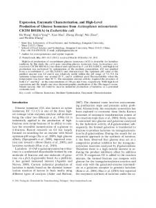
RESULTS Overall Structure of Spr1479/SapH—We initially found crystals of full-length Spr1479 (SapH-FL) but could not optimize them to a high diffraction quality. Limited proteolysis combined with LC-MS enabled us to define a relatively stable region of residues 1–262 (termed SapH). We overexpressed this region and subjected it to crystallization, and the resulting crystals were diffracted to a resolution of 1.90 Å. An asymmetric unit contains a dimer of SapH with an interface area of ⬃1700 Å2. Met-3–Leu-260 in subunit A and Met3–His-252 in subunit B are well fitted in the final model. The two subunits are very similar, with an overall root mean square deviation (r.m.s.d.) of 0.15 Å over 210 C␣ atoms (Fig. 1A). Sizeexclusion chromatography also confirmed the existence of SapH-FL and SapH as a dimer in solution (data not shown). The two five-stranded -sheets of subunit A are aligned antiparallel to their counterparts in subunit B, forming two continuous 10-stranded mixed -sheets, which are perpendicular to the dimer-related non-crystallographic 2-fold axis (Fig. 1A). Each subunit adopts a compact core structure with a four-layer ␣␣-sandwich fold: two five-stranded -sheets sandwiched by helices ␣1 and ␣2 on one side and helix ␣6 on the opposite side (Fig. 1B). One -sheet is composed of strands 4 –8, whereas the facing -sheet consists of strands 1–3, 9, and 10 (Fig. 1B). Beyond the core structure, SapH has a cap of five ␣-helices (␣3–␣5, ␣7, and ␣8) packing against the active site. Among these five ␣-helices, ␣3–␣5 (Asn-66 –His-109) form one side wall of the active site, whereas the other two ␣-helices ␣7 and ␣8 (Val-225–Asn-249) at the C terminus might function as a scaffold. A Dali search using the five-helix subdomain
35908 JOURNAL OF BIOLOGICAL CHEMISTRY
revealed no hit of a similar domain. Compared with the core domain, the secondary structural elements of this additional subdomain appear to be more variable in length and structure, which may be responsible for the versatile substrate specificities among the MPP superfamily members. The core structures of subunits A and B are packed against each other to form a dimer interface, which consists mainly of the backbones of strands 7 and 10 from both subunits. Specifically, the interface is composed of hydrogen bonds between Val-214 O␣ and Arg-220⬘ N␣, Met-216 N␣ and Phe-218⬘ O␣, Met-216 O␣ and Phe-218⬘ N␣, Phe-218 N␣ and Met-216⬘ O␣, Phe-218 O␣ and Met-216⬘ N␣, and Arg-220 N␣ and Val-214⬘ O␣ from strand 10 and between Leu-170 N␣ and Leu-170⬘ O␣ and Leu-170 O␣ and Leu-170⬘ N␣ from strand 7 (the residues in subunit B are labeled with a prime) (Fig. 1C). In addition to these backbone hydrogen bonds, the carboxyl group of Asp-209 (Asp-209⬘) forms two hydrogen bonds with N1 and N2 of Arg-200⬘ (Arg-200), further stabilizing the dimer. Binuclear Metals at the Active Site—During the model building and refinement process, two electron density peaks were outstanding in the Fo ⫺ Fc Fourier difference map (at the 30 level), indicating the presence of metals. The brown color of concentrated protein in solution indicated the presence of iron. Atomic absorption spectroscopy showed that iron and manganese were present at molarity ratios to the protein of 0.7 and 0.8, respectively. X-ray diffraction experiments at various wavelengths were done to confirm the chemical identity of these metals. We collected data at a wavelength of 1.74 Å and found two strong anomalous peaks of 31.0 and 30.7 in the structure model-phased anomalous Fourier difference maps. This result excluded the presence of zinc, copper, nickel, and cobalt, which have a theoretical absorption K-edge below 1.61 Å. To further VOLUME 286 • NUMBER 41 • OCTOBER 14, 2011
Downloaded from http://www.jbc.org/ by guest on November 4, 2015
SapH-KI
Streptococcal Nucleotide and Phosphodiester Hydrolase SapH
distinguish iron from manganese, we collected data at a wavelength of 1.80 Å and assigned peaks of 16.9 to iron and 25.0 to manganese. (The theoretical absorption K-edges for iron and manganese are 1.74 and 1.90 Å, respectively.) A significant anomalous signal of the iron site indicates that this site might be also mixed with manganese. Thus, the two sites should have mixed occupancies of Fe3⫹ and Mn2⫹, respectively (Fig. 2A). The following activity assays also confirmed that the binuclear metals are Fe3⫹ and Mn2⫹. The two metals are 3.4 and 3.3 Å away from each other in subunits A and B, respectively. Fe3⫹ and coordinating residues Asp-11, His-13, Asp-39, and His-166, together with Mn2⫹ and coordinating residues Asp-39, Asn-66, His-128, and His-164, constitute an octahedral structure of the active site (Fig. 2A). A planar water molecule (designated Wat1) forms coordination bonds with Fe3⫹ and Mn2⫹ and a hydrogen bond with the carbonyl oxygen of His-164. This binuclear active site is located at the interface between the core structure and the cap of ␣-helices. Comparative Structure Analysis—Overall structure comparison of SapH with structures in the Protein Data Bank using the OCTOBER 14, 2011 • VOLUME 286 • NUMBER 41
JOURNAL OF BIOLOGICAL CHEMISTRY
35909
Downloaded from http://www.jbc.org/ by guest on November 4, 2015
FIGURE 1. Overall structure of SapH. A and B, schematic representations of the dimer and monomer, respectively. Subunits A and B are shown in cyan and orange, respectively. The secondary structural elements are labeled sequentially. The metal ions at the active site are shown in purple for Mn2⫹ and orange for Fe3⫹. C, dimer interface. The residues are shown as cyan and orange sticks for subunits A and B, respectively. The hydrogen bonds are denoted as dashed lines. Residues and strands from subunit B are labeled with a prime.
Dali server gave 188 hits for 41 unique proteins with a Z-score higher than 10.0; these proteins were all members of the MPP superfamily. The top hit was a hypothetical protein from Pyrococcus furiosus (Protein Data Bank code 1NNW; Z-score of 25.7, r.m.s.d. of 2.6 Å over 234 C␣ atoms), followed by E. coli phosphodiesterase YfcE (code 1SU1) (32) and M. jannaschii phosphodiesterase MJ0936 (code 1S3M) (33). The other hits included human VPS29 (code 1W24) (34) and Ser/Thr phosphatase 2B (code 1AUI) (35), symmetric Ap4A hydrolases from Trypanosoma brucei (code 2QJC) and Shigella flexneri (code 2DFJ) (36), and cyclic nucleotide phosphodiesterase Rv0805 from M. tuberculosis (code 2HY1) (37). Most of these proteins have similar molecular functions (phosphodiesterase, phosphatase, nuclease, or nucleotidase), although two are symmetric Ap4A hydrolases. Moreover, the binuclear active site of SapH can be closely superimposed onto that of M. tuberculosis cyclic nucleotide phosphodiesterase Rv0805 (Fig. 2B) (37), despite the fact that SapH and Rv0805 have a sequence identity of only 17% and markedly different structures, with an r.m.s.d. of 3.3 Å over 149 C␣ atoms. The highly conserved ␣␣-sandwich core structure and binuclear active site strongly implied that SapH might act as a phosphodiesterase, phosphatase, or Ap4A hydrolase. Phosphodiesterase Activity—We first tested the enzymatic activity of SapH for generic phosphomonoesterase or phosphodiesterase substrates (pNPP or bis-pNPP) in the presence of 0.2 mM Fe3⫹ and 0.2 mM Mn2⫹. No activity for pNPP was detected (data not shown). By contrast, both SapH-FL and SapH showed considerable activity for bis-pNPP. The Km and kcat values were 1.76 ⫾ 0.02 mM and 5.70 ⫾ 0.12 s⫺1, respectively, for SapH-FL and 2.46 ⫾ 0.15 mM and 5.51 ⫾ 0.22 s⫺1, respectively, for SapH (supplemental Table S1). These results suggested that SapH is a phosphodiesterase. We further systematically screened all of the reported physiological substrates of metallophosphodiesterase. These substrates fall into three groups: cyclic nucleotides, nucleic acids, and phospholipids. We tested the following representative substrates of phospholipases: 2⬘,3⬘-cAMP (Sigma), 3⬘,5⬘-cAMP (Sigma), double- or single-stranded DNA and RNA, and the generic substrate p-nitrophenylphosphorylcholine (Sigma). However, SapH-FL showed no catalytic activity for any of the above substrates. This indicates that SapH-FL most likely performs a novel phosphodiesterase activity for a unique physiological substrate, or the activity of SapH-FL for the above-listed substrates needs the assistance of an unknown partner. Similar cases of phosphodiesterases with unknown physiological substrate have also been reported previously (32, 33). The metal-coordinating residues have a crucial role in SapH activity. Mutation of Asp-39, His-13, or His-128 to Ala completely abolished the phosphodiesterase activity. Activity was elevated by the addition of 0.2 mM Fe3⫹ and/or Mn2⫹ and somewhat inhibited by the addition of Mg2⫹, Ni2⫹, Ca2⫹, Co2⫹, Zn2⫹, or Fe2⫹ individually (supplemental Fig. S1). Moreover, the addition of 5 mM EDTA did not change the activity (supplemental Fig. S1), indicating that the co-purified metals have a very high affinity for SapH.
Streptococcal Nucleotide and Phosphodiester Hydrolase SapH
Nucleotide/Derivative Hydrolase Activity—In addition to phosphodiesterases, the Dali search returned two hits of ApaHrelated symmetric Ap4A hydrolases from T. brucei and S. flexneri (r.m.s.d. of 3.1 Å over 177 C␣ atoms and 3.0 Å over 184 C␣ atoms and sequence identities of 21 and 20%, respectively). Thus, we tested the hydrolase activity of SapH-FL for Ap4A, as well as Ap3A and Ap5A (Sigma). The results showed that SapH hydrolyzed all three molecules asymmetrically, producing ADP and AMP from Ap3A, ATP and AMP from Ap4A, and Ap4 and AMP from Ap5A. The kcat values of SapH-FL for the three substrates are comparable, whereas the Km values are 0.28 mM for Ap3A, 2.09 mM for Ap4A, and 1.04 mM for Ap5A (Table 2). It is worth noting that the Km value for Ap3A is comparable with that of T4 bacteriophage NudE.1 (0.41 mM) and E. coli Orf186 (0.15 mM), both of which are Nudix proteins (38, 39). However, the molarity ratio of the product AMP is obviously higher than that of ATP in the reaction with Ap4A, suggesting that ATP might be further hydrolyzed to AMP. Assays showed that both SapH-FL and SapH had relatively low activity of hydrolyzing ATP to AMP (supplemental Fig. S2). This gave us the idea that SapH-FL should have a broad spectrum of substrate specificity. In consequence, several nucleotides and derivatives (Sigma), including ADP, ATP, ADP-ribose, NAD(H), and NADP(H), were tested. SapH-FL could somewhat hydrolyze ATP, NAD(H), and ADP-ribose but not ADP or NADP(H). The relative activities are 24% (ATP), 13% (ADPribose), 10% (NADH), and 2% (NAD⫹) of that of the activity for Ap3A (supplemental Table S2). Furthermore, nucleotides GTP, CTP, and UTP were also tested. SapH-FL could hydrolyze GTP and CTP at a comparable rate to ATP, whereas little activity for UTP was detected (Table 2 and supplemental Table S2). Despite the relatively lower activity of SapH-FL for these substrates, the Km value for ATP is ⬃2.37 mM, which is in the range of intracellular ATP concentrations (1–5 mM) (40). Thus, SapH might function as an ATP pyrophosphatase in vivo.
35910 JOURNAL OF BIOLOGICAL CHEMISTRY
TABLE 2 Kinetic parameters of SapH-FL Substrate
Km mM
bis-pNPP Ap3A Ap4A Ap5A ATP GTP CTP
1.76 ⫾ 0.02 0.28 ⫾ 0.02 2.09 ⫾ 0.10 1.04 ⫾ 0.10 2.37 ⫾ 0.17 2.42 ⫾ 0.32 3.00 ⫾ 0.18
kcat s
⫺1
5.70 ⫾ 0.12 15.95 ⫾ 0.90 14.89 ⫾ 0.35 11.29 ⫾ 0.27 9.91 ⫾ 0.24 7.58 ⫾ 0.47 23.74 ⫾ 1.28
kcat/Km s⫺1 mM⫺1
3.24 ⫾ 0.10 57.94 ⫾ 2.48 7.14 ⫾ 0.24 10.96 ⫾ 1.05 4.21 ⫾ 0.28 3.17 ⫾ 0.25 7.92 ⫾ 0.07
AMP-binding Pocket—The electrostatic potential surface of SapH revealed an extended pocket of two clefts perpendicular to each other, with binuclear metals at the corner. To reveal the substrate-binding mode, we attempted to prepare crystals of SapH in the presence of inorganic phosphate, AMP, and ATP by either crystal soaking or co-crystallization. We also tried crystallizing the inactive W67H mutant of SapH in complex with Ap4A. At the end, we obtained only the phosphate- and AMP-complexed structures (termed SapH-PO4 and SapHAMP, respectively) by soaking SapH crystals with 500 mM NaH2PO4 or 30 mM AMP. In SapH-PO4, the inorganic phosphate bridges the two metals at the active site and makes hydrogen bonds with the metal-coordinating residues Asn-66, His164, His-13, and His-166, in addition to Trp-67 and the planar water molecule Wat1 (Fig. 3A). The binding mode is very similar to that of other enzymes in the MPP superfamily, suggesting that SapH also adopts the common catalytic mechanism (9, 12, 41). In SapH-AMP, one molecule of AMP is deeply buried in the active-site pocket, with the adenine moiety in an anti-conformation and the ribose ring in a C4 exo-conformation (Fig. 3B). The adenine and ribose ring are frozen in the inner cleft between loop 10 (between 5 and ␣6) and loop 14 (between 8 and 1) via hydrogen bonds and hydrophobic interactions. The O4⬘ of ribose forms a hydrogen bond with Arg-137 N2, whereas O3⬘ and O2⬘ form two hydrogen bonds with His-252 N␦1. The adenine is stabilized in an anti-conformation via a VOLUME 286 • NUMBER 41 • OCTOBER 14, 2011
Downloaded from http://www.jbc.org/ by guest on November 4, 2015
FIGURE 2. Binuclear active site. A, Mn2⫹- and Fe3⫹-coordinating residues. The residues are shown as sticks, and the planar water molecule Wat1 is shown as a red sphere. B, superposition of the active sites of SapH (cyan) and cyclic nucleotide phosphodiesterase Rv0805 (yellow).
Streptococcal Nucleotide and Phosphodiester Hydrolase SapH
Downloaded from http://www.jbc.org/ by guest on November 4, 2015
FIGURE 3. AMP-binding pocket of SapH. A, inorganic phosphate-binding site in SapH-PO4. The residues are shown as sticks. The hydrogen bonds are shown as dashed lines. B, AMP-binding site. The binding pocket is between loops 10 and 14. The binding residues are shown as cyan sticks and AMP as green sticks. The metals and Wat1 are rendered as spheres. C, docking model of Ap3A (yellow for its C atoms) in the active-site pocket.
- stacking interaction against Phe-189 and a hydrogen bond with His-141 N␦1. The phosphate group is bound to the active site in the same manner as the inorganic phosphate in SapH (Fig. 3B). To reveal the Ap3A-binding mode, we docked Ap3A onto SapH using the RosettaDock program (42). Restraints were used to fix the AMP moiety of Ap3A in a position similar to that in the complex of SapH-AMP. In the model, the inner and outer clefts accommodate the AMP (the first adenine nucleotide and P1-phosphate group) and ADP (the second adenine nucleotide and P2- and P3-phosphate groups) moieties of Ap3A, respectively (Fig. 3C). The AMP moiety in the inner cleft could be well superimposed with that of SapH-AMP. The ADP moiety is staOCTOBER 14, 2011 • VOLUME 286 • NUMBER 41
bilized in the outer cleft via two hydrogen bonds between the P2-phosphate group and Arg-137. The adenosine moiety is stabilized by - stacking against Trp-135 and two hydrogen bonds with Asn-134 O␦1 and Ser-70 O␥, respectively (Fig. 3C). Multiple-sequence alignment revealed that Ser-70, Asn-134, Trp-135, and Arg-137 at the outer cleft are highly conserved in streptococci. It is worth noticing that the active-site residues Asn-66, Trp-67, and Ser-70 are all from helix ␣3 of the fivehelix cap. The phosphate in SapH-PO4 and SapH-AMP could be closely superimposed with the P1-phosphate group of the SapH-Ap3A model. Moreover, this phosphate could be superimposed with that of protein analogs such as dAMP in nuclease JOURNAL OF BIOLOGICAL CHEMISTRY
35911
Streptococcal Nucleotide and Phosphodiester Hydrolase SapH TABLE 3 Kinetic parameters of SapH-FL mutants for bis-pNPP, ATP, Ap3A, and Ap4A bis-pNPP Enzyme W67A W67H a
ATP/Ap3A/Ap4A
Km
kcat
kcat/Km
Km
kcat
kcat/Km
mM
s⫺1
s⫺1 mM⫺1
mM
s⫺1
s⫺1 mM⫺1
ND ND
ND ND
0.53 ⫾ 0.05 1.08 ⫾ 0.04 2.06 ⫾ 0.13 NDa 11.79 ⫾ 1.15 2.56 ⫾ 0.20 0.22 ⫾ 0.01 ND
ND, not detectable.
Mre11 from P. furiosus (9) and AMP in cyclic nucleotide phosphodiesterase Rv0805 (6), despite the binding modes being very different. These results suggest that the metal-bound phosphate group is under attack from the planar water molecule, driven by the binuclear metals. As for SapH, the inner cleft is complementary to an AMP (but not ADP or ATP) moiety, leaving the P1-phosphate group at the elbow exposed to the nucleophilic water molecule. Furthermore, protein fluorescence spectrometry assays revealed that the addition of GTP and CTP to the apo-form SapH could trigger a comparable decrease in flu-
Downloaded from http://www.jbc.org/ by guest on November 4, 2015
FIGURE 4. Multiple-sequence alignment. A, the binuclear active site is conserved in Gram-positive bacteria. B, the AMP-binding residues are conserved in streptococci. The metal-coordinating residues are indicated by blue triangles, whereas the AMP- and Ap3A-binding residues are indicated by red triangles.
35912 JOURNAL OF BIOLOGICAL CHEMISTRY
VOLUME 286 • NUMBER 41 • OCTOBER 14, 2011
Streptococcal Nucleotide and Phosphodiester Hydrolase SapH orescence intensity to that of ATP. The equilibrium dissociation constants (Kd) for these three substrates are 443 ⫾ 12 M (ATP), 198 ⫾ 64 M (GTP), and 283 ⫾ 10 M (CTP). These results indicate that CTP and GTP could also bind to the AMP pocket.
OCTOBER 14, 2011 • VOLUME 286 • NUMBER 41
Acknowledgments—We thank Wei Zhao (University of Science and Technology of China) for docking analysis. We are grateful to all the developers of the CCP4 Suite, ESPript, TreeView, and PyMOL. We thank the staff of the Shanghai Synchrotron Radiation Facility.
REFERENCES 1. Hausdorff, W. P., Bryant, J., Paradiso, P. R., and Siber, G. R. (2000) Clin. Infect. Dis. 30, 100 –121 2. Hoskins, J., Alborn, W. E., Jr., Arnold, J., Blaszczak, L. C., Burgett, S., DeHoff, B. S., Estrem, S. T., Fritz, L., Fu, D. J., Fuller, W., Geringer, C., Gilmour, R., Glass, J. S., Khoja, H., Kraft, A. R., Lagace, R. E., LeBlanc, D. J., Lee, L. N., Lefkowitz, E. J., Lu, J., Matsushima, P., McAhren, S. M., McHenney, M., McLeaster, K., Mundy, C. W., Nicas, T. I., Norris, F. H., O’Gara, M., Peery, R. B., Robertson, G. T., Rockey, P., Sun, P. M., Winkler, M. E., Yang, Y., Young-Bellido, M., Zhao, G., Zook, C. A., Baltz, R. H., Jaskunas, S. R., Rosteck, P. R., Jr., Skatrud, P. L., and Glass, J. I. (2001) J. Bacteriol. 183, 5709 –5717 3. Tettelin, H., Nelson, K. E., Paulsen, I. T., Eisen, J. A., Read, T. D., Peterson, S., Heidelberg, J., DeBoy, R. T., Haft, D. H., Dodson, R. J., Durkin, A. S., Gwinn, M., Kolonay, J. F., Nelson, W. C., Peterson, J. D., Umayam, L. A., White, O., Salzberg, S. L., Lewis, M. R., Radune, D., Holtzapple, E., Khouri, H., Wolf, A. M., Utterback, T. R., Hansen, C. L., McDonald, L. A., Feldblyum, T. V., Angiuoli, S., Dickinson, T., Hickey, E. K., Holt, I. E., Loftus, B. J., Yang, F., Smith, H. O., Venter, J. C., Dougherty, B. A., Morrison, D. A., Hollingshead, S. K., and Fraser, C. M. (2001) Science 293, 498 –506 4. Wizemann, T. M., Heinrichs, J. H., Adamou, J. E., Erwin, A. L., Kunsch, C., Choi, G. H., Barash, S. C., Rosen, C. A., Masure, H. R., Tuomanen, E., Gayle, A., Brewah, Y. A., Walsh, W., Barren, P., Lathigra, R., Hanson, M., Langermann, S., Johnson, S., and Koenig, S. (2001) Infect. Immun. 69, 1593–1598 5. Polissi, A., Pontiggia, A., Feger, G., Altieri, M., Mottl, H., Ferrari, L., and Simon, D. (1998) Infect. Immun. 66, 5620 –5629 6. Podobnik, M., Tyagi, R., Matange, N., Dermol, U., Gupta, A. K., Mattoo, R., Seshadri, K., and Visweswariah, S. S. (2009) J. Biol. Chem. 284, 32846 –32857 7. Bateman, A., Birney, E., Cerruti, L., Durbin, R., Etwiller, L., Eddy, S. R., Griffiths-Jones, S., Howe, K. L., Marshall, M., and Sonnhammer, E. L. (2002) Nucleic Acids Res. 30, 276 –280 8. Voegtli, W. C., White, D. J., Reiter, N. J., Rusnak, F., and Rosenzweig, A. C. (2000) Biochemistry 39, 15365–15374 9. Hopfner, K. P., Karcher, A., Craig, L., Woo, T. T., Carney, J. P., and Tainer, J. A. (2001) Cell 105, 473– 485 10. Klabunde, T., Sträter, N., Fröhlich, R., Witzel, H., and Krebs, B. (1996) J. Mol. Biol. 259, 737–748 11. Knöfel, T., and Sträter, N. (2001) J. Mol. Biol. 309, 239 –254 12. Schenk, G., Gahan, L. R., Carrington, L. E., Mitic, N., Valizadeh, M., Ham-
JOURNAL OF BIOLOGICAL CHEMISTRY
35913
Downloaded from http://www.jbc.org/ by guest on November 4, 2015
DISCUSSION SapH Is a Novel Member of the MPP Superfamily—To date, all nucleotide/derivative hydrolases of known structure belong to the Nudix hydrolase superfamily (43). However, the core structure of SapH adopts an ␣␣-sandwich fold rather than an ␣␣-sandwich fold with a Nudix signature motif (17, 19, 43). The ␣␣-sandwich fold and binuclear active site of SapH are highly conserved in members of the MPP superfamily, which share a low sequence identity (⬍20%) with SapH. Among these MPP superfamily members, only Bacillus subtilis PrpE (44) and E. coli ApaH (13) were found to hydrolyze Ap4A. However, PrpE is a Tyr-specific phosphatase and an asymmetric Ap4A hydrolase of unknown three-dimensional structure. E. coli ApaH was proved to be a symmetric Ap4A hydrolase. The structure of its 100% sequence-identical homolog from S. flexneri (Protein Data Bank code 2DFJ) also has an ␣␣-sandwich fold with two layers of five-stranded -sheets sandwiched by ␣-helices on the two sides, respectively (36). Superposition of SapH onto S. flexneri ApaH revealed an r.m.s.d. of 3.0 Å over 184 C␣ atoms. The core ␣␣-sandwich structure of SapH is quite similar to that of ApaH. Among the helices of the additional subdomain, three helices (␣3–␣5) of SapH exhibit structural similarity to the counterparts of ApaH, despite that helix ␣4 (Pro-84 –Glu-99) of SapH is 10 residues longer (supplemental Fig. S3). By contrast, the other two ␣-helices (␣7 and ␣8) at the C terminus of SapH are not found in ApaH. Alternatively, ApaH has an insertion of three additional ␣-helices (␣7–␣9, Asp-128 –Arg-182) located at the opposite side of the active site (supplemental Fig. S3). Moreover, the active site of ApaH is coordinated by two Mn2⫹ ions. Therefore, SapH represents the first structure of a novel nucleotide/derivative hydrolase in the MPP superfamily. SapH Is a Bifunctional Enzyme Conserved Exclusively in Streptococci—In addition to its phosphodiesterase activity, SapH demonstrates nucleotide/derivative hydrolase activity for ATP and ApnA. This dual function of SapH might be a gain of function due to subtle substitutions at the active site that were proposed to explain the diverse substrate specificity of the MPP superfamily. For instance, the metal and substrate specificities of Clostridium thermocellum polynucleotide kinase/phosphatase can be dramatically altered by substitution of the activesite residues (45– 49). Based on the analysis of the phosphatebinding residue His-98 in Rv0805, mutation of the corresponding Cys-74 to His in E. coli phosphodiesterase YfcE results in a gained 2⬘,3⬘-cAMP phosphodiesterase activity (48). In SapH, the corresponding residue Trp-67 also forms a hydrogen bond with the phosphate in SapH-PO4 and SapH-AMP. Compared with wild-type SapH-FL, the W67A mutant has a lower Km and kcat and comparable activity (kcat/Km) for bispNPP. By contrast, the W67H mutation increases the Km for bis-pNPP by ⬃7-fold, resulting in activity that is about onefifteenth that of the wild-type protein (Table 3). However, both
the W67A and W67H mutants have no detectable activity for ATP, Ap3A, or Ap4A (Table 3). These results indicate that Trp-67 of SapH-FL is indispensable for hydrolysis of ATP, Ap3A, and Ap4A. To find the evolutionary hints of the bifunctionality of SapH, we performed a multiple-sequence alignment. The binuclear active-site residues Asp-11, His-13, Asp-39, Asn66, His-128, His-164, and His-166 are highly conserved in Gram-positive bacteria (Fig. 4A). However, in addition to Trp-67, the other AMP-binding residues Arg-137, His-141, Phe-189, and His-252 are conserved exclusively in streptococci (Fig. 4B). Thus, we propose that the emergence of Trp67, together with substitutions of residues at the substratebinding pocket, resulted in a gain of nucleotide/derivative hydrolase function for streptococcal SapH, in addition to its phosphodiesterase activity.
Streptococcal Nucleotide and Phosphodiester Hydrolase SapH
13. 14. 15.
16. 17. 18. 19. 20. 21. 22. 23.
25. 26. 27. 28. 29.
35914 JOURNAL OF BIOLOGICAL CHEMISTRY
30. 31. 32. 33. 34. 35.
36. 37. 38. 39. 40. 41. 42. 43.
44. 45. 46. 47. 48. 49.
X., Murray, L. W., Arendall, W. B., 3rd, Snoeyink, J., Richardson, J. S., and Richardson, D. C. (2007) Nucleic Acids Res. 35, W375–W383 Laskowski, R., Macarthur, M., Moss, D., and Thornton, J. (1993) J. Appl. Crystallogr. 26, 283–291 DeLano, W. L. (2002) The PyMOL Molecular Graphics System, DeLano Scientific LLC, San Carlos, CA Miller, D. J., Shuvalova, L., Evdokimova, E., Savchenko, A., Yakunin, A. F., and Anderson, W. F. (2007) Protein Sci. 16, 1338 –1348 Chen, S., Yakunin, A. F., Kuznetsova, E., Busso, D., Pufan, R., Proudfoot, M., Kim, R., and Kim, S. H. (2004) J. Biol. Chem. 279, 31854 –31862 Wang, D., Guo, M., Liang, Z., Fan, J., Zhu, Z., Zang, J., Zhu, Z., Li, X., Teng, M., Niu, L., Dong, Y., and Liu, P. (2005) J. Biol. Chem. 280, 22962–22967 Kissinger, C. R., Parge, H. E., Knighton, D. R., Lewis, C. T., Pelletier, L. A., Tempczyk, A., Kalish, V. J., Tucker, K. D., Showalter, R. E., and Moomaw, E. W. (1995) Nature 378, 641– 644 Wang, Q. H., Hu, W. X., Gao, W., and Bi, R. C. (2006) Proteins 65, 1032–1035 Shenoy, A. R., Capuder, M., Draskovic, P., Lamba, D., Visweswariah, S. S., and Podobnik, M. (2007) J. Mol. Biol. 365, 211–225 O’Handley, S. F., Frick, D. N., Dunn, C. A., and Bessman, M. J. (1998) J. Biol. Chem. 273, 3192–3197 Xu, W., Gauss, P., Shen, J., Dunn, C. A., and Bessman, M. J. (2002) J. Biol. Chem. 277, 23181–23185 Traut, T. W. (1994) Mol. Cell. Biochem. 140, 1–22 Bellinzoni, M., Wehenkel, A., Shepard, W., and Alzari, P. M. (2007) Structure 15, 863– 872 Gray, J. J., Moughon, S., Wang, C., Schueler-Furman, O., Kuhlman, B., Rohl, C. A., and Baker, D. (2003) J. Mol. Biol. 331, 281–299 Mildvan, A. S., Xia, Z., Azurmendi, H. F., Saraswat, V., Legler, P. M., Massiah, M. A., Gabelli, S. B., Bianchet, M. A., Kang, L. W., and Amzel, L. M. (2005) Arch. Biochem. Biophys. 433, 129 –143 Iwanicki, A., Herman-Antosiewicz, A., Pierechod, M., Séror, S. J., and Obuchowski, M. (2002) Biochem. J. 366, 929 –936 Martins, A., and Shuman, S. (2005) RNA 11, 1271–1280 Keppetipola, N., and Shuman, S. (2006) RNA 12, 73– 82 Keppetipola, N., and Shuman, S. (2006) J. Biol. Chem. 281, 19251–19259 Keppetipola, N., and Shuman, S. (2007) Nucleic Acids Res. 35, 7721–7732 Keppetipola, N., and Shuman, S. (2008) J. Biol. Chem. 283, 30942–30949
VOLUME 286 • NUMBER 41 • OCTOBER 14, 2011
Downloaded from http://www.jbc.org/ by guest on November 4, 2015
24.
ilton, S. E., de Jersey, J., and Guddat, L. W. (2005) Proc. Natl. Acad. Sci. U.S.A. 102, 273–278 Guranowski, A., Jakubowski, H., and Holler, E. (1983) J. Biol. Chem. 258, 14784 –14789 Guranowski, A. (2000) Pharmacol. Ther. 87, 117–139 Jeyakanthan, J., Kanaujia, S. P., Nishida, Y., Nakagawa, N., Praveen, S., Shinkai, A., Kuramitsu, S., Yokoyama, S., and Sekar, K. (2010) Acta Crystallogr. D Biol. Crystallogr. 66, 116 –124 Swarbrick, J. D., Bashtannyk, T., Maksel, D., Zhang, X. R., Blackburn, G. M., Gayler, K. R., and Gooley, P. R. (2000) J. Mol. Biol. 302, 1165–1177 Swarbrick, J. D., Buyya, S., Gunawardana, D., Gayler, K. R., McLennan, A. G., and Gooley, P. R. (2005) J. Biol. Chem. 280, 8471– 8481 Bailey, S., Sedelnikova, S. E., Blackburn, G. M., Abdelghany, H. M., Baker, P. J., McLennan, A. G., and Rafferty, J. B. (2002) Structure 10, 589 – 600 Iwai, T., Kuramitsu, S., and Masui, R. (2004) J. Biol. Chem. 279, 21732–21739 Bessman, M. J., Frick, D. N., and O’Handley, S. F. (1996) J. Biol. Chem. 271, 25059 –25062 Gabelli, S. B., Bianchet, M. A., Bessman, M. J., and Amzel, L. M. (2001) Nat. Struct. Biol. 8, 467– 472 Otwinowski, Z., and Minor, W. (1997) Methods Enzymol. 276, 307–326 Brodersen, D. E., de La Fortelle, E., Vonrhein, C., Bricogne, G., Nyborg, J., and Kjeldgaard, M. (2000) Acta Crystallogr. D Biol. Crystallogr. 56, 431– 441 Adams, P. D., Afonine, P. V., Bunkóczi, G., Chen, V. B., Davis, I. W., Echols, N., Headd, J. J., Hung, L. W., Kapral, G. J., Grosse-Kunstleve, R. W., McCoy, A. J., Moriarty, N. W., Oeffner, R., Read, R. J., Richardson, D. C., Richardson, J. S., Terwilliger, T. C., and Zwart, P. H. (2010) Acta Crystallogr. D Biol. Crystallogr. 66, 213–221 Vagin, A., and Teplyakov, A. (2010) Acta Crystallogr. D Biol. Crystallogr. 66, 22–25 Collaborative Computational Project No. 4. (1994) Acta Crystallogr. D Biol. Crystallogr. 50, 760 –763 Murshudov., G. N., Vagin., A. A., and Dodson., E. J. (1997) Acta Crystallogr. D Biol. Crystallogr. 53, 240 –255 Emsley, P., and Cowtan, K. (2004) Acta Crystallogr. D Biol. Crystallogr. 60, 2126 –2132 Davis, I. W., Leaver-Fay, A., Chen, V. B., Block, J. N., Kapral, G. J., Wang,
SUPPLEMENTARY INFORMATION FIGURE LEGENDS Fig. S1. The enzymatic activity of SapH-FL towards bis-pNPP in the presence of various metal ions (0.2 mM) or 5 mM EDTA. The assays were performed in the presence of 100 nM enzyme and 1 mM bis-pNPP. Fig. S2. ATP hydrolysis assays of SapH-FL and SapH. Fig. S3. Stereo representation of structural comparison between SapH (cyan) and S. flexneri ApaH (yellow, PDB code 2DFJ).
Fig. S1
Fig. S2
Fig. S3
Table S1. Kinetic parameters of SapH-FL and SapH towards bis-pNPP. bis-pNPP Enzyme
Km (mM)
kcat (s-1)
kcat/Km (s-1mM-1)
SapH-FL
1.76 ± 0.02
5.70 ± 0.12
3.24 ± 0.10
SapH
2.46 ± 0.15
5.51 ± 0.22
2.24 ± 0.04
Table S2. Substrate specificity of SapH-FL. Substrate
Relative activity (%)
Ap3A
100
Ap4A
90
Ap5A
69
ADPR
13
NAD+
2
NADH
10
ATP
24
GTP
11
CTP
17
UTP
2
All substrates were present at a concentration of 2 mM.
Protein Structure and Folding: Structural and Enzymatic Characterization of the Streptococcal ATP/Diadenosine Polyphosphate and Phosphodiester Hydrolase Spr1479/SapH Yong-Liang Jiang, Jun-Wei Zhang, Wei-Li Yu, Wang Cheng, Chen-Chen Zhang, Cecile Frolet, Anne-Marie Di Guilmi, Thierry Vernet, Cong-Zhao Zhou and Yuxing Chen
Access the most updated version of this article at doi: 10.1074/jbc.M111.228585 Find articles, minireviews, Reflections and Classics on similar topics on the JBC Affinity Sites. Alerts: • When this article is cited • When a correction for this article is posted Click here to choose from all of JBC's e-mail alerts
Supplemental material: http://www.jbc.org/content/suppl/2011/08/23/M111.228585.DC1.html This article cites 48 references, 22 of which can be accessed free at http://www.jbc.org/content/286/41/35906.full.html#ref-list-1
Downloaded from http://www.jbc.org/ by guest on November 4, 2015
J. Biol. Chem. 2011, 286:35906-35914. doi: 10.1074/jbc.M111.228585 originally published online August 23, 2011