to characterize the structural properties of six fixation meth- ods used to fix a ... methods and the best of the prominent fixation methods to determine if the ...
0363-5465/99/2727-0035$02.00/0 THE AMERICAN JOURNAL OF SPORTS MEDICINE, Vol. 27, No. 1 © 1999 American Orthopaedic Society for Sports Medicine
Structural Properties of Six Tibial Fixation Methods for Anterior Cruciate Ligament Soft Tissue Grafts Hugh E. Magen,* MS, Stephen M. Howell,†‡§ LT COL, USAFR, MC, and Maury L. Hull,*† PhD From the *Biomedical Engineering Graduate Group and the †Department of Mechanical Engineering, University of California at Davis, Davis, California, and the ‡Clinical Investigation Facility, David Grant Medical Center, Travis Air Force Base, California
ABSTRACT This study compared the stiffness (K), yield load (YL), and slippage (SL) of six tibial fixation methods. These properties were determined from load-to-failure and cyclic tests of double-looped tendon grafts fixed to both animal and young human tissue. Tandem washers (K = 259 N/mm, YL = 1159 N, SL = 0.5 mm) and the Washerloc (K = 248 N/mm, YL = 905 N, SL = 2.0 mm) were the two best fixations. At 500 N of load, which is the estimated daily tension of an anterior cruciate ligament graft during intensive rehabilitation, slippage was significantly greater in either of the other two methods for sutures tied to a post (4.9 mm), double staples (3.3 mm), and a 20-mm spiked metal washer (3.5 mm). Interference screw fixation performed well in animal tissue (YL = 776 N), but was significantly worse in young human tissue (YL = 350 N), with 57% of the fixations failing before 500 N of load. Animal tissue should not be used to estimate the performance of interference screw fixation in human tissue. Because 57% of the interference screw fixations using human tissue failed at loads below 500 N, their ability to provide adequate fixation during intensive rehabilitation should be questioned. However, both the Washerloc and tandem washers and screws provide fixation structural properties in young human tibia that should be appropriate for intensive rehabilitation.
§ Address correspondence and reprint requests to Stephen M. Howell, MD, 8100 Timberlake Way, Suite F, Sacramento, CA 95823. One author has a commercial affiliation with a product used in this study. See “Acknowledgments” for funding information.
The method used to fix an ACL graft must be stiff enough to restore the load-displacement response (that is, stability:) of the knee to normal, strong enough to avoid failure, and secure enough to resist slippage under cyclic loading during the first 1 to 2 months after reconstruction.15 Fix-ation methods are more likely to be subjected to worst-case loads during intensive rehabilitation when high cyclic loads are produced in the graft before the conversion from mechanical to biologic fixation.” Any fixation method with poor structural properties of either stiffness, strength, or slippage has the potential to compromise the clinical outcome. A study of the structural properties of different methods for fixing soft tissue grafts may be of interest to the sur-geon because of the increased use of hamstring grafts as a tissue to replace the torn ACL. Because the fixation method. Especially on the tibial side, is generally the weak link of the initial graft-fixation method complex,2 this study was designed to characterize the structural properties of six fixation methods used to fix a double-looped tendon graft to the tibia. Because of the scarcity of young human tissue, the first objective of our study was to compare the stiffness, yield load, and slippage of two low-profile fixation methods designed to avoid pain and irritation caused by prominent hardware by fixing the graft within the tibial tunnel, and four prominent fixation methods that attach the graft to the anterior cortex of the tibia using animal tissue, The second objective was to reevaluate, in young human tissue, both low-profile fixa-tion methods and the best of the prominent fixation methods to determine if the structural properties of these fixation methods are similar. The final objective was to determine if the source of tissue chosen to evaluate a fixation method has a
36
Magen, et at,
significant effect on the fixation structural properties MATERIALS AND METHODS Specimen Preparation Six tibial fixation methods were evaluated using freshfrozen animal tissue stored at -20˚C. Common digital extensor tendons were harvested from 84 bovine forelimbs. Bovine tendons were used for the graft because the stiffness and viscoelastic behavior at higher initial stresses (20 MPa) are not significantly different from a human double-looped semitendinosus and gracilis graft.9 The bifurcated tendon was divided into two halves. A double-looped bovine tendon graft was prepared by placing the two tendons side-by-side, folding them in half, and thinning them until the graft just passed through an 8-mm diameter cylinder. A No. 1 suture (Ethibond, Ethi-con Inc., Somerville, New Jersey) was used to sew 4 cm of both ends of each tendon using a crisscrossing stitch. The cross-sectional area of the double-looped bovine tendon graft was calculated by averaging cross-sectional area measurements obtained at 15, 45, and 75 mm from the looped end of the graft using an area micrometer.12 Porcine tibias were used in this study because they are readily available, free from disease, inexpensive, and have been used in the only other study that measured slippage under cyclic loading to failure.8 The 84 porcine tibias were prepared by removing the patella, fibula, and soft tissue, potting them in polymethyl methacrylate inside a metal cylinder, and drilling a tibial tunnel that was 8 mm in diameter and 40mm in length. The tests using human tissue required 14 human gracilis and semitendinosus tendons obtained from donors with an average age of 47 years (range, 18 to 67), and 7 pairs of human tibias obtained from donors with an average age of 35 years (range, 18 to 48). The preparation and measurement of the cross-sectional area of the double-looped semitendinosus and gracilis graft were similar to the techniques used for the double-looped bovine tendon graft; however, the graft diameter was measured without being thinned. The preparation of the human tibias was similar to that of the porcine tibias, with the exception that the diameter of the tibial tunnel was drilled to match the diameter of the double-looped semitendinosus and gracilis graft, which ranged from 7.0 to 9.0 mm.
American Journal of Sports Medicine
dons be sewn together using a modified baseball stitch. The diameter of the tibial tunnel was drilled to within 0.5 mm of the diameter of the graft in accordance with the manufacturer’s recommenda-tion. A 9 x 25 mm soft tissue interference screw was advanced over a guide pin, and inserted between the graft and the posterior wall of the tibial tunnel until it was contained within the tibial tunnel. The Washerloc was countersunk into a 21-mm diameter counterbore drilled perpendicular to the posterior wall of the tibial tunnel (Fig. 1). The Washerloc was 20 mm in diameter, had four 11-mm long peripheral spikes that straddled the graft, and nineteen 6-mm long centrally placed spikes that penetrated the graft in multiple locations. The Washerloc was placed on top of the graft, driven into the posterior wall of the tibial tunnel within the counterbore, and compressed with a 4.5-mm diameter cortical screw that purchased the posterior cortex of the tibia. The four prominent fixation methods that were evaluated included sutures tied to a post (No. 5 Ethibond), two staples (regular fixation staples, Smith & Nephew Richards, Memphis, Tennessee), a 20-mm spiked metal washer (Linvatec, Largo, Florida), and two soft tissue washers (Synthes, Paoli, Pennsylvania). A larger No. 5 suture was sewn to each of the four limbs of the graft in place of the No. 1 suture and tied around a 4.5-mm diameter bicortical screw14,17 placed 15 mm distal to the tibial tunnel. The length of suture bridging the graft to the post was 25 mm. Two 7.9 x 25.4 mm soft tissue staples were used to fix the graft to the tibial cortex using the “belt-buckle” technique.4,5 The four limbs of the graft were stapled to the tibial cortex, the graft was folded back over the staple, and the second staple was applied. A 20-mm diameter metal washer with twelve 1.3-mm length spikes was used to compress the graft to the tibial cortex
Description of Fixation Methods Six tibial fixation methods were evaluated: two were low in profile because they were recessed inside the tibial tunnel, and four were prominent because they were applied to the anteromedial cortex of the tibia. The two low-profile fixation methods consisted of the soft tissue interference screw (standard interference screw, Smith & Nephew DonJoy, Carlsbad; California) and the Washerloc (Washerloc, Arthrotek, Inc., Ontario, Canada). Fixation with the interference screw required that the four free limbs of the double-looped ten-
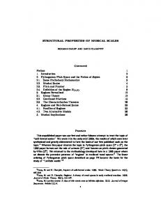
Figure 1. Diagram depicting the method for determining the total graft length (XT), the length of the intraarticular portion of the graft (X1), and the length of the graft from the articular surface of the tibia to the point of tibial fixation (X2) using the low-profile Washerloc as an example. The double-looped graft was looped around the bar attached to the materials testing machine.
Vol. 27, No. 1, 1999
Structural Properties of Tibial Fixation Methods 37
with a 4.5-mm diameter bicortical screw. Two limbs of the graft were wrapped 180° counterclockwise around the screw, while the other two limbs were wrapped clockwise. Fixation with the two washers required two 4.5mm diameter bi-cortical screws and two 13.5mm diameter plastic spiked washers spaced 15 mm apart in tandem.14,17 Two limbs from different tendons were wrapped in an “S-shaped” configuration around both screws and the other two limbs were wrapped in the opposite direction. Experimental Apparatus Structural tests of the grafts and the graft-fixation method-tibia complexes were administered using a materials testing machine (Instron 5566 Materials Testing Machine, Instron Corp., Canton, Massachusetts) that applied a strain rate of 5% of the total graft length per second. Tensile loads were measured with a 5 kN load cell attached to the crosshead. Elongation was measured with a customdesigned extensometer incorporating a linear variable differential transducer (lOOO-DCD, Schaevitz Engineering, Pennsauken, New Jersey). A computer was used both to control the tests and to acquire load and elongation data (Instron Series IX Software, Instron Corp.). The tibia, cemented in the metal cylinder, was attached to the crosshead of the materials testing machine using a custom-designed fixture that allowed the tibial tunnel and graft to be loaded in alignment with the motion axis of the actuator (Fig. 2). Testing Using Animal Tissue Three different tests were used to obtain the stiffness of the double-looped bovine tendon graft, the stiffness of each fixation method, and the stiffness, yield load, and slippage of the graft-fixation method-tibia complexes. First, the stiffness of each double-looped bovine tendon graft was determined by looping it over a bar attached to the cross-head of the materials testing machine, evenly tensioning the four limbs of the graft, and clamping them with a freeze clamp bolted to the base of the materials testing machine. The distance between the bar and freeze clamp was individualized for each fixation method to match the length of the graft as if it was implanted in a reconstructed knee. A length of 50 mm (X1) was chosen to replicate the intraarticular portion of the graft (30 mm) and the section within the femoral tunnel (20 mm). The total length of the graft (XT) was calculated by adding this length to the distance from the articular surface of the tibia to the point of tibial fixation (X2), which differed for each of the six fixation methods (see Table 1; Fig. 1). Because the average failure load of the double-looped bovine tendon is 2817 ± 298 N,9 the maximum load was limited to 1000 N to avoid plastic deformation. The double-looped bovine tendon was cycled 11 times, and stiffness was determined from the load-elongation curve of the last cycle. A load-to-failure test was performed to determine the stiffness and yield load of the graft-fixation method-tibia com-
Figure 2. Experimental apparatus with animal tissue and tandem washer fixation as an example. The potted tibia was set in a customdesigned fixture attached to the load cell in the crosshead of the materials testing machine. The fixture has three rotational and two translational degrees of freedom to allow alignment of the tibial tunnel and graft with the motion axis of the materials testing machine. The tendon was wrapped around a rigid post, attached to the base of the materials testing machine, pulled through the tibial tunnel, and attached to the tibia. An extensometer, built with a linear variable differential transducer (LVDT), measured graft elongation.
plex. The cylinder containing the potted tibia was mounted in a specially designed testing apparatus that allowed the limbs of the graft to be tensioned while each fixation method was applied. The double-looped bovine tendon graft of known stiffness was looped around the bar attached to the jig, and the four limbs of the graft were passed through the tibial tunnel. The distance from the bar to the articular surface of the tibial plateau was kept at 50 mm. The four limbs of the double-looped bovine tendon graft were tensioned and secured to the tibia using a fixation method selected at random. The graft-fixation method-tibia complex was transferred to the materials testing machine. The looped end of the double-looped bovine tendon graft was passed around a bar attached to the base of the materials testing machine and the cylinder containing the tibia was bolted to the fixture attached to the crosshead of the materials testing machine. The extensometer was attached to the bar of the materials testing machine and to a pin drilled into the lateral cortex
38 Magen, et at,
of the tibia, level with the point of fixation. The graft-fixation method-tibia complex was loaded to failure. Each of the six fixation methods was evaluated seven times using fresh grafts and tibias. Slippage of the graft-fixation method-tibia complex was determined using additional grafts and tibias by applying progressively higher loads (in 50-N increments) under load control for subsequent cycles until failure. Computed as the difference between the length after each complete loading cycle (at 10 N of load) and the original length (at 10 N of load), the residual displacement measured the combined effects of tendon graft stretch and fixation slippage.8 Fixation slippage was the primary cause of any differences in length between fixation methods because graft stretch was assumed to be constant at a specified load. Slippage was compared at 250 and 500 N of load within the elastic region, and well below the failure load of the double-looped bovine tendon (2817 N9) and double-looped semitendinosus and gracilis (4213 N18) grafts. Each of the six fixation methods was evaluated seven times. Testing Using Young Human Tissue Based on findings from the tests using animal tissue, the testing procedures using human tissue were modified from those using the animal tissue to conserve scarce, young, fresh-frozen, human tissue. The evaluation of the fixation methods was limited to a comparison between the two lowprofile fixation methods and the best prominent fixation method, which was the tandem washers. In addition, the load-to-failure test was eliminated because the yield load obtained from the residual displacement test was more conservative, being either the same or less than the load-to-failure test for the six fixation methods. Two different total graft lengths were used to test fixation with the tandem washers. “Ideal” fixation was achieved by shortening the distance from the bar to the articular surface of the tibia from 50 to 25 mm so that all four limbs of the double-looped semitendinosus and gracilis graft were long enough to be compressed under the more distal washer (see Table 3). “Typical” fixation resulted when the distance was left at 50 mm and the tendons were secured as best as possible to the distal screw. In six of seven tests, the sutures sewn in each limb of the gracilis tendon were tied around the distal screw because the tendon was too short. One low-profile and one prominent fixation method were randomly selected to be tested in a tibia using the same double-looped semitendinosus and gracilis graft, and the remaining two fixation methods were tested in the opposite, paired tibia. To avoid possible carry-over effects from refixing the same double-looped semitendinosus and gracilis graft within a tibia, the more distal prominent fixation method was tested first. Then the distance from the bar to the articular surface of the tibial plateau was shortened from 50 to 35 mm for the low-profile fixation method so that the graft was not gripped
American Journal of Sports Medicine
in the same location twice. A smaller Washerloc 16 mm in diameter with 13 instead of 19 short spikes, was used for the tests in human tissue. Data Analysis A simple regression was used to derive stiffness from the slope of the linear region of the load-displacement curve for the grafts and graft-fixation method-tibia complexes. For testing using animal tissue, the stiffness of the graft-fixation method-tibia complex was derived from the load-to- failure test. For testing using human tissue, the stiffness of the complex was measured from the residual displacement test because the load-to-failure test was not performed. A compromise was required in selecting the load cycle for measuring stiffness. Stiffness could not be measured from load cycles below 350 N because the load-displacement curves were predominantly nonlinear because of the “toe-in” region. For this reason, the seventh cycle (load to 350 N) was chosen to measure stiffness, even though two of seven of the interference screws had failed by this cycle. The stiffness of the fixation method alone (KF) was calculated knowing the stiffness of the graft (KG) and the stiffness of the graft-fixation method-tibia complex (KGFC) and using the equation KF = KGFC * KG/(KG - KGFC). This equation was derived by considering the graft-fixation method-tibia complex to be represented by a springs-in-series model.l8 The yield load was determined from the load-displacement curve obtained from both the load-to-failure and residual displacement tests. A line was extended from the linear portion of the load-displacement curve until it intersected the x-axis. The yield load was defined as the load where a second line, drawn parallel but intersecting the x-axis at 0.5% greater strain than the first line, crossed the load-displacement curve. The yield load for the residual displacement test was reported for the first cycle to exhibit yield. To determine the best fixation method using animal tissue and then using human tissue (first and second objectives), the stiffness of the graft-fixation method-tibia complex, calculated stiffness of the fixation method, and yield load were compared for each fixation method in animal and human tissue using an analysis of variance. The slippage for each fixation method in animal and human tissue was compared using a KruskalWallis analysis of variance on ranks. All pairwise comparisons were performed using Duncan’s multiple range test. To evaluate whether the tissue source affected the structural properties (third objective), the yield load and slippage from the residual displacement tests were compared between animal and human tissue for each fixation method using an unpaired t-test. Also, the stiffness of the graft-fixation methodtibia complex and calculated stiffness of the fixation method from the load-to-failure test in the animal tissue were compared with the corresponding stiffness from the residual displacement test in human tissue using an unpaired t-test. Differences were considered significant when P < 0.05.
Vol. 27, No. 1, 1999
Structural Properties of Tibial Fixation Methods 39
RESULTS Properties of Fixation Methods Using Animal Tissue The mean (± standard deviation) stiffness for the graftfixation method-tibia complex was not significantly differ-ent for the interference screw (226 ± 56 N/mm), Washerloc (200 ± 76 N/mm), and tandem washers (203 ± 42 N/mm); however, these fixation methods were significantly stiffer than fixation with sutures, staples: and a 20-mm washer (Table 1). Similarly, the mean stiffness of just the fixation method alone without the graft was not significantly different for the interference screw (476 ± 251 N/mm), tandem washers (420 ± 180 N/mm), and Washerloc (429 ± 269 N/mm); however, these were significantly greater than the other three fixation methods. Yield load for the tandem washers was 1375 ± 213 N for the load-to-failure test and 1269 ± 456 N for the residual displacement test, and both yield loads were significantly greater than those of the other fixation methods evaluated within each test. Mean slippage at 250 N of load was 0.2 ± 0.2 mm for the Washerloc and 0.3 ± 0.1 mm for the interference screw, and both values of slippage were significantly less than those of the other fixation methods (Table 2). However, at 500 N of load, three of seven interference screw fixations had failed. The least slippage at 500 N occurred with the Washerloc (0.8 ± 0.6 mm) and tandem washers (l.2 ± 0.5 mm), and both devices had slippage values that were significantly less than the three other fixation methods. Properties of Fixation Methods Using Human Tissue The mean stiffness for the graft-fixation method-tibia complex was not significantly different for the interference screw (248 ± 52 N/mm), the Washerloc (273 ± 56 N/mm), and ideal fixation with tandem washers (259 ± 23 N/mm), but the stiffness of all three methods was significantly greater than typical fixation with tandem washers (181 ± 39 N/mm)
TABLE 2 Slippage at 250 and 500 N of Load for Six Tibial Fixation Methods Using Animal Tissue (Mean ± SD) Slippage (mm) Fixation device Interference screw Washerloc Sutures/post Staples 20-mm washer Tandem washers
250 N
500 N
0.25 ± 0.15 0.23 ± 0.15a 1.67 ± 0.98b 1.01 ± 0.39b 1.12 ± 0.64b 0.49 ± 0.23c a
0.72 ± 0,42b,c* 0.81 ± 0.61a 4.87 ± 1.59b 3.31 ± 1.29b 3.52 ± 2.14b 1.23 ± 0.53a,c
a Fixations within this group are not significantly different from each other, and have lower values than those in other groups. b Fixations within this group are not significantly different from each other, but have higher values than those in groups a and c. c Fixations within this group are not significantly different from each other but have higher values than those in group a. * Three fixations within this group had failed at this or a lower load.
in human tissue (Table 3). The mean stiffness of the fixation was highest for the Washerloc (506 ± 197 N/mm) and ideal fixation with tandem washers (414 ± 57 N/mm). The mean stiffness of the Washerloc was significantly greater than typical fixation with tandem washers (318 ± 112 N/mm) and the interference screw (340 ± 84 N/mm). The mean yield load was significantly greater for the ideal fixation with tandem washers (1159 ± 221 N) and Washerloc (905 ± 291 N) compared with fixation with the interference screw (350 ± 134 N). The mean yield load of the ideal fixation with tandem washers was significantly greater than the typical fixation with tandem washers (768 ± 293 N). The mean slippage for tandem washer fixations-both ideal (0.1 ± 0.0 mm) and typical (0.3 ± 0.3 mm)-was significantly less than for the interference screw (1.8 ± 3.1 mm), although only the ideal configuration was significantly less than the
TABLE 1 Graft Length, Cross-Sectional Area, Height, Stiffness, and Yield Load for Six Tibial Fixation Methods Using Animal Tissue Evaluated With Both the Load-to-Failure and Residual Displacement Tests (Mean ± SD) Load-to-failure test Fixation device
Interference screw Washerloc Sutures/post Staples 20-mm washer Tandem washers
Graft lengtha XT (mm)
Cross-sectional area (mm2)
Fixation height (mm)
65 80 80 95 95 95
42 ± 2.6 41 ± 2.6 41 ± 2.5 42 ± 2.2 40 ± 3.2 41 ± 2.1
0 ± 0C 2.0 ± l.8f 5.0 ± 1.3e 7.0 ± 2.4d 7.7 ± 1.4d 7.5 ± 0.7d
Stiffness (N/mm) G-FM-Tb complex
Fixation method
Yield load (N)
Residual displacement test Yield load (N)
226 ± 56d 200 ± 76d 60 ± 14f 118 ± 47e 126 ± 26e 203 ± 42d
476 ± 251d 429 ± 269d 70 ± 19e 174 ± 92e 192 ± 61e 420 ± 180d
776 ± 155e 821 ± 193e 830 ± 187e 705 ± 174e 930 ± 323d 1375 ± 213d
598 ± 167e,f 903 ± 178e 442 ± 67f 785 ± 273e 724 ± 284e,f 1269 ± 456d
XT is the graft test length, set as the distance from the peg of the materials testing machine to the tibial fixation device. Graft-fixation method-tibia. c Fixations in this group are not significantly different from each other, and have the lowest values compared with those in other groups. d Fixations within this group are not significantly different from each other, but have higher values than those in groups c, e, and f. e Fixations within this group are not significantly different from each other but. have higher values than those in group c and f. f Fixations within this group are not significantly different from each other, and have lower values than those in groups d and e, but not c. a
b
40 Magen, et at,
American Journal of Sports Medicine
TABLE 3 Graft Length, Cross-Sectional Area, Yield Load and Stiffness of Four Tibial Fixation Methods Using Human Tissue Evaluated With the Residual Displacement Test (Mean ± SD) Graft lengtha (MM)
Stiffness (N/mm)
Fixation device
XT
X1
X2
Cross-sectional area mm2
Yield load (N)
G-FM-Tb complex
Fixation method
Interference screw Washerloc Tandem washers Ideal Typical
50 65
35 35
15 30
46 ± 6.4 43 ± 8.4
350 ± 134c 905 ± 291d,e
248 ± 52d 273 ± 56d
340 ± 84c 506 ± 197d
70 95
25 50
45 45
46 ± 6.7 43 ± 8.6
1159 ± 221d 768 ± 293e
259 ± 23d 181 ± 39e
414 ± 57d,e 318 ± 112e
a XT, graft test length, set as the distance from the peg of the materials testing machine to the tibial fixation device (XT = Xi + X2) (Fig. 1); Xi, distance from the peg of the materials testing machine to the tibial articulating surface; X2, distance from the tibial articulating surface to the edge of the fixation device. b Graft-fixation method-tibia. c Fixations within this group are not significantly different from each other, and have lower values than those in other groups. d Fixations within this group are not significantly different from each other, but have higher values than those in groups c and e. e Fixations within this group are not significantly different from each other but have higher values than those in group c.
TABLE 4 Slippage at 250 and 500 N of Load for Four Tibial Fixation Methods Using Human Tissue (Mean ± SD) Slippage (mm) Fixation device Interference screw Washerloc Tandem washers Ideal Typical
250 N
500 N
1.80 ± 3.10 * 0.55 ± 0.43a,b
3.67 ± l.12a† 1.95 ± 1.28b
0.15 ± 0.04c 0.30 ± 0.30b,c
0.52 ± 0.l0c 0.86 ± 0.30b*
a
a Fixations within this group are not significantly different from each other, but have higher values than those in groups b and c. b Fixations within this group are not significantly different from each other but have higher values than those in group c. c Fixations within this group are not significantly different from each other. and have lower values than those in other groups. * One fixation within this group had failed at this or a lower load. † Four fixations within this group had failed at this or a lower load.
Washerloc (0.6 ± 0.4 mm) at 250 N of load (Table 4). At 500 N of load, the mean slippage of ideal fixation with tandem washers (0.5 ± 0.1 mm) was significantly less than the typical fixation with tandem washers (0.9 ± 0.3 mm) and the Washerloc (2.0 ± 1.3 mm), which were all significantly less than fixation with the interference screw (3.7 ± 1.1 mm), One of seven typical fixations with tandem washers failed at 500 N of load. One of seven interference screw fixations failed at 250 N and four of seven fixations failed at 500 N of load. Comparison of Properties of Fixation Methods Between Animal and Human Tissue The source of tissue had a significant effect on some of the structural properties of the interference screw, the Washerloc, and the ideal fixation with tandem washers (Table 5). The yield load for interference screw fixation was significantly lower in human tissue than in animal tissue (P
TABLE 5 Comparison of Structural Properties of Fixation Methods Between Human and Animal Tissue (P Values)a Fixation method Structural property Interference Washerloc Tandem screw washers Yield load 0.010 Stiffness G-FM-T complex NS 0.507 Fixation NS 0.278 Slippage 250 N 0.003 500 N NA
NS 0.988
NS 0.574
0.062 NS 0.552
0.009 NS 0.933
NS 0.190 0.028
0.011 0.006
a G-FM-T, graft-fixation method-tibia; NS, not significant; NA, not available.
= 0.010), and slippage was significantly greater in human tissue at 250 N load (P = 0.003). A comparison of slippage at 500 N of load could not be made with interference screw fixation because four of seven fixations using human tissue had failed by this load level. Slippage at 500 N for the Washerloc was significantly greater in human tissue (P = 0.028). Stiffness of the graft-fixation method-tibia complex for ideal fixation with tandem washers was significantly greater in human tissue (P = 0.0091, and slippage at 250 and 500 N of load was significantly less in human tissue (P = 0.011 and P = 0.006, respectively). DISCUSSION The goals of this study were to compare the structural properties of six tibial fixation methods in animal tissue, reevaluate three of the fixation methods in young human tissue, and determine if the source of tissue chosen to evaluate a fixation method had a significant effect on the structural properties. Before discussing the findings from this study, a critical examination of the experimental techniques is warranted to determine how the methods may have affected the
Vol. 27, No. 1, 1999
interpretation of the results. Methods Issues The comparisons of structural properties between fixation methods may have been affected by the axis used to load the graft. Loading the graft physiologically by translating the tibia anterior on the femur with the knee in 20° of flexion angles the graft, resulting in friction at the intra-articular rim of the tibial tunnel. Friction between the graft and edge of the drill hole can stress shield the more distal site of graft fixation.” In this study, the graft was loaded along the axis of the tibial tunnel, which would result in less friction at the tunnel edge. However, loading along the tunnel axis allowed higher forces to be directed to the fixation site, representing a worstcase scenario for analyzing a fixation technique.” The friction at the tunnel rim must, be quantified to determine the effect of stress shielding of the fixation. The reduction in tension across the rim of the femoral tunnel during 0° to 120° of motion is less than 10%.19 Less angulation can be expected for the same arc of motion at the tibial tunnel so a 10% reduction represents a worst-case analysis. A 500-N tension applied to the graft using a “physiologic” axis would result in 450 N of tension at the site of fixation instead of 500 N when the graft is loaded along the axis of the tibial tunnel. However, since the reduction in tension would occur at the edge of the tibial tunnel, and the point of fixation for all the tested methods was distal to the tunnel edge, the reduction in tension would be systematic and hence would not affect the comparisons of structural properties between fixation methods. Because the interference screw, Washerloc, staple, and washer provide fixation by compressing the graft against either the cortical bone or wall of the bone tunnel, non-uniformity in the cross-sectional area of the graft either between fixation methods or between animal and human tendons could have affected the grip on the graft and biased the comparisons. In this study, however, there were no significant differences in the cross-sectional area of the graft between fixation methods and tissue source (Tables 1 and 3); therefore, the comparisons of structural properties in this study were valid. The reason for individualizing the graft length for each fixation method was to reproduce the length of the graft as if it was fixed 20 mm inside a femoral tunnel to simulate a reconstructed knee. Although using grafts of different lengths did affect the stiffness of the graft, it did not affect either the conclusions regarding the stiffness comparisons of the graft-fixation method-tibia complex or the calcu-lated stiffness of the fixation method, both of which were the structural properties of interest in this study. The results indicated that the stiffness of the graft-fixation method-tibia complex was influenced more by the stiffness of the fixation method rather than by the stiffness of the graft. For example, the shorter, stiffer graft of 80 mm used with suture fixation resulted in
Structural Properties of Tibial Fixation Methods 41
a complex stiffness of only 60 N/mm, and the longer, more elastic graft of 95 mm used with tandem washer fixation provided a significantly higher complex stiffness of 203 N/mm in animal tissue (Table 1). The springs-in-series analysis factored out the variability in graft stiffness from the stiffness of the graft-fixation method-tibia complex and allowed the stiffness of just the fixation method to be calculated. The comparison of stiffness between fixation methods was valid because the confounding effect of variability in graft stiffness was eliminated. Interpretation of Results The structural properties of stiffness and slippage were included in our study in addition to yield load because they each may affect the ability of a ligament replacement to restore and maintain stability of the reconstructed knee, especially during intensive rehabilitation. A graft-fixation method-tibia complex less stiff than the normal ACL (182,12 296,22 303,16 N/mm) requires a higher pre-tension to restore normal laxity. Tension in a patellar tendon graft, with an estimated stiffness of 18% to 28% of the normal ACL (51 N/mm),16 was threefold greater than in the intact ACL when it was pretensioned to restore normal anteroposterior laxity.10 The higher forces in the graft required to restore normal laxity may exceed the ability of a fixation method to resist slippage when its stiffness is less than that of the normal ACL. In animal tissue, fixation methods with lower stiffness, including sutures tied to a post, double staples, and the 20-mm washer, were found to have significantly more slippage than the higher stiffness fixation methods of the Washerloc and tandem washers at 500 N of load. Predicting the vulnerability of a fixation method to slippage during intensive rehabilitation requires an estimate of the tension in the graft before biologic fixation. The daily tensile loads of a normal ACL are believed to be, at most. 20% of its failure capacity.1,3,6 Thus, the loads in the ACL during daily activities can be expected to be about 500 N since the failure load of the normal ACL in young adults is approximately 2500 N.3 The load in an ACL graft may be even greater if the graft is overtensioned.10 However, it can be speculated that strains and the resultant forces in an ACL graft may be lower in the early postoperative period when reconstructed knees are restricted from vigorous use because of pain.3 With the current understanding, it seems reasonable to conclude that a fixation method should function to loads of at least, 500 N if a reconstructed knee is to be intensively rehabilitated, assuming that the graft is not overtensioned. The fixation method that functioned best at loads above 500 N in young human tibia using the double-looped semitendinosus and gracilis graft was ideal fixation with tandem washers. When both tendons were securely compressed under the distal washer, the stiffness (259 ± 23 N/mm) was similar to that of the normal ACL, slippage averaged only 0.5 mm at 500 N of load, and the yield load averaged 1159 N. The
42 Magen, et at,
structural properties were significantly worse, however, when the gracilis tendon was too short and sutures sewn to the graft had to be tied to the distal screw. This typical fixation, defined as suture fixation of the gracilis tendon to the distal screw, was required in six of seven specimens. One of the fixations failed by 500 N, the stiffness was 69% less (181 N/mm), slippage was 1.6 times greater (0.9 mm at 500 N of load), and yield load was 66% less (768 N) than ideal fixation with tandem washers. Therefore, it may be sensible to consider using another fixation method if the reconstructed knee is to be intensively rehabilitated when the gracilis tendon is too short to be compressed under the distal screw. The Washerloc provided significantly higher stiffness and yield load than typical fixation with tandem washers in young human tibia using the double-looped semitendinosus and gracilis graft. The low-profile Washerloc, countersunk in the distal end of the tibial tunnel, adequately fixed shorter gracilis tendons because the point of fixation was closer to the joint. Stiffness of the Washerloc was similar to that of the normal ACL (273 ± 56 N/mm), slippage averaged 2.0 mm at 500 N of load, and the average yield load was 904 N. Other than the structural property of slippage, there were no significant differences in the stiffness and yield load between the Washerloc and ideal fixation with tandem washers. One of the important results from this study was that the structural properties of a fixation method may not be the same in animal and human tissue. Interference screw fixation performed significantly worse in human tissue compared with animal tissue. The average yield load was only 350 ± 134 N in young human tibia and compared favorably with the maximum pull-out force found in an investigation by the manufacturer of the interference screw of 336 N 59 N using human knees.” The yield loads in both of these studies were significantly less than the yield load of 776 and 598 N using animal tissue for the load-to-failure and residual displacement tests, respectively. The property of slippage was even more affected by the tissue source. At 250 N of load, fixation with the interference screw had the least slippage (0.2 mm) in animal tissue but the greatest slippage (1.8 mm) in human tissue of all fixation methods tested. A possible explanation for these differences may be that the interference screw purchases solely in cancellous bone, which could vary in density between tissue sources. Another example where fixation properties were overestimated by testing in animal bone instead of human bone is in a study that used bovine tibia to evaluate interference screw fixation of a three-stranded semitendinosus graft.20 The selection of bovine tibia was based on the study by Brown et al.,2 who found that bovine knees provide a more clinically relevant model of a young human knee than do elderly human cadaveric knees. In bovine tibia, the pull-out force of the three-stranded semitendinosus graft fixed with a round, threaded: titanium cannulated interference screw (RCI [round cannulated interference screw], Smith & Nephew
American Journal of Sports Medicine
DonJoy) was 419 ± 77 N, with a range from 316 to 558 N. In young human tibia, the average yield load for a doublelooped semitendinosus and gracilis graft with interference screw fixation was lower at 350 ± 134 N, the minimal yield load was only 222 N, and the range was broader, from 222 to 682 N compared with the tests in bovine tibia. Surgeons should not assume that the structural properties of interference screw fixation of soft tissue grafts determined in animal tissue predict its performance in human knees. Recently, it has been shown that the fixation level of an ACL graft has a significant influence on anterior knee stability and that anatomic graft fixation at the original ACL insertion site (aperture fixation) is the most preferable.7,11,20 The principle behind this concept is that the stiffness of the graftfixation method-bone complex is increased by keeping the length of the graft as short as possible. The findings from this study do not support this principle with currently available fixation methods. The stiffness of the graft-fixation method-tibia complex is influenced more by the stiffness of the fixation method than by the length of the graft.18 For example, in human tissue there was no significant difference in the stiffness of the graft-fixation method-tibia complex for the interference screw, Washerloc, and ideal fixation with tandem washers even though the site of fixation for the Washerloc and tandem washers was 15 and 20 mm more distal than the site of interference screw fixation. The reason the stiffness was similar for these three fixation complexes in spite of different graft lengths was that the stiffnesses of the fixation methods of the Washerloc (506 N/mm) and tandem washers (414 N/mm) were significantly greater than those of interference screw fixation (340 N/mm). Surgeons interested in improving the stiffness of the graft-fixation method-bone complex can be just as effective using fixation methods of higher stiffness as by securing the graft close to the joint. Furthermore, a greater yield load and less slippage can be realized by the fixation device purchasing in cortical bone more distally in the tibial tunnel (that is, Washerloc) or on the tibial cortex (that is, tandem washers) than by purchasing in the less dense cancellous bone inside the tibial tunnel with an interference screw. Although the results from studies that have evaluated interference screw fixation of bone-patellar tendon-bone graft can be compared directly with the findings in this study, the stiffness and slippage of the tibia-graft-interference screw fixation complex must be estimated because two sites of fixation (tibia and femur)8,16 were tested simultaneously instead of the single site (tibia) as in this study. Assuming that both sites of interference screw fix-ation contribute equally to the structural properties, the slippage at a single fixation site would be half of the total, the stiffness of a single fixation site would be double that of the total ( derived from the springs-in-series analysis18, and the yield load would remain the same because failure usually occurs at one site of fixation. In young human knees, the average failure load of inter-
Vol. 27, No. 1, 1999
ference screw fixation of bone-patellar tendon-bone was 412 N and the average stiffness was 51 N/mm (doubled = 102 N/mm).16 In porcine knees, slippage averaged 3.8 mm (1.9 mm per fixation) at 500 N of load.’ In contrast, fixation of a double-looped semitendinosus and gracilis graft in a young human femur over a post with bone compaction had an average failure load of 1126 N and an average stiffness of 225 N/mm.18 Fixation of a double-looped semitendinosus and gracilis graft in young human bone using the best fixation in the tibia and femur should provide a fivefold increase in stiffness, either a fourfold decrease or no difference in slippage, and a two- to threefold increase in yield strength compared with interference screw fixation of bone-patellar tendon-bone. Surgeons should no longer be concerned that soft tissue fixation of a double-looped semitendinosus and gracilis graft is inferior to interference screw fixation of a bonepatellar tendon-bone graft. In fact, the opposite is true. The functional importance of stiffness, slippage, and yield load associated with any method of attachment of a graft to bone has not been extensively studied.3 Wide variability was observed in the three structural properties between fixation methods used to attach soft tissue grafts to the tibia in our study. Future studies are required to determine whether these different fixation properties affect the clinical outcome. Differences in clinical outcome may become more apparent if the fixation methods are subjected to loads imposed by intensive rehabilitation. ACKNOWLEDGMENTS The authors are grateful to the institutions and companies that supported this study, including the United States Air Force for providing the laboratory and assisting in the funding of this project (grant no. SGO 96-186); Smith & Nephew DonJoy; Arthrotek, Inc.; the University of California at Davis Donated Body Program; the National Disease Research Interchange; and the Musculoskeletal Transplant Foundation. The authors are also grateful to Dr. Neil Willitz for his statistical advice and to Dr. Neil Sharkey for his guidance. REFERENCES 1. Beynnon BD, Fleming BC, Johnson RJ, et al: Anterior cruciate ligament strain behavior during rehabilitation exercises in vivo Am J Sports Med 23:24-34, 1995 2. Brown CH Jr, Hecker AT, Hipp JA, et al: The biomechanics of Interference screw fixation of patellar tendon anterior cruciate ligament grafts. Am J Sports Med 21 880-886, 1993
Structural Properties of Tibial Fixation Methods 43 3. Frank CB, Jackson DW: The science of reconstruction of the anterior cruciate ligament. J Bone Joint Surg 79A: 1556-1576, 1997 4. Good L, Tarlow SD, Odensten M, et al: Load tolerance, security, and failure modes of fixation devices for synthetic knee ligaments. Clin Orthop 253: 190-196, 1990 5. Gottlieb DJ, Pyne Jl, Beynnon BD, et al: Evaluation of tendon fixation techniques. Orthop Trans 16: 455, 1992 6. Holden JP, Grood ES, Korvick DL, et al: In vivo forces in the anterior cructate ligament: Direct measurements during walking and trotting in a quadruped. J Biomech 27: 517-526, 1994 7. lshibashi Y, Rudy TW, Livesay GA, et al: The effect of anterior cruciate ligament graft fixation site at the tibia on knee stability: Evaluation using a robotic testing system. Arthroscopy 13: 177-182, 1997 8. Liu SH, Kabo JM, Osti L: Biomechanics of two types of bone-tendonbone graft for ACL reconstruction. J Bone Joint Surg 77B: 232-235, 1995 9. Magen HE, Hull ML, Howell SM: Comparison of structural and mechanical properties of bovine and human tendons, Proceedings of the Third World Congress of Biomechanics. Sapporo, Japan, August 1998, p 126 10. Markolf KL, Burchfield DM, Shapiro MM, et al: Biomechanical consequences of replacement of the anterior cruciate ligament with a patellar ligament allograft Part II: Forces in the graft compared with forces in the intact ligament. J Bone Joint Surg 78A: 1728-1734, 1996 11. Morgan CD, Kalmam VR Grawl DM: lsometry testing for anterior cruciate ligament reconstruction revisited. Arthroscopy 11: 647-659, 1995 12. Noyes FR, Butler DL, Grood ES, et al: Biomechanical analysis of human ligament grafts used in knee-ligament repairs and reconstructions. J Bone Joint Surg 66A: 344-352, 1984 13. Pflaster D: Pullout test, Pincewski endoscopic hamstring technique utilizing the DonJoy RCI ACL fixation screw. Report: Biomechanics Research Laboratory Smith and Nephew DonJoy, Carlsbad, California, 1994 14. Robertson DB, Daniel DM, Biden E: Soft tissue fixation to bone. Am J Sports Med 14: 398-403, 1986 15. Rodeo SA, Arnoczky SP, Torzilli PA, et al: Tendon-healing in a bone tunnel: A biomechanical and histological study in the dog J Bone Joint Surg 75A: 1795-1803, 1993 16.Rowden NJ, Sher D, Rogers GJ, et al: Anterior cruciate ligament graft fixation, Initial comparison of patellar tendon and semitendinosus autografts in young fresh cadavers. Am J Sports Med 25: 472-476, 1997 17. Steiner ME, Hecker AT, Brown CH Jr, et al: Anterior cruciate ligament graft fixation. Comparison of hamstring and patellar tendon grafts. Am J Sports Med 22: 240-246, 1994 18.To J, Howell S Hull M: Factors affecting the stiffness of anterior cruciate ligament replacements at implantation. Arthroscopy, in press, 1999 19. Ventura C, Wolchok J Hull ML. et al: An Implantable transducer for measuring tension in an anterior cructate ligament graft. J Biomech Eng 120: 327-333, 1998 20. Weller A, Hoffman RFG, Stahelin AC, et al: Hamstring tendon fixation using interference screws: A biomechanical study in calf tibial bone. Arthroscopy 14: 29-37, 1998 21. Wetter A, Windhagen HJ, Rashke MJ, et al: Biodegradable interference screw fixation exhibits pull-out force and stiffness similar to titanium screws. Am J Sports Med 26: 1 19-128, 1998 22. Woo SL-Y, Adams DJ: in Daniel DM, Akeson WH, O’Connor JJ (eds): Knee Ligaments. New York, Raven Press, 1990, pp 279-289