JPEG Y'CbCr colorspace. The Y'CbCr ECG image is transformed using various transforms, all but the largest transform coefficients are set to zero, and then ...
International Conference on Recent Advances and Future Trends in Information Technology (iRAFIT2012) Proceedings published in International Journal of Computer Applications® (IJCA)
Study of some Non-Linear Transforms for ECG Image Compression Vibha Aggarwal
Manjeet Singh Patterh Electronics &
Electronics & Communication Engineering COEM, Punjabi University Neighborhood Campus Rampura Phul, Punjab, India
Communication Engineering UCOE, Punjabi University Patiala, Punjab, India
ABSTRACT Out of the many non-linear transform methods the paper is making use of the (i) Essentially Non-Oscillatory cell-average decomposition (ENOCA), (ii) Max-lifting morphological wavelet transform (Maxlift), (iii) Med-lifting morphological wavelet transform (Medlift) and (iv) Morphological Haar wavelet transform (Mhaar) for ECG image compression. This study aims at recreating the image using inverse transforms after compressing the ECG image by making the use of the above mentioned transforms. There is an inversely proportionate relationship between image quality and image compression. This study has been undertaken not for the purpose of quantization and encoding rather an effort is being made to compare the different transform methods for ECG image compression.
General Terms Digital image processing and ECG image compression
Keywords Essentially Non-Oscillatory cell-average decomposition (ENOCA), Max-lifting morphological wavelet transform (Maxlift), Med-lifting morphological wavelet transform (Medlift), Morphological Haar wavelet transform (Mhaar) and ECG image compression
1. INTRODUCTION The aim of compression is to represent an ECG image with the smallest possible number of bits [1]. Traditionally there were various methods to compress an image such as Fourier transform, Cosine transform etc [2]. But a new tool wavelet transform (WT) provides a novel approach for the analysis of the image. This is due to fact that WT supports features like progressive image transmission (by quality, by resolution), ease of compressed image manipulation, region of interest coding etc [3]. Wavelets are non-adaptive sachems. Hence large coefficients always appear at discontinuities when wavelets are used. In images, edges constitute discontinuity in the data. The edges in an image are vital information and it is necessary to preserve them while efficiently representing the image [4]. Hence essentially nonoscillatory (ENO) interpolation technique avoids the discontinuities and large coefficients are not appearing at edges and provide better compression capabilities. The ENO interpolatory technique [5] is a data dependent, nonlinear technique which can eliminate the Gibbs phenomenon [6]. In
this paper, ENO cell average (ENOCA) decomposition is used for ECG compression. A major impulse to the development of nonlinear wavelet transform has been given by the introduction of lifting schemes by Sweldens [7]. Lifting amounts to modifying the analysis and synthesis operators in such a way that the properties of the modified scheme are “better” than those of original one. Here, “better” can be interpreted in different ways. For example, in the linear case, it may mean that the number of vanishing moments is larger. Lifting can be used to construct wavelet decompositions for signals that are defined on arbitrary domains, or to construct nonlinear coupled or uncoupled wavelet decompositions. In Mhaar scheme has two “vanishing moments” as opposed to the morphological Haar wavelets that has only one [7]. By one “vanishing moment” mean that a constant input signal produces a zero detail signal, whereas by two “vanishing moments” means that a linear signal produces a zero detail signal. Max-lifting scheme preserves the numbers and shapes of flat regions in a piecewise constant signal. This scheme preserves local maxima and moreover it does not create new ones. It is therefore expected that max-lifting will preserve, over a range of scales, the number and shapes of regions of constant signal value. Max-lifting decomposition produces one scaled image and three detail image (a horizontal, vertical, and diagonal detail image). The detail signals are zero at areas of smooth gray level variations, and that sharp gray level variations are mapped to negative (black) detail signal values [7]. In this paper, Maxlift, Medlift and Mhaar non linear transform methods [7] are also used ECG image compression. Section II describes the performance metrics. Section III presents the methodology. Finally, results and concluding remarks are given in section IV and V respectively.
2. PERFORMANCE METRICS For the performance analysis, the metrics like number of approximation components and Peak signal to noise ratio (PSNR) are used. Image approximation is the problem of representing image with as few components as possible. This is similar to lossy image compression, but ignoring the problems of quantization and encoding. PSNR is the comparison between an original image and a reconstructed image. Typical value for the PSNR in lossy image are between 30 and 50 dB, where higher is better.
25
International Conference on Recent Advances and Future Trends in Information Technology (iRAFIT2012) Proceedings published in International Journal of Computer Applications® (IJCA)
3. METHODOLOGY First, the input ECG image is converted from RGB to the JPEG Y’CbCr colorspace. The Y’CbCr ECG image is transformed using various transforms, all but the largest transform coefficients are set to zero, and then inverse transformed. The inverse transformed ECG image is converted into Y’CbCr to the RGB. The following steps have to be incorporated for ECG image compression: (i) ECG image is converted from RGB to JPEG Y’CbCr colorspace. (ii) Transform the Y’CbCr to another domain. (iii) Apply a threshold. (iv) Inverse-transform (v) Calculate the PSNR (vi) Converted from Y’CbCr JPEG to RGB colorspace ECG image.
4. RESULTS AND DISCUSSION The efficiency of the various transforms is tested by experimentation on the ECG image database [8]. The ECG image is transformed using ENOCA with two stage and five degree interpolation. Level used for decomposition is 2 in case of Maxlift, Medlift and Mhaar. The results are presented in Table 1 and Table 2. Table 1 and Table 2 conclude that the ENOCA and Maxlift transform are better from other transforms. ENOCA for high number of approximation components gives high PSNR value. And Maxlift for low number of approximation components gives high PSNR value. Figure 1-2 shows the original ECG images. Further, Figures 3-10 represents the reconstructed ECG images after compression with various transforms.
8:1 9:1 10:1
29.9 29.3 28.6
29.0 27.8 26.7
26.9 26.0 25.1
27.4 26.3 25.2
Table 2. Comparative performance of various transforms on ecg-exigency-003-b (800*160) ecg-exigency-003-b (800*160) Transform ENOCA Maxlift Medlift Number of PSNR PSNR PSNR approximation components 3:1 39.7 40.8 40.5 4:1 38.0 40.0 39.3 5:1 36.3 38.5 37.4 6:1 34.9 36.9 35.6 7:1 33.8 35.4 33.9 8:1 32.7 33.9 32.2 9:1 31.8 32.1 30.7 10:1 31.0 30.4 29.1
Mhaar PSNR
40.5 39.8 38.4 36.7 34.7 32.3 30.4 28.8
image 121(800x416)
5. CONCLUSION This paper represents the comparative analysis of ENOCA, Maxlift, Medlift and Mhaar transforms for ECG image compression. In case of, image_121 using ENOCA at 3:1 gives PSNR of 44.5 db while at 10:1 gives PSNR of 28.6 db. Maxlift at 3:1 gives PSNR of 58.1 db while at 10:1 gives PSNR of 26.7 db. Medlift at 3:1 gives PSNR of 51.1 db while at 10:1 gives PSNR of 25.1 db. Mhaar at 3:1 gives PSNR of 56.6 db while at 10:1 gives PSNR of 25.2 db. The above sited results justify the statement that high number of approximation components of the image gives a poor PSNR and hence the poor quality. Also the image quality of ENOCA (high number of approximation components i.e 10:1) and Maxlift (low number of approximation components i.e 3:1) is better than the other transforms.
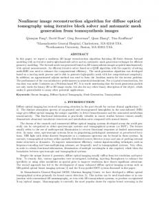
Fig 1: Original ECG image of image_121 (800*416).
ecg-exigency-003-b(800x160)
Table 1. Comparative performance of various transforms on image_121 (800*416) Transform Number of approximation components 3:1 4:1 5:1 6:1 7:1
image_121 (800*416) ENOCA Maxlift Medlift PSNR PSNR PSNR
Mhaar PSNR Fig 2: Original ECG image of ecg-exigency-003-b (800*160).
44.5 34.9 32.8 31.5 30.7
58.1 40.1 34.7 31.9 30.2
51.1 35.4 31.5 29.5 28.1
56.6 38.2 33.3 30.7 28.9
26
International Conference on Recent Advances and Future Trends in Information Technology (iRAFIT2012) Proceedings published in International Journal of Computer Applications® (IJCA)
ENOCA,5:1 approximation,PSNR,32.8 dB
Maxlift,5:1 approximation,PSNR,38.5 dB
Fig 3: Reconstructed ECG image of image_121 (800*416) using ENOCA
Fig 6: Reconstructed ECG image of ecg-exigency-003-b (800*160) using Maxlift
Medlift,5:1 approximation,PSNR,31.5 dB
ENOCA,5:1 approximation,PSNR,36.3 dB
Fig 4: Reconstructed ECG image of ecg-exigency-003-b (800*160) using ENOCA
Fig 7: Reconstructed ECG image of image_121 (800*416) using Medlift
Maxlift,5:1 approximation,PSNR,34.7 dB
Maxlift,5:1 approximation,PSNR,38.5 dB
Fig 5: Reconstructed ECG image of image_121 (800*416) using Maxlift
Fig 8: Reconstructed ECG image of ecg-exigency-003-b (800*160) using Medlift
27
International Conference on Recent Advances and Future Trends in Information Technology (iRAFIT2012) Proceedings published in International Journal of Computer Applications® (IJCA)
6. REFERENCES Mhaar,5:1 approximation,PSNR,33.3 dB
[1] Kazi Rafiqul Islam, Md. Anwarul Abedin, Masuma Akter, and Rupam Debm. 2011 High Speed ECG Image Compression Using Modified SPIHT. International Journal of Computer and Electrical Engineering, Vol. 3, No. 3, pp 398-402. [2] Anil K. Jain. 1989 Fundamental of Digital Image Processing. Prentice-Hall Inc. [3] Andra, K., Chakrabarti, C., and Acharya, T. 2002 A VLSI architecture for lifting-based forward and inverse wavelet transform. IEEE Transactions on Signal Processing, Vol. 50, No.4, pp 966-977. [4] Gandhi, S. 2005 ENO interpolation compression. Thesis of Master of Science.
Fig 9: Reconstructed ECG image of image_121 (800*416) using Mhaar
image
[5] Gatreuer, P. and Meyer, F. G. 2008 ENO multiresolution schemes with general discretizations. SIAM J. NUMER. ANAL. Vol. 46, no. 6, pp 2953-2977. [6]
Mhaar,5:1 approximation,PSNR,38.4 dB
for
Chan, T. F. and Zhou, H.-M. 2001 ENO-wavelet transforms and some applications. Acdemic Press, Inc., pp 1-34.
[7] Heijmans and Goutsias 2000 Nonlinear multiresolution signal decomposition schemes – Part II: Morphological Wavelets. IEEE Transactions on Image Processing, Vol. 9, no. 11, pp 1897-1913.
Fig 10: Reconstructed ECG image of ecg-exigency-003-b (800*160) using Mhaar
28