Biochemical Journal Immediate Publication. Published on 15 Jan 2014 as manuscript BJ20131567
Submitted to Biochemical Journal, revised version, January 9, 2014
Substrate interaction dynamics and oxygen control in the active site of thymidylate synthase ThyX
t
Hubert F. Becker*,†,‡, Kamel Djaout*,†, Isabelle Lamarre*,†, Jonathan E. Ulmer*,†, Delphine Schaming§, Véronique Balland§, Ursula Liebl*,†, Hannu Myllykallio*,† & Marten H. Vos*,†,1
nu
To whom correspondence should be addressed (email
[email protected])
ce
pte d
Ma
Thymidylate synthase ThyX, required for DNA synthesis in many pathogenic bacteria, is considered a promising anti-microbial target. It binds FAD and three substrates, producing deoxythymidine-5’-monophosphate from deoxyuridine-5’monophosphate (dUMP). However, ThyX proteins also act as NADPH oxidase by directly reacting with oxygen. We investigated the dynamic interplay between the substrates and their role in competing with this wasteful and potentially harmful oxidase reaction in catalytically efficient ThyX from Paramecium bursaria Chlorella virus 1. dUMP binding accelerates the oxygen-insensitive half-reaction between NADPH and FAD by over four orders of magnitude to ~30 s-1. Thus, although dUMP does not have a direct role in FAD reduction, any turnover with molecular oxygen requires its presence. Inversely, NADPH accommodation accelerates dUMP binding ~3 fold and apparently precedes dUMP binding under physiological conditions. In the oxidative half-reaction, excess CH2H4folate was found to re-oxidize FADH2 within 1 ms, thus very efficiently competing with FADH2 oxidation by O2, (1.5 s-1 under aerobic conditions). The resulting reaction scheme points out how the interplay between the fast reactions with the native substrates, while not rate-limiting overall catalysis, avoids NADPH oxidase activity in aerobic micro-organisms, including many pathogens. These observations also explain why ThyX proteins are also present in aerobic micro-organisms. Keywords: enzyme kinetics, stopped flow, flavin, time-resolved spectroscopy, thymidylate synthase, molecular evolution
Ac
THIS IS NOT THE VERSION OF RECORD - see doi:10.1042/BJ20131567
1
sc rip
*Laboratory for Optics and Biosciences, CNRS Ecole Polytechnique, 91128 Palaiseau, France † INSERM U696, 91128 Palaiseau, France ‡ UPMC Univ Paris 06, 75005 Paris, France § Laboratoire d'Electrochimie Moléculaire, UMR CNRS 7591, Université Paris Diderot, Sorbonne Paris Cité, 15, rue Jean-Antoine de Baïf, 75205 Paris Cedex 13, France.
Short title: Substrate interaction dynamics and O2 control in the active site of ThyX Summary statement: Thymidylate synthase ThyX is required for synthesis of thymine in many pathogenic bacteria. Using time-resolved spectroscopy of the flavin cofactor, we unraveled the interplay between the three native substrates and show how harmful oxidase activity is avoided in aerobic environments. Abbreviations used: dTMP: 2’-deoxythymidine-5’-monophosphate; dUMP: deoxyuridine-5’-monophosphate; PBCV: Paramecium bursaria Chlorella virus 1 Licenced copy. Copying is not permitted, except with prior permission and as allowed by law. © 2014 The Authors Journal compilation © 2014 Biochemical Society
2’-
Biochemical Journal Immediate Publication. Published on 15 Jan 2014 as manuscript BJ20131567
INTRODUCTION
sc rip
nu
Ma
pte d
ce
Ac
THIS IS NOT THE VERSION OF RECORD - see doi:10.1042/BJ20131567
t
Nature has established two structurally unrelated enzyme families, dubbed ThyA and ThyX, that form 2’-deoxythymidine-5’-monophosphate (dTMP), an essential DNA precursor [1]. The key mechanistic difference between these non-orthologous enzyme families lies in their redox chemistry. ThyA proteins catalyze reductive methylation of 2’-deoxyuridine-5’monophosphate (dUMP) to dTMP using N5,N10-methylene-5,6,7,8-tetrahydrofolate (CH2H4folate) as both, carbon donor and reductant. In contrast, ThyX proteins from prokaryotes and viral sources are flavoproteins that use the NADPH/FADH2 couple as hydride source to reduce a methylene moiety of CH2H4folate to form dTMP [2-5]. As FADH2 is sensitive to the presence of molecular dioxygen [6] and ThyX proteins have substantial NADPH oxidase activity [3, 7, 8], it has been speculated that ThyX proteins are preferentially used under anaerobic or microaerophilic conditions [9]. However, we have recently discovered that the majority (≈ 65%) of ThyX-carrying species are either strict or facultative aerobes, raising the possibility that ThyX proteins use to date unexplored strategies to prevent or hinder turnover with molecular oxygen to favor the physiologically relevant dTMP forming reaction. This is of fundamental importance, as turnover with oxygen would result in formation of hydrogen peroxide and activation of the microbial defense against oxidative stress, particularly in actively dividing cells. Very little is known regarding the transient interplay of the different substrates and the FAD cofactor at the active site of ThyX proteins. Such interactions are of high interest as substrate binding brings about essential conformational changes during ThyX catalysis that may influence oxygen reactivity or turnover of ThyX proteins under aerobic conditions. From measurements at equilibrium performed using ThyX proteins from either Paramecium bursaria Chlorella virus (PBCV-1) or Thermotoga maritima, consensus emerged that the early chemical step in the ThyX reaction cycle is the reduction of FAD to FADH2 by hydride transfer from NADPH (reductive half-reaction) [2, 3, 10, 11]. Subsequently, CH2H4 folate binds close to FADH2 and triggers the transfer of a methylene moiety from CH2H4folate to dUMP [5, 12, 13]. The transferred methylene is subsequently converted to a methyl group by hydride transfer from FADH2 (oxidative half-reaction), resulting in flavin oxidation and dTMP formation. Whereas dUMP presumably undergoes chemical modification only after FAD reduction, the binding order and kinetics of the nucleotide substrates remain controversial. Previous work has suggested that dUMP either accelerates FAD reduction by NADPH [3] or, alternatively, may only participate in the FAD reoxidation reaction [7]. Turnover rates and catalytic efficiencies of ThyX proteins are substantially lower than the corresponding rates measured for ThyA proteins. The highest kcat value reported for ThyX proteins of 0.25 s-1 (PBCV-1 enzyme) is an order of magnitude smaller than reported for bacterial ThyA proteins [14]. However, as this value was measured under normoxic conditions and the degree of reoxidation of FADH2 by O2 (instead of CH2H4folate) was not accounted for, the actual rate limiting steps of ThyX catalysis remain poorly characterized. In the present work we take advantage of spectral features of ThyX from PBCV-1 that make this viral protein suitable for rapid-mixing time-resolved experiments to investigate ThyX ligand binding and redox chemistry. ThyX from PBCV-1 provides a robust model for understanding the functioning of more fragile ThyX proteins from pathogenic species. We use stopped flow transient absorption spectroscopy to investigate the transient dynamics of flavinsubstrate binding and flavin reduction and reoxidation under anaerobic and aerobic conditions. Our new findings reveal a dual role of dUMP, first as essential activator (reductive half cycle) and then as substrate (oxidative half cycle). This work also clearly establishes that a complex interplay of dUMP and NADPH in the vicinity of the FAD cofactor is necessary for formation of a transient ternary complex (dUMP-NAPDH-ThyX) that enables FAD
2 Licenced copy. Copying is not permitted, except with prior permission and as allowed by law. © 2014 The Authors Journal compilation © 2014 Biochemical Society
Biochemical Journal Immediate Publication. Published on 15 Jan 2014 as manuscript BJ20131567
sc rip
t
reduction. We also show that flavin reoxidation in the presence of CH2H4folate intrinsically occurs fast (< 1 ms), and efficiently competes with flavin oxidation by molecular oxygen, thus implying that the redox chemistry of ThyX proteins is not rate-limiting even under aerobic conditions. These kinetic measurements therefore provide a highly feasible explanation why ThyX proteins are not eliminated from aerobic micro-organisms. Our observations have important implications for the ThyX reaction mechanism, and more generally point at the important role of substrate-substrate and substrate-cofactor interactions in positioning the enzyme complex for its catalytic function.
Ma
nu
Sample preparation The PBCV-1 ThyX enzyme was expressed and purified following a previously described protocol [3] and stored at -80°C in 30 mM HEPES pH 8, 150 mM NaCl, 10% glycerol. The flavin content of the ThyX protein was estimated from the FAD absorbance maximum at 448 nm and the protein concentration by the Bradford assay. As in all isolated ThyX solutions, the FAD content was substoichiometric, for the PBCV-1 enzyme typically 0.7 per tetramer. No external flavin was added. Samples were prepared in 30 mM HEPES buffer, pH 8, 150mM NaCl, 1% glycerol. Unless stated differently, all experiments were performed at 20°C. kcat values were determined by following the release of tritium from the [5-3H]dUMP stock (Moravek), as described in Ref. [3], under aerobic and anaerobic (as below) conditions.
ce
pte d
Spectroscopy Most equilibrium and stopped-flow spectroscopic experiments were performed under anaerobic conditions. To achieve this, all solutions were prepared in vials that were sealed with gastight stoppers and degassed by several cycles of vacuum pumping and equilibration with argon. During this stage, the solutions contained glucose oxidase from Aspergillus niger and catalase from bovine liver, at a concentration of 0.3 U/µl. Glucose solutions were degassed separately and added with a gastight syringe to the reaction solutions to a final concentration of 10 mM, completing the glucose oxidase/catalase/glucose oxygen scavenger cocktail. Equilibrium absorption spectra were taken in gastight 1 mm optical path length cells using a Shimadzu UV-Vis 1700 spectrometer. Equilibrium dUMP titration spectra were corrected for scattering by baseline subtraction. Stopped-flow transient spectrally-resolved absorption experiments were performed using a Biologic SFM 300 instrument equipped with a J&M Tidas diode array detector. The optical path length of the measuring cell was 0.8 mm. The dead time of the instrument was ~1 ms and spectra were recorded every 0.8 ms. For the experiments under anaerobic conditions, the instrument was extensively flushed with gaseous nitrogen prior to use. For the stopped-flow experiments, after mixing, the final concentration of flavin-containing active sites was ~50 µM. For the MTHF mixing experiments, the intensity of the measuring light was lowered to avoid light-induced effects on the timescale of the measurements; therefore the S/N was somewhat lower for these experiments.
Ac
THIS IS NOT THE VERSION OF RECORD - see doi:10.1042/BJ20131567
EXPERIMENTAL
Spectroelectrochemistry UV-visible mediated spectroelectrochemical titration of ThyX was performed in solution using a home-made bulk electrolysis cell described elsewhere [15]. The working electrode was a gold grid, the auxiliary electrode was a Pt wire and the reference electrode was a DRIREF-2 Ag/AgCl/KCl 3M (World Precision Instruments, E° = 0.20 V vs. NHE, T=20°C). All potentials are given vs. NHE. The electrochemical cell was maintained under
3 Licenced copy. Copying is not permitted, except with prior permission and as allowed by law. © 2014 The Authors Journal compilation © 2014 Biochemical Society
Biochemical Journal Immediate Publication. Published on 15 Jan 2014 as manuscript BJ20131567
sc rip
nu
RESULTS
Ma
With the exception of the data presented in Fig. 3C, all experiments were performed under anaerobic conditions (see Materials and Methods). This allows monitoring kinetics under single turnover conditions.
ce
pte d
Kinetics of dUMP binding to oxidized ThyX Using spectroscopy at equilibrium we observed that binding of dUMP to the oxidized ThyX enzyme leads to substantial changes in the flavin absorption spectrum, including significant red shifts of the peaks at 370 and 450 nm (Fig. 1A). Note that dUMP itself does not absorb in this spectral range. This finding is in agreement with X-ray crystallographic studies of the T. maritima enzyme [16] that dUMP binds in close proximity to flavin, and with observations of dUMP-induced spectral changes in the oxidized form of that enzyme [17]. Titration of dUMP allows determining an apparent Kd value of 0.12±0.02 mM (Fig. 1A, inset). We note that this relatively high value may reflect the possibility that dUMP also efficiently binds to active sites not containing flavin. We exploited this spectral change to study the binding kinetics of dUMP. Fig. 1B shows an example of the spectral changes observed on the millisecond timescale. Three clear isosbestic points are also observed at 370, 405 and 480 nm, implying that no intermediate forms of the flavin-dUMP complex are populated. Accordingly, the kinetics could be uniformly described well by single exponential functions. Fig. 1C shows that both the kinetics and the amplitude (cf. Fig. 1A, inset) of the spectral changes depend on the dUMP concentration in the millimolar range. The rate varies linearly with [dUMP] (Fig. 1D) and a bimolecular binding constant (kon) of 19(±1).103 M-1s-1 was determined (Fig. 2). The off rate (koff) was determined from the intercept as 11±2 s-1. Hence the affinity (Kd) for dUMP in the absence of other substrates is 0.58±0.11 mM. This value is of the same order of magnitude as the apparent Kd value determined from the amplitudes of the final dUMP-binding induced absorption changes, 0.12±0.02 mM (Fig. 1A, inset). The difference between the two values may be due to minor relaxation processes not giving rise to strong spectral changes, although it cannot be excluded that it is also related to a minor extend to the more extensive [dUMP] range used in the kinetic experiments.
Ac
THIS IS NOT THE VERSION OF RECORD - see doi:10.1042/BJ20131567
t
argon pressure during the entire experiment and anaerobicity of the cell was verified by checking the stability of the reduced enzyme in the absence of an applied potential over a few minutes. Spectral changes were monitored at 449 nm on a SPECORD S600 diode-array spectrometer (Analytik Jena). After each potential step, the solution was left to equilibrate until two identical UV-visible spectra were recorded. Redox titrations were conducted on 1.5 mL of a protein sample containing 12.5 µM of FAD cofactor in 30 mM HEPES, 300 mM NaCl buffer at pH 8. For the experiment conducted in the presence of the dUMP, 2 mM dUMP was added to the solution. The redox titrations were performed in the presence of equimolar amounts (with respect to the FAD cofactor) of each of the following mediators: hexaamineruthenium, 2-hydroxy-1,4-naphtoquinone, anthraquinone-2-sulfonate and methylviologen. The data were analyzed using the Nernst equation for a single redox process: fraction of oxidized FAD =(exp[Eappl- E°’]nF/RT + 1)-1, where n is the number of electrons, Eappl is the applied potential and E°’ is the apparent standard potential of interest (F = 96 500 C mol-1, R = 8.31 J mol-1 K-1). n values ranging from 1.8 to 1.95 a posteriori validated the hypothesis of a single 2-electron redox process FAD FADH2.
4 Licenced copy. Copying is not permitted, except with prior permission and as allowed by law. © 2014 The Authors Journal compilation © 2014 Biochemical Society
Biochemical Journal Immediate Publication. Published on 15 Jan 2014 as manuscript BJ20131567
sc rip
nu
Ma
pte d
ce
Kinetics of dUMP binding and flavin reduction in NADPH-bound ThyX As detailed before, the reduction of flavin by NADPH is strongly accelerated by dUMP, suggesting simultaneous interactions of the flavin cofactor with both substrates. This is in agreement with previous studies under equilibrium conditions indicating formation of a sequential dUMP-NADPH-ThyX complex during ThyX catalysis [3, 11]. We therefore set out to investigate the influence of the presence of NADPH on the binding of dUMP. To minimize flavin reduction by NADPH prior to dUMP binding, in these experiments ThyX and NADPH were mixed just prior to adding dUMP in the stopped flow apparatus. Fig. 4A shows an example of the spectral evolution under these conditions. The red-shift of the flavin band reflecting dUMP binding is seen to occur within a few milliseconds; this spectral phase is immediately followed by the disappearance of the FAD band by reduction by NADPH. The overall process is essentially completed within ~100 ms, which is much faster than flavin reduction when the binding order of dUMP and NADPH are inversed (Fig. 3, Fig. 4C, open circles). Figs. 4B shows the kinetics at 506 nm that highlights the appearance of the red-shift and subsequent disappearance of flavin absorption whereas Figure 4C follows absorption changes at 480 nm (isosbestic point for dUMP binding) essentially reflecting flavin reduction. These experiments were performed at different dUMP concentrations. After ~50 ms, the kinetics at 480 nm are similar to the decay at 506 nm, confirming that the time constant of the decay at 506 nm reflects flavin reduction and that of the rise at 506 nm dUMP binding.
Ac
THIS IS NOT THE VERSION OF RECORD - see doi:10.1042/BJ20131567
t
Kinetics of single turnover reduction of FAD by NADPH In the absence of other substrates, reduction of FAD to FADH2 by NADPH occurs on the timescale of minutes and is incomplete (Fig. 3B, inset). This extremely low NADPH oxidation activity is consistent with previous measurements under aerobic, multiple turnover, conditions [3]. In the presence of dUMP prior to mixing with NADPH, the reduction rate drastically speeds up as is shown in Fig. 3. The FAD absorption in the visible range is seen to disappear on the millisecond time range concomitant with a decrease of the NADPH absorption that has a maximum at 340 nm (Fig. 3A), close to the FAD/FADH2 isosbestic point [2]. The kinetics at all wavelengths >315 nm can be fit with two main components with time constants of ~25 ms and ~250 ms (rates 40 and 4 s-1). Whereas both phases have the same sign, the amplitude ratio varies somewhat over the spectrum. At the 450-nm maximum of the FAD-band the relative amplitude of the fast phase is ~35%, whereas at the extreme red side the fast phase dominates the decay (Fig. 3B); at 505 nm the fast phase represents 75% of the decay. The origin of this heterogeneity will be discussed in the Discussion section. Within the investigated NADPH concentration range (0.3-3 mM) we observed no significant difference in the flavin reduction dynamics (Fig. 3B). This implies that the observed dynamics reflect intrinsic reduction kinetics within the NADPH-bound ThyXdUMP complex. The rate of NADPH binding is thus faster than 40 s-1 at a NADPH concentration of 0.3 mM, implying that the bimolecular NADPH binding constant is higher than 0.13.106 M-1s-1. For comparison, similar experiments were performed under aerobic conditions (Fig. 3C), with air-equilibrated reactants (~280 μM O2). Even in the presence of large excess of O2, FADH2 is formed near-stoichiometrically with initial NADPH oxidation, with kinetics (rates of 10-40 s-1, somewhat decreasing with [NADPH] at concentrations < 0.3 mM) on the same time scale as in the absence of O2. Under single turnover conditions, FADH2 reoxidation by O2 occurs with a time constant of ~0.7 s, substantially slower than its reduction. Consistently, under multiple turnover conditions, FADH2 is maintained for seconds if sufficient NADPH is present (and much longer when the NADPH concentration exceeds the O2 concentration), and the rate of NADPH oxidation after the first fast phase is ~1.5 s-1 per flavin.
5 Licenced copy. Copying is not permitted, except with prior permission and as allowed by law. © 2014 The Authors Journal compilation © 2014 Biochemical Society
Biochemical Journal Immediate Publication. Published on 15 Jan 2014 as manuscript BJ20131567
sc rip
nu
ce
pte d
Ma
dUMP binding does not modulate the redox potential of ThyX To investigate the possibility that modification of the flavin apparent standard potential by dUMP is responsible for the drastic change in FAD reduction yield and rate by NADPH, we performed redox titrations of the PBCV-1 ThyX protein. Fig. 5 shows that the titration curves are identical within experimental error in the absence and presence of nearsaturating (2 mM) concentrations of dUMP, both with a E°’(FAD/FADH2) of ~-275 mV vs. NHE at pH 8.0. We conclude that the dUMP-induced gating of the reduction of FAD by NADPH is not due to modulation of the redox potential of the FAD/FADH2 couple in the presence of dUMP. The potential value obtained is quite similar to the one reported for the FMN/FMNH2 redox couple in solution (ca. 240 mV at pH 8.0 [18]) which indicates that the protein environment, as dUMP, has only a minor influence on the redox properties of the flavin cofactor. We note that the FAD/FADH2 apparent potential value in ThyX lies between those of both physiological redox partners, ie. the NADP+/NADPH redox couple (E°’ = 354 mV at pH 8.0) and the estimated CH2/CH3 H4folate redox couple (E°’ ≈ 200 mV at pH 7.0 [19]) fully consistent with the catalytic role of the flavin as hydride transfer agent between NADPH and the methylene moiety. Kinetics of MTHF binding and flavin re-oxidation The ThyX reaction cycle is closed by adding MTHF to the reduced enzyme in the presence of dUMP. To investigate the kinetics of the MTHF binding and flavin reoxidation process, the enzyme, as reduced by excess NADPH in the presence of dUMP, was mixed with MTHF. Fig. 6 shows that with excess MTHF, the flavin was reoxidized within the instrument deadtime, ~1 ms. This implies that the intrinsic rate of flavin oxidation by MTHF is > 103 s-1. At lower MTHF concentration, a fraction of the oxidation occurs with a rate of ~4 s-1 and the amount of oxidized flavin after this phase depends on the MTHF concentration (Fig. 6, inset). The first observation can be ascribed to MTHF binding to non-flavin-binding sites followed by migration to flavin-binding sites in ~250 ms. The second observation can be understood in terms of competition between MTHF and NADPH for the same site. Indeed, re-oxidized
Ac
THIS IS NOT THE VERSION OF RECORD - see doi:10.1042/BJ20131567
t
Equilibration with dUMP occurs significantly faster than in the absence of NADPH. This is due to both faster on and off rates: from the fitted rate constants as a function of [dUMP] we deduce a kon of 54(±2).103 M-1s-1 and a koff of 88±2s-1. This finding indicates that NADPH can bind to ThyX in the absence of dUMP and that this binding facilitates both the binding and nonproductive dissociation of dUMP. Thus NADPH binding appears to create or open up an exchange channel for dUMP with the environment. Our conclusion that ThyX accommodates NADPH in the absence of dUMP is supported by microcalorimetric NADPH titrations (see SI). The absorption at 480 nm (isosbestic for dUMP binding) (Fig. 4C) reflects the presence of oxidized FAD. The initial lag phase in the decay can be ascribed to dUMP binding, required for flavin to be efficiently reduced by NADPH. The subsequent decay kinetics are almost single exponential, with >90% of the decay occurring with a rate in the order of 30 s-1. This rate is similar to that of the fastest phase of flavin reduction when dUMP is bound first (open circles, Fig. 4C), but the dominant slower (~4 s-1) phase is strongly diminished. This indicates that NADPH stabilizes the flavin in a configuration favourable for its reduction once dUMP is bound. This stabilization occurs in ~250ms. The flavin reduction kinetics also modestly depend on the dUMP concentration (Figs. 2, 4C). This finding may reflect an effect of dUMP binding to non-flavin-containing active sites (see Experimental Section) on those that contain a flavin cofactor.
6 Licenced copy. Copying is not permitted, except with prior permission and as allowed by law. © 2014 The Authors Journal compilation © 2014 Biochemical Society
Biochemical Journal Immediate Publication. Published on 15 Jan 2014 as manuscript BJ20131567
flavin can be reduced in a following cycle by NADPH and dUMP provided NADPH can bind competitively to the flavin.
sc rip
t
Catalysis kcat values were measured under air and under anaerobic conditions and found to be 0.28 s-1, in excellent agreement with previous work [3], and 0.55 s-1, respectively. These results are in agreement with the notion that the flavin redox chemistry of ThyX proteins occurs substantially faster than the ensemble of chemical reactions required for dTMP synthesis.
ce
pte d
Ma
nu
The ménage à trois of ThyX substrates is very complex. We have performed experiments that aimed at isolating and time resolving individual reaction steps under single turnover conditions and at establishing the interplay between the different substrates and the flavin cofactor. We made use of the spectroscopic properties of the flavin, which not only monitor the redox states, but are also exquisitely sensitive to the environment. Our main experimental findings will be discussed in the framework of the reaction scheme of Fig. 7. The sheer finding that the flavin spectrum shifts when only the dUMP substrate is present implies that dUMP binding in the active site does not require NADPH to be present, consistent with crystallographic observations of the T. maritima enzyme [16, 20, 21]. dUMP binding and dissociation in the absence of NADPH was found to occur with rather low rates (19(±1).103 M-1s-1 and 11±2 s-1, respectively) and an apparent Kd as high as ~150 µM. The very low rate of flavin reduction by NADPH in the absence of dUMP in PBCV-1 ThyX is physiologically insignificant, but greatly facilitated our study on the influence of NADPH on the dUMP binding process. Both the kon and the koff for dUMP binding substantially increased in the presence of NADPH. This finding has several implications. First, it suggests that NADPH can be accommodated by the protein in the absence of dUMP (Fig. 7). This suggestion is supported by microcalorimetric NADPH binding studies (Fig. S1). In the absence of dUMP, NADPH presumably does not bind in close contact with FAD, as a) FAD reduction by hydride transfer from NADPH occurs with an extremely low rate and b) spectral changes of FAD are not observed upon addition of NADPH (prior to flavin reduction). Second, it indicates that the presence of NADPH in ThyX substantially facilitates exchange of dUMP between the active site and the environment. The flavin reduction kinetics are heterogeneous when dUMP is bound first. A more red absorbing fraction of the flavins appears to be reduced faster by NADPH. As dUMP binding to oxidized ThyX leads to a red shift of the FAD spectrum, this finding suggests that the fraction that binds dUMP closest to the flavin cofactor is most efficiently reduced. This heterogeneity observed in real-time in the dUMP binding mode is generally consistent with a high flexibility of FAD in the active site of ThyX proteins [17]. An alternative explanation of the spectral dependence of the kinetics could be the formation of an intermediate FADNADPH complex with a different spectral shape. We cannot fully exclude this possibility, but the observation that in the experiments of Fig. 3 there is only one, coinciding (at 315 nm), isosbestic point for both kinetic phases (not shown) indicates that FAD spectral heterogeneity is a more likely explanation. Also, analysis of the kinetics over the NADPH absorption band yields no indication for formation of a red-shifted covalent NADPH adduct, as recently reported as an intermediate in enzymatic hydride transfer [22]. We further note that apart from changes in the flavin and NADPH absorption bands, no indications for absorption bands at >~520 nm from charge transfer transitions involving a formation of an FAD/NADP+ complex
Ac
THIS IS NOT THE VERSION OF RECORD - see doi:10.1042/BJ20131567
DISCUSSION
7 Licenced copy. Copying is not permitted, except with prior permission and as allowed by law. © 2014 The Authors Journal compilation © 2014 Biochemical Society
Biochemical Journal Immediate Publication. Published on 15 Jan 2014 as manuscript BJ20131567
sc rip
nu
Ma
pte d
ce
Ac
THIS IS NOT THE VERSION OF RECORD - see doi:10.1042/BJ20131567
t
are observed. This is different from what has been previously suggested for the T. maritima enzyme [8]. Whereas the flavin reduction kinetics are heterogeneous when dUMP is bound first, they are faster and almost monophasic when NADPH is allowed to enter the protein first. This finding also indicates that NADPH accommodates within the protein prior to dUMP binding. However, dUMP binding leads to repositioning of NADPH for efficient electron transfer to FAD to take place. Our redox titrations indicate that this is not due to modulation of the driving force of this reaction, as the redox potential of the FAD/FADH2 couple are identical in the presence and absence of dUMP (Fig. 5). Altogether, a complex picture of the interplay between NADPH and dUMP in the active site of ThyX emerges. Although dUMP itself does not participate in the chemistry of the reductive half reaction, it acts as an essential activator that positions the NADPH reductant with respect to the N5-atom of the FAD and thereby activates (‘gates’) the reduction process. NADPH associates with the protein with a kon exceeding 0.13.106 M-1s-1 and then needs a few hundred milliseconds to be stabilized in the optimal position for reducing FAD, intrinsically at ~30 s-1 (provided dUMP is present). Reversely, NADPH opens an exchange pathway for dUMP, thus favouring rapid equilibration. In summary, our kinetic data reveal that, although, both NADPH and dUMP could a priori initiate the ThyX reaction cycle by binding to the oxidized active site, the reaction scheme where NADPH binding precedes that of dUMP is kinetically favoured (Fig. 7, bold arrows). Very recently, a high functional flexibility of the active site of the T. maritima ThyX enzyme was highlighted. Ultrafast spectroscopic experiments uncovered extremely high, and substrate-dependent, flexibility of the active site [17], that was interpreted in terms of a binding mechanism governed by very rapid conformational sampling (cf. Ref. [23]). Also, crystallographic studies identified important conformational changes associated with substrate binding [13]. The complex interplay between the substrates that we reveal here is in general agreement with these studies. It appears that the stacking interactions between the dUMP substrate and the isoalloxazine moiety of the flavin cofactor play a crucial role in positioning NADPH, thus accelerating the first step of the catalytic process. This finding is in general agreement with findings in other nucleotide-processing enzymatic reactions [24, 25]; the specific additional feature in ThyX is that this stacking effect also promotes a reaction not involving the dUMP substrate itself. The requirement for dUMP to be present to allow NADPH oxidation by FAD can be regarded as a mechanism to initiate enzymatic activity only if dUMP is available, thus limiting wasteful and potentially harmful turnover with molecular oxygen. Indeed, the dUMP role as an essential activator limits the build-up of the intermediate FADH2 state that is reactive with molecular oxygen. In this context it is of interest to note that the rate of NADPH reduction in the absence of dUMP is much higher in ThyX from the anaerobic bacterium T. maritima, where this competing reaction does not play a role in vivo [7]. In this enzyme the activation by dUMP in the NADPH oxidase reaction is only ~5-fold [7], whereas it is >104 for the reductive reaction in the PBCV-1 enzyme (Fig. 3B). Strong inhibition of NADPH oxidation by flavin in the absence of aromatic substrates is a well-known protective mechanism in functional monooxygenases [26]. The assessment of such a mechanism in ThyX, where O2 is not a functional substrate, but where its presence cannot be avoided in aerobic organisms, strongly suggests that this mechanism actually functions in a wide variety of flavoenzymes. We have also demonstrated that fully reduced FADH2 is oxidized to the FAD form within 1 ms upon binding of CH2H4 folate to the active site (Figure 6). As this reaction involves both CH2H4 folate and dUMP (flavin reoxidation does not occur in the absence of
8 Licenced copy. Copying is not permitted, except with prior permission and as allowed by law. © 2014 The Authors Journal compilation © 2014 Biochemical Society
Biochemical Journal Immediate Publication. Published on 15 Jan 2014 as manuscript BJ20131567
sc rip
nu
Ma
pte d
ce
Ac
THIS IS NOT THE VERSION OF RECORD - see doi:10.1042/BJ20131567
t
dUMP [2]), this indicates that the folate binding, rearrangement of the FAD-dUMPCH2H4 folate ternary complex and efficient hydride transfer from FADH2 all occur within 1 ms. Our finding is consistent with a mechanism where flavin reoxidation occurs prior to (or concerted with) methylene transfer between as proposed for T. maritima ThyX by Kohen and co-workers in ref. [5]. However it is inconsistent with that proposed by the same groups in a more recent paper [12] in which flavin oxidation was hypothesized to occur after transmethylation (itself thought to occur on the 100 ms-timescale). Under aerobic conditions in the presence of dUMP, we observed an NADPH oxidase activity of 1.5 s-1 for the PBCV-1 enzyme (Fig. 3C), similar to that of the T. maritima enzyme [7]. This value corresponds to a rate of ~5.103 M-1s-1, well above the reaction rate of O2 with reduced flavin in solution (250 M-1s-1 [26, 27]), implying that ThyX also moderately catalyzes the wasteful and peroxide-producing reaction of the dUMP-complexed FADH2 with O2. To compete with this reaction, fast reoxidation of the flavin complex by the CH2H4 folate substrate is therefore also relevant for organisms evolving in O2-containing environments. Indeed, the > 103 s-1 reaction rate assessed here compares favourably with the NADPH oxidase activity on the order of 100 s-1. Thus, whereas the absence of dUMP essentially blocks formation of the FADH2 complex (see above), NADPH oxidase activity only takes place when CH2H4folate is scarce. In actively dividing ThyX containing bacteria this is not expected to be the case [28]. Our observations are analogous to methylenetetrahyrofolate reductase (MetF) that also catalyzes the reduction of CH2H4 folate by (NADH-reduced) FADH2. In the initial characterization of this enzyme [29], the flavin oxidation reaction was reported to be much faster than the ~30 s-1 flavin reduction reaction [although the MetF reaction mechanism may be dependent on experimental conditions like temperature and enzyme concentration [30]]. This situation is similar to the one we report here for ThyX. Therefore, efficient and kinetically optimized folate reduction by FAD may be a general property for CH2H4 folate dependent enzymes and especially useful in aerobic environments. In conclusion, we have dissected the early reaction steps of the ThyX reaction kinetics in a detailed manner. Our results have revealed that complex substrate-substrate and substrate-cofactor interactions position NADPH for optimal reactivity at the highly flexible [13, 17] active site of this class of enzymes. The role of dUMP as an essential activator of NADPH oxidation activity and the rapidity of FADH2 oxidation by CH2H4 folate ensure efficient turnover of ThyX proteins even in the presence of molecular oxygen. These observations therefore provide a mechanistic explanation for the unexpected presence of ThyX proteins in a large number of facultative or strict aerobic micro-organisms. Our data has also revealed that substrate binding and the redox chemistry of ThyX proteins does not limit overall ThyX catalysis, and underlines the fact that further characterization of rate limiting reaction steps and/or chemical intermediates of ThyX catalysis should help in understanding the limits of prokaryotic genome evolution (12). As ThyX proteins are a very promising antimicrobial target [31, 32] and ThyX inhibitors are currently actively being identified [31, 3335], the characterization of the dynamics of transient substrate interactions has also provided valuable information that may help in fighting against the increasing problem of antimicrobial resistance [36].
FUNDING
This work was supported by Agence National de la Recherche [ANR-09-PIRI-0019].
9 Licenced copy. Copying is not permitted, except with prior permission and as allowed by law. © 2014 The Authors Journal compilation © 2014 Biochemical Society
Biochemical Journal Immediate Publication. Published on 15 Jan 2014 as manuscript BJ20131567
REFERENCES
sc rip
nu
Ma
pte d
ce
Ac
THIS IS NOT THE VERSION OF RECORD - see doi:10.1042/BJ20131567
t
1 Myllykallio, H., Skouloubris, S., Grosjean, H. and Liebl, U. (2009) Folate Dependent Thymidylate Forming Enzymes: Parallels between DNA and RNA Metabolic Enzymes and Evolutionary Implications. In DNA and RNA Modification Enzymes: Structure, Mechanism, Function and Evolution (Grosjean, H., ed.), Landes Bioscience, Austin 2 Gattis, S. G. and Palfey, B. A. (2004) Direct Observation of the Participation of Flavin in Product Formation by thyX-Encoded Thymidylate Synthase. J. Am. Chem. Soc. 127, 832833 3 Graziani, S., Xia, Y., Gurnon, J. R., Van Etten, J. L., Leduc, D., Skouloubris, S., Myllykallio, H. and Liebl, U. (2004) Functional Analysis of FAD-dependent Thymidylate Synthase ThyX from Paramecium bursaria Chlorella Virus-1. J. Biol. Chem. 279, 5434054347 4 Griffin, J., Roshick, C., Iliffe-Lee, E. and McClarty, G. (2005) Catalytic Mechanism of Chlamydia trachomatis Flavin-dependent Thymidylate Synthase. J. Biol. Chem. 280, 5456-5467 5 Koehn, E. M., Fleischmann, T., Conrad, J. A., Palfey, B. A., Lesley, S. A., Mathews, I. I. and Kohen, A. (2009) An unusual mechanism of thymidylate biosynthesis in organisms containing the thyX gene. Nature. 458, 919-923 6 Mattevi, A. (2006) To be or not to be an oxidase: challenging the oxygen reactivity of flavoenzymes. Trends Biochem. Sci. 31, 276-283 7 Chernyshev, A., Fleischmann, T., Koehn, E. M., Lesley, S. A. and Kohen, A. (2007) The relationships between oxidase and synthase activities of flavin dependent thymidylate synthase (FDTS). Chem. Comm., 2861-2863 8 Wang, Z., Chernyshev, A., Koehn, E. M., Manuel, T. D., Lesley, S. A. and Kohen, A. (2009) Oxidase activity of a flavin-dependent thymidylate synthase. FEBS J. 276, 2801-2810 9 Giladi, M., Bitan-Banin, G., Mevarech, M. and Ortenberg, R. (2002) Genetic evidence for a novel thymidylate synthase in the halophilic archaeon Halobacterium salinarum and in Campylobacter jejuni. FEMS Microbiol. Lett. 216, 105-109 10 Agrawal, N., Lesley, S. A., Kuhn, P. and Kohen, A. (2004) Mechanistic Studies of a Flavin-Dependent Thymidylate Synthase. Biochemistry. 43, 10295-10301 11 Graziani, S., Bernauer, J., Skouloubris, S., Graille, M., Zhou, C. Z., Marchand, C., Decottignies, P., van Tilbeurgh, H., Myllykallio, H. and Liebl, U. (2006) Catalytic mechanism and structure of viral flavin-dependent thymidylate synthase ThyX. J. Biol. Chem. 281, 24048-24057 12 Mishanina, T. V., Koehn, E. M., Conrad, J. A., Palfey, B. A., Lesley, S. A. and Kohen, A. (2012) Trapping of an Intermediate in the Reaction Catalyzed by Flavin-Dependent Thymidylate Synthase. J. Am. Chem. Soc. 134, 4442-4448 13 Mathews, I. I. (2013) Flavin-Dependent Thymidylate Synthase as a Drug Target for Deadly Microbes: Mutational Study and a Strategy for Inhibitor Design. J. Bioterr. Biodef. S12, 004 14 Escartin, F., Skouloubris, S., Liebl, U. and Myllykallio, H. (2008) Flavin dependent thymidylate synthase X limits chromosomal DNA replication Proc. Natl. Acad. Sci. U.S.A. 105, 9948-9952 15 Balland, V., Hureau, C., Cusano, A. M., Liu, Y., Tron, T. and Limoges, B. (2008) Oriented Immobilization of a Fully Active Monolayer of Histidine-Tagged Recombinant Laccase on Modified Gold Electrodes. Chemistry, Eur. J. 14, 7186-7192 16 Mathews, I. I., Deacon, A. M., Canaves, J. M., McMullan, D., Lesley, S. A., Agarwalla, S. and Kuhn, P. (2003) Functional Analysis of Substrate and Cofactor Complex Structures of a Thymidylate Synthase-Complementing Protein. Structure. 11, 677-690
10 Licenced copy. Copying is not permitted, except with prior permission and as allowed by law. © 2014 The Authors Journal compilation © 2014 Biochemical Society
Biochemical Journal Immediate Publication. Published on 15 Jan 2014 as manuscript BJ20131567
sc rip
nu
Ma
pte d
ce
Ac
THIS IS NOT THE VERSION OF RECORD - see doi:10.1042/BJ20131567
t
17 Laptenok, S. P., Bouzhir-Sima, L., Lambry, J.-C., Myllykallio, H., Liebl, U. and Vos, M. H. (2013) Ultrafast real time visualization of the active site flexibility of the flavoenzyme thymidylate synthase ThyX. Proc. Natl. Acad. Sci. U.S.A. 110, 8924-8929 18 Mayhew, S. G. (1999) The effects of pH and semiquinone formation on the oxidation– reduction potentials of flavin mononucleotide. Eur. J. Biochem. 265, 698-702 19 Maden, B. E. H. (2000) Tetrahydrofolate and tetrahydromethanopterin compared: functionally distinct carriers in C1 metabolism. Biochem. J. 350, 609-629 20 Sampathkumar, P., Turley, S., Ulmer, J. E., Rhie, H. G., Sibley, C. H. and Hol, W. G. J. (2005) Structure of the Mycobacterium tuberculosis Flavin Dependent Thymidylate Synthase (MtbThyX) at 2.0 Å Resolution. J. Mol. Biol. 352, 1091-1104 21 Zhang, X., Zhang, J., Guo, G., Mao, X., Hu, Y. and Zou, Q. (2012) Crystal structure of a flavin-dependent thymidylate synthase from Helicobacter pylori strain 26695. Protein Pept Lett. 19, 1225-1130 22 Rosenthal, R. G., Ebert, M.-O., Kiefer, P., Peter, D. M., Vorholt, J. A. and Erb, T. J. Direct evidence for a covalent ene adduct intermediate in NAD(P)H-dependent enzymes. Nat. Chem. Biol. in press 23 Vogt, A. D. and Di Cera, E. (2013) Conformational Selection Is a Dominant Mechanism of Ligand Binding. Biochemistry. 52, 5723-5729 24 Kaukinen, U., Lonnberg, H. and Perakyla, M. (2004) Stabilisation of the transition state of phosphodiester bond cleavage within linear single-stranded oligoribonucleotides. Org. Biomol. Chem. 2, 66-73 25 Pecsi, I., Leveles, I., Harmat, V., Vertessy, B. G. and Toth, J. (2010) Aromatic stacking between nucleobase and enzyme promotes phosphate ester hydrolysis in dUTPase. Nucl. Acids Res. 38, 7179-7186 26 Massey, V. (1994) Activation of molecular oxygen by flavins and flavoproteins. J. Biol. Chem. 269, 22459-22462 27 Massey, V., Palmer, G. and Ballou, D. P. (1973) In Oxidases and related systems (King, T., Mason, H. and Morrison, M., eds.). pp. 25-43, University Park Press, Baltimore 28 Leduc, D., Escartin, F., Nijhout, H. F., Reed, M. C., Liebl, U., Skouloubris, S. and Myllykallio, H. (2007) Flavin-Dependent Thymidylate Synthase ThyX Activity: Implications for the Folate Cycle in Bacteria. J. Bacteriol. 189, 8537-8545 29 Sheppard, C. A., Trimmer, E. E. and Matthews, R. G. (1999) Purification and Properties of NADH-Dependent 5,10-Methylenetetrahydrofolate Reductase (MetF) from Escherichia coli. J. Bacteriol. 181, 718-725 30 Trimmer, E. E., Ballou, D. P. and Matthews, R. G. (2001) Methylenetetrahydrofolate Reductase from Escherichia coli: Elucidation of the Kinetic Mechanism by Steady-State and Rapid-Reaction Studies. Biochemistry. 40, 6205-6215 31 Basta, T., Boum, Y., Briffotaux, J., Becker, H., Lamarre-Jouenne, I., Lambry, J.-C., Skouloubris, S., Liebl, U., Graille, M., van Tilbeurgh, H. and Myllykallio, H. (2012) Mechanistic and structural basis for inhibition of thymidylate synthase ThyX Open Biol. 2, 2046-2441 32 Myllykallio, H., Lipowski, G., Leduc, D., Filee, J., Forterre, P. and Liebl, U. (2002) An Alternative Flavin-Dependent Mechanism for Thymidylate Synthesis. Science. 297, 105107 33 Kögler, M., Vanderhoydonck, B., De Jonghe, S., Rozenski, J., Van Belle, K., Herman, J., Louat, T., Parchina, A., Sibley, C., Lescrinier, E. and Herdewijn, P. (2011) Synthesis and Evaluation of 5-Substituted 2'-deoxyuridine Monophosphate Analogues As Inhibitors of Flavin-Dependent Thymidylate Synthase in Mycobacterium tuberculosis. J. Med. Chem. 54, 4847-4862
11 Licenced copy. Copying is not permitted, except with prior permission and as allowed by law. © 2014 The Authors Journal compilation © 2014 Biochemical Society
Biochemical Journal Immediate Publication. Published on 15 Jan 2014 as manuscript BJ20131567
sc rip
nu Ma pte d ce Ac
THIS IS NOT THE VERSION OF RECORD - see doi:10.1042/BJ20131567
t
34 Önen, F. E., Boum, Y., Jacquement, C., Spanedda, M. V., Jaber, N., Scherman, D., Myllykallio, H. and Herscovici, J. (2008) Design, synthesis and evaluation of potent thymidylate synthase X inhibitors. Bioorg. Med. Chem. Lett. 18, 3628-3631 35 Parchina, A., Froeyen, M., Margamuljana, L., Rozenski, J., De Jonghe, S., Briers, Y., Lavigne, R., Herdewijn, P. and Lescrinier, E. (2013) Discovery of an Acyclic Nucleoside Phosphonate that Inhibits Mycobacterium tuberculosis ThyX Based on the Binding Mode of a 5-Alkynyl Substrate Analogue. ChemMedChem. 8, 1373-1383 36 Copeland, R. (2005) Evaluation of Enzyme Inhibitors in Drug Discovery: A Guide for Medicinal Chemists and Pharmacologists. Wiley, Hoboken 37 Copeland, R. (2004) Enzymes: A Practical Introduction to Structure, Mechanism, and Data Analysis. John Wiley & Sons
12 Licenced copy. Copying is not permitted, except with prior permission and as allowed by law. © 2014 The Authors Journal compilation © 2014 Biochemical Society
Biochemical Journal Immediate Publication. Published on 15 Jan 2014 as manuscript BJ20131567
FIGURE LEGENDS
sc rip
t
Fig. 1. dUMP binding to ThyX in the absence of NADPH. A: Equilibrium absorption spectra of ThyX in the absence (black) and presence of 0.5 mM dUMP. Inset: Titration of the amplitude of the spectral change at 440 nm as a function of [dUMP]. The fit to the tight binding formalism [37] yields a Kd of.0.12 ± 0.02 mM. B: spectra at selected times after mixing ThyX (final flavin concentration ~50 µM) with dUMP (final concentration 0.4 mM), showing the red-shift of both maxima of the FAD spectrum upon dUMP binding. The arrows indicate the three isosbestic points. C: kinetics of absorption changes at 500 nm at final dUMP concentrations of 0.2 and 3.2 mM. The lines are fits to single exponential rises. D: normalized 500-nm kinetics.
Ma
nu
Fig. 3. Kinetics of the reduction of ThyX (final flavin concentration ~50 µM) by NADPH in the presence of dUMP (450 µM final concentration). A: anaerobic conditions; spectra at selected times after mixing with NADPH (final concentration 0.3 mM). B: normalized kinetics of flavin reduction monitored at 450 nm (red) and 505 nm (blue) and NADPH oxidation at 340 nm (black, vertically offset for clarity), corresponding to the arrows in panel A. The 450-nm kinetics at a final 1.5 mM NADPH concentration (green) are overlaid and virtually identical. Inset: kinetics of flavin reduction by NADPH (final concentration 0.5 mM) in the absence of dUMP. Note the different time scales. C: air-equilibrated conditions. Upper traces: single turnover conditions ([NADPH]:[FAD] 0.3). Lower traces: multiple turnover conditions ([NADPH]:[FAD] 6). Solid lines are fits. Traces are shifted to the same initial level.
ce
pte d
Fig. 4 Kinetics of dUMP binding and flavin reduction in the presence of NADPH, as determined from double mixing experiments. ThyX was mixed with NADPH and after a waiting time of 2 s mixed with dUMP. A: spectra at selected times after mixing the ThyX-NADPH mixture (final NADPH concentration 0.5 mM) with dUMP (final concentration 2 mM). The arrows indicate the wavelength of which the kinetics are shown in panels B (506 nm) and C (480 nm) at various dUMP concentrations. The blue open symbols in panel C correspond to the kinetics under the conditions of Fig. 3 (NADPH mixed with ThyX-dUMP complex), with a final dUMP concentration of 0.5 mM. Fig. 5. Fraction of oxidized ThyX determined from the absorbance variation monitored at 449 nm as a function of the applied potential. The redox titration was performed in the absence (closed circles) and presence (open circles) of 2 mM dUMP. The line is the fitting of the Nernst equation to the experimental data obtained in the absence of dUMP (E°’ = 279 ± 10 mV, n=1.95). Similar values were obtained in the presence of dUMP (E°’ = 275 ± 10 mV, n=1.8).
Ac
THIS IS NOT THE VERSION OF RECORD - see doi:10.1042/BJ20131567
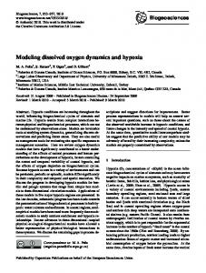
Fig. 2 Rates of dUMP binding in the absence (data of Fig. 1) and presence (data of Fig. 3) of NADPH, as well as FAD reduction by NADPH (data of Fig. 4) as a function of dUMP concentration. The slopes of the solid lines correspond to bimolecular rates of 19.103 and 54.103 M-1s-1 in the absence and presence of NADPH, respectively.
Fig. 6 Flavin re-oxidation by MTHF. ThyX reduced by NADPH (final concentration 0.5 mM) in the presence of dUMP (final concentration 0.5 mM) was mixed with MTHF. Solid and dashed curves are spectra taken 1 ms and 500 ms after mixing with MTHF (final concentration 2 mM). The dotted curve is obtained 1 ms after mixing with buffer. Inset: kinetics at 450 nm after mixing with MTHF to various final concentrations. 13 Licenced copy. Copying is not permitted, except with prior permission and as allowed by law. © 2014 The Authors Journal compilation © 2014 Biochemical Society
Biochemical Journal Immediate Publication. Published on 15 Jan 2014 as manuscript BJ20131567
sc rip nu Ma pte d ce Ac
THIS IS NOT THE VERSION OF RECORD - see doi:10.1042/BJ20131567
t
Fig. 7 Proposed substrate binding and reaction scheme. See text for details.
14 Licenced copy. Copying is not permitted, except with prior permission and as allowed by law. © 2014 The Authors Journal compilation © 2014 Biochemical Society
Biochemical Journal Immediate Publication. Published on 15 Jan 2014 as manuscript BJ20131567
0.05
A
0.04
405
480
370
0.02
sc rip
0.02
0.01
0.01 0.00
0.25
0.50
0.75
1.00
[dUMP] (mM)
0.00
0.00 300
350
400
450
500
300
550
350
400
450
500
550
wavelength (nm)
Wavelength (nm)
0.008
C
3.2 mM
1.0
0.006
0.8
nu
A 500
t
0.03
0.03
A 440 nm
Absorbance
0.04
D
0.6
0.004
0.4
Ma
0.002
0.000
200 uM 400 uM 800 uM 1600 uM 3200 uM
0.2
0.2 mM
0.0
0
20
40
60
80
100
120
Time (ms)
160
180
200
0
50
100
150
200
250
Time (ms)
ce
pte d
Figure 1
140
Ac
THIS IS NOT THE VERSION OF RECORD - see doi:10.1042/BJ20131567
1 ms 10 ms 30 ms 70 ms 150 ms 310 ms
B
0.05
15 Licenced copy. Copying is not permitted, except with prior permission and as allowed by law. © 2014 The Authors Journal compilation © 2014 Biochemical Society
300
350
400
Biochemical Journal Immediate Publication. Published on 15 Jan 2014 as manuscript BJ20131567
200
160
dUMP binding w/o NADPH dUMP binding with NADPH Flavin reduction
sc rip
rate (s-1)
140 120 100 80 60
nu
40 20 0 0.0
0.5
1.0
1.5
2.0
2.5
3.0
3.5
Ma
dUMP (mM)
ce
pte d
Figure 2
Ac
THIS IS NOT THE VERSION OF RECORD - see doi:10.1042/BJ20131567
t
180
16 Licenced copy. Copying is not permitted, except with prior permission and as allowed by law. © 2014 The Authors Journal compilation © 2014 Biochemical Society
Biochemical Journal Immediate Publication. Published on 15 Jan 2014 as manuscript BJ20131567
1 ms 30 ms 60 ms 120 ms 300 ms 1.6 s
0.12
0.08
0.04
0.02
0.00 300
320
340
360
380
400
420
440
460
480
500
520
540
A (norm)
0.6
nu
0.04
0.02
Ma
B
0.8
Absorbance 450 nm
0.3mM 450nm 1.5mM 450nm 0.3mM 505nm 0.3mM 340nm
1.0
0.00 0
60
120
180
Time (min)
0.4
0.0 0.0
0.2
pte d
0.2
0.4
0.6
0.8
1.0
1.2
1.4
1.6
Time (s)
C
0.00
ce
-0.02
-0.04
Ac
A
THIS IS NOT THE VERSION OF RECORD - see doi:10.1042/BJ20131567
wavelength (nm) 1.2
t
A
0.06
sc rip
Absorbance
0.10
340 nm low NADPH 450 nm low NADPH 340 nm high NADPH 450 nm high NADPH
-0.06
0.0
0.2
0.4
0.6
0.8
1.0
Time (s)
Figure 3
17 Licenced copy. Copying is not permitted, except with prior permission and as allowed by law. © 2014 The Authors Journal compilation © 2014 Biochemical Society
Biochemical Journal Immediate Publication. Published on 15 Jan 2014 as manuscript BJ20131567
0.030
A
0.025
0.015
sc rip
Absorbance
0.020
0.010
0.005
0.000 420
440
460
480
500
520
540
nu
wavelength (nm) 0.005
B
Ma
0 dUMP 2 mM 0.5 mM 0.125 mM
0.003
0.002
0.001
0.000 0
50
100
150
200
C 0 mM 2 mM 0.5 mM 0.125 mM 0.5mM/NADPH first
ce
0.020
pte d
Absorbance 506 nm
0.004
0.015
0.010
Ac
Absorbance 480 nm
THIS IS NOT THE VERSION OF RECORD - see doi:10.1042/BJ20131567
400
t
2 ms 5 ms 11 ms 20 ms 31 ms 47 ms 85 ms 185 ms
0.005
0.000 0
50
100
150
200
Time (ms)
Figure 4
18 Licenced copy. Copying is not permitted, except with prior permission and as allowed by law. © 2014 The Authors Journal compilation © 2014 Biochemical Society
Biochemical Journal Immediate Publication. Published on 15 Jan 2014 as manuscript BJ20131567
t sc rip
0.75
0.50
0.25
nu
Fraction of oxidized FAD
0.00 -0.4
-0.3
-0.2
-0.1
Ma
-0.5
E (V) vs. NHE
ce
pte d
Figure 5
Ac
THIS IS NOT THE VERSION OF RECORD - see doi:10.1042/BJ20131567
1.00
19 Licenced copy. Copying is not permitted, except with prior permission and as allowed by law. © 2014 The Authors Journal compilation © 2014 Biochemical Society
Biochemical Journal Immediate Publication. Published on 15 Jan 2014 as manuscript BJ20131567
0.03
2 mM 0.04
t 0.8 mM
sc rip
Abs 450 nm
Absorbance
0.02
0.01
0.33 mM
0.02
0 mM
0.00
100 200 300 400 500 600 700 800
0.00 400
nu
Time (ms)
500
600
Ma
wavelength (nm)
ce
pte d
Figure 6
Ac
THIS IS NOT THE VERSION OF RECORD - see doi:10.1042/BJ20131567
0
20 Licenced copy. Copying is not permitted, except with prior permission and as allowed by law. © 2014 The Authors Journal compilation © 2014 Biochemical Society
700
sc rip
t
Biochemical Journal Immediate Publication. Published on 15 Jan 2014 as manuscript BJ20131567
nu Ma pte d ce Ac
THIS IS NOT THE VERSION OF RECORD - see doi:10.1042/BJ20131567
Fig. 7
21 Licenced copy. Copying is not permitted, except with prior permission and as allowed by law. © 2014 The Authors Journal compilation © 2014 Biochemical Society