JBC Papers in Press. Published on January 8, 2015 as Manuscript M114.612069 The latest version is at http://www.jbc.org/cgi/doi/10.1074/jbc.M114.612069 Substrate binding and specificity of fungal aa transporters
The Aspergillus nidulans proline permease as a model for understanding the factors determining substrate binding and specificity of fungal amino acid transporters Christos Gournas1,3, Thomas Evangelidis2, Alexandros Athanasopoulos1, Emmanuel Mikros2 and Vicky Sophianopoulou1,3 *Running title: Substrate binding and specificity of fungal aa transporters To whom correspondence should be addressed: V. Sophianopoulou, Microbial Molecular Genetics Laboratory, Institute of Biosciences and Applications, NCSR “Demokritos”, Agia Paraskevi, 15310, Athens, Greece. e-mail:
[email protected] C. Gournas, Microbial Molecular Genetics Laboratory, Institute of Biosciences and Applications, NCSR “Demokritos”, Agia Paraskevi, 15310, Athens, Greece. e-mail:
[email protected]
1 Copyright 2015 by The American Society for Biochemistry and Molecular Biology, Inc.
Downloaded from http://www.jbc.org/ at ULB on January 9, 2015
Keywords: Amino acid transport, Aspergillus, Membrane transporters, Molecular modeling, Sitedirected mutagenesis, In-gel fluorescence, Ligand docking, Gating, Proline, L-azetidine-2-carboxylic acid. Background: PrnB, differently from Put4p, its substitutions in variable residues. Put4pSaccharomyces cerevisiae orthologue, is a mimicking substitutions in TMS3 (S130C), highly specific L-proline transporter of TMS6 (F252L, S253G), TMS8 (W351F) and Aspergillus nidulans. TMS10 (T414S) broadened the specificity of Results: Identification of 12 residues important PrnB, enabling it to recognize more efficiently for PrnB activity and/or specificity. Put4pL-alanine, L-azetidine-2-carboxylic acid and mimicking mutants of PrnB recognize other glycine without significantly affecting the Put4p substrates. apparent Km for L-proline. S253G and Conclusion: Residues variable in the Yeast W351F could transport L-alanine, while Amino acid Transporter (YAT) family T414S, despite displaying reduced proline determine transporter specificity. uptake, could transport L-alanine and glycine, Significance: Understanding the molecular a phenotype suppressed by the S130C mechanisms underlying amino acid recognition mutation. Combination of all five Put4pand translocation through YATs. ressembling substitutions resulted in a functional allele that could also transport LABSTRACT alanine and glycine, displaying a specificity Amino acid uptake in fungi is mediated by profile impressively similar to that of Put4p. general and specialized members of the Yeast Our results support a model where residues Amino acid Transporter (YAT) family, a in these positions determine specificity by branch of the Amino acid Polyamine interacting with the substrates, acting as organoCation (APC) transporter superfamily. gating elements, altering the flexibility of the PrnB, a highly specific L-proline transporter, substrate binding core or affecting conformational changes of the transport cycle. only weakly recognizes other Put4p substrates, its Saccharomyces cerevisiae Amino acid supply in cells depends on orthologue. Taking advantage of the high specialized transmembrane transporter proteins. sequence similarity between the two The majority of these proteins belong to the transporters, we combined molecular ubiquitous Amino acid – Polyamine modeling, induced-fit docking, genetic and organoCation (APC) superfamily, one of the biochemical approaches to investigate the largest families of secondary transporter (1, 2). molecular basis of this difference and identify The APC includes members that function as residues governing substrate binding and solute/cation symporters and solute/solute specificity. We demonstrate that L-proline is antiporters and are found in bacteria, archeae, recognized by PrnB via interactions with fungi, protists, plants and animals (1, 3). Fungal residues within TMS1 (G56, T57), TMS3 APC members belong to the Yeast Amino acid (E138) and TMS6 (F248), that are Transporter (YAT) family (4). Numerous YAT evolutionary conserved in YATs, while transporters can be identified in all sequenced specificity is achieved by subtle amino acid genomes of fungi. Characterized members
Substrate binding and specificity of fungal aa transporters
EXPERIMENTAL PROCEDURES Homology modeling- Multiple threading alignments were created with LOMETS metaserver (38) using the full-length sequence of PrnB from A. nidulans or Put4p from S. cerevisiae (Uniprot accession codes P18696 and P15380, respectively). The alignments of the PrnB and Put4p sequences with that of AdiC (PDB ID: 3L1L), shown in Figure 2, were obtained by TM-Coffee (39) and modified manually on the basis of the multiple alignment of YAT sequences shown in Figure 1. The fulllength sequences of PrnB and Put4p were submitted to TOPCONS meta-server (40) for consensus membrane protein topology prediction. 2000 models of PrnB residues 38515 and 2000 models of Put4p residues 107-590 were constructed in total using the Rosetta threading protocol with the membrane specific scoring function terms (41–43). The secondary structure of the protein, as predicted by SAM server (44), as well as the consensus membrane protein topology prediction made by TOPCONS meta-server, were also taken into account in the form of restraints during model construction. 3mer and 9-mer peptide fragments for modeling were created locally using the SAM-predicted secondary structure of PrnB and Put4p. Unfolded and broken structures were excluded by retaining only the 90% top-scores models. A random model among them was superimposed on the crystal structure of AdiC using the combinatorial extension (CE) algorithm (45),
2
Downloaded from http://www.jbc.org/ at ULB on January 9, 2015
an attempt to identify substrate binding residues and specificity determinants of YATs. More precisely, we provide further evidence that residues G56 and T57 of TMS1 contribute to substrate binding and also identify new substrate interacting residues in TMS3 (E138), TMS6 (F248), TMS8 (W351) and TMS10 (T414). Moreover, we show that PrnB is a highly specific L-proline transporter compared to Put4p, which is also known to transport L-alanine, glycine, GABA and the toxic proline analogue L-azetidine-2-carboxylic acid (AZC) (32–37). Taking advantage of the high primary sequence similarity and the specificity differences of PrnB and Put4p, we identify residues in TMSs 3 (S130), 6 (F252, S253), 8 (W351) and 10 (T414) that contribute to the determination of the specificity profile of proline transporters. Based on these results, we discuss the potential mechanisms governing specificity determination in fungal amino acid transporters.
include 15 out of the 16 characterized amino acid transporters of Saccharomyces cerevisiae (5, 6), two proteins of Aspergillus nidulans, PrnB and AgtA (7, 8), as well as transporters from Candida glabrata, Neurospora crassa and Penicillium chrysogenum (9–11). These proteins display different specificities and are subject to tight transcriptional and post-translational regulation (12–14). The prnB gene encoding the main proline transporter of A. nidulans is part of the prn gene cluster mediating transport and utilization of proline as a sole carbon and/or nitrogen source (7, 15, 16). The prnB gene has been extensively used as a model for transcriptional and post-transcriptional regulation of gene expression (15–20). The PrnB transporter has been used as a model to address structure-function relationships in YATs, by mutational analysis and cysteine scanning mutagenesis (21–23). Earlier computational studies suggested that APC members possess the so called “5+5” fold (24), commonly found in the crystal structures of several prokaryotic transporters belonging to evolutionary distinct protein families with different substrate specificities (25–28). By using a sensitive homology threading approach, a 3D model of PrnB was previously generated, using as template the crystal structure of the LeuT transporter of Aquifex auelicus (23). This model predicted that PrnB, as well as other members of the APC superfamilly and the YAT family, share a common structural core (23). This core consists of two intertwined, antiparallel V-shaped repeating units of five transmembrane segments each (TMS1-5 and TMS6-10) connected by a relatively long loop and is followed by two additional helices (TMS11 and 12). Along with mutational evidence, the model suggested that the substrate binding pocket of APCs would be located in the vicinity of the unwound part of two kinked helices (TMS1 and TMS6) of each V-shaped repeated unit of the transporter. These predictions were confirmed by the subsequent crystal structures of three bacterial APC members: the arginine/agmatine (AdiC) and glutamate/γ-aminobutyric acid (GadC) antiporters and the ApcT, a broad-specificity proton/amino acid symporter (29–31). In this study, using the PrnB transporter, we combine molecular modeling and induced-fit docking along with comparative sequence analyses, genetic and biochemical approaches, in
Substrate binding and specificity of fungal aa transporters The alleles were confirmed by sequencing (VBC Genomics, Austria). Linearized sequences of these plasmids were used to transform, as previously described (51), strain prn397. Transformants were isolated and analyzed by Southern blotting for the sequence of the prnB gene, ensuring that all strains used in this study carry single copy in-locus prnB alleles derived from homologous integration events. A ~1100 bp SmaI fragment, including part of the promoter and ORF of prnB, from plasmid pA4 was used as a prnB-specific probe. The ~1500bp 5’ and 3’ flanking sequences of gabA were PCR amplified using primers 1-8 of Table 2 digested with KpnI/XbaI and XbaI/NotI and subsequently cloned into a KpnI/Not1 linearized pBlueScript II SK(+)vector. In the unique XbaI site of the resulting plasmid, the AfpyrG gene, PCR amplified using primers 9 and 10 from plasmid p1439 (52, 53) and XbaI digested was cloned. The resulting cassette was PCR amplified using primer pairs 1 and 4 and ~2 µg of the products were used to transform strain nkuA∆, selecting for uracil/uridine prototrophy. Single copy gabA∆::AfpyrG strains were checked by Southern analysis, using as probes the sequences of AfpyrG and the 5’ flanking sequence of gabA. Plasmid preparation from E. coli strains and DNA bands purified from agarose gels were performed with the Nucleospin Plasmid kit and the Nucleospin ExtractII kit, respectively, according to the manufacturer’s instructions (Macherey-Nagel, Lab Supplies Scientific SA, Hellas, Greece). DNA probes for Southern analysis (54) were labeled with [32P]dCTP (3000 Ci/mmol, Institute of Isotopes Co. Ltd, Miklόs, Hungary) using a random hexanucleotide primer kit following the supplier’s instructions (Takara Bio, Lab Supplies Scientific SA) and purified on MicroSpinTM S-200 HR columns following the supplier’s instructions (Roche Diagnostics). Restriction enzymes used were from Takara Bio. Epifluorescence microscopy- Samples were prepared as previously described (55, 56). 20 mM L-proline was used for induction of prnB expression, at 25° C for 12-14 h. Samples were observed using a Plan-Apochromat 100× 1.40 NA oil immersion objective lens on an Axioplan 2 fluorescence microscope (Carl Zeiss, Inc.) with appropriate filters and the resulting images were acquired with a ZeissMRC5 digital camera using the AxioVs40 V4.40.0 software. Images were then processed with ImageJ (NIH) and annotated with the Photoshop CS5 software (Adobe).
3
Downloaded from http://www.jbc.org/ at ULB on January 9, 2015
and the putative substrate binding cavities of PrnB and Put4p were determined by selecting every residue that was poised within 8 Å from the arginine ligand. Trajectories were assembled from the remained low-energy models and were clustered with GROMACS Tools v4.6.1 (46) using the backbone heavy atoms and Cβ carbon of the putative substrate binding pocket. The algorithm used for clustering was “gromos” with cutoff 1.5 Å. Root Mean Square Flunctuation (RMSF) of the backbone atoms was calculated with g_rmsf module of GROMACS Tools. Induced Fit Docking- Protein preparation using OPLS2005 force field (47) and molecular docking was performed with the Schrödinger Suite 2014, as described in our previous study (48). Substrates were docked on the most dominant cluster representative receptor structures, using the induced fit docking (IFD) protocol (Schrödinger Suite 2014 Induced Fit Docking protocol), which is intended to circumvent the inflexible binding site and accounts for the side chain and backbone movements upon ligand binding (49). Strains, media and growth conditions- Strains used in this study are summarized in Table 1. All carry the veA1 mutation. Chemicals were from Sigma (St Louis, MO, USA) and AppliChem GmbH. Standard, previously described MM and CM media for A. nidulans, as well as medium supplements, were used (described in the Fungal Genetics Stock Centre, FGSC, http://www.fgsc.net). Assessment of the functionality of PrnB alleles was performed in MM using 1% glucose or 1% glycerol as carbon sources. Urea, amino acids and GABA have been supplemented at a final concentration of 5 or 10 mM, unless otherwise indicated. D-serine was used at a final concentration of 0.1 mg/ml, using urea as sole nitrogen source. For membrane enriched protein extracts, strains were incubated 3-4 days at 37° C in solid CM. Conidiospores were harvested, filtered through miracloth and used to inoculate flasks containing 200 mL liquid MM, appropriately supplemented, using 0,1% fructose and 10 mM NaNO3 as carbon and nitrogen sources, respectively. Cultures were incubated 18 h at 25° C and prnB induction was achieved by the addition of 20 mM L-proline for the last 4 h of growth. Molecular manipulations and transformationprnB alleles have been generated by in-vitro site-directed mutagenesis of plasmid pA4 (50), using Kapa-HiFi (Kapa Biosystems) DNA polymerase and the primers 11 – 32 of Table 2.
Substrate binding and specificity of fungal aa transporters using a cut-off filter of 515 nm, by increasing the exposure time until fluorescent bands were clearly visible (10 - 60 sec). The gel was subsequently stained with Coomassie staining solution. Images were processed with ImageJ (NIH) and annotated with the Adobe Photoshop CS5 software. RESULTS To identify the topology of PrnB and Put4p residues that are essential or crucial for proline binding and transport, we constructed the threedimensional structural models of the transporters, using a robust comparative modeling approach (Figure 3). The models were based on the outward-facing occluded structure of AdiC (PDB accession 3L1L). In that structure, AdiC was crystallized with its substrate, arginine, in an intermediate conformation, that enables the identification of most of the amino acid residues involved in substrate binding (29). Two thousand structural models of PrnB and Put4p were built and divided into clusters, based on the structural similarity of their substrate binding sites. The three largest clusters of each protein were used for docking. These clusters represent 28%, 17.5% and 6.7% of the total model population in PrnB and 28%, 26.4% and 12.4% of that in Put4p, respectively. Figure 3A shows a representative structure of the largest cluster of each of the two transporters. The overall models of the two permeases are, as expected, very similar (Figure 3B). A notable difference was observed in the positioning of TMS6a and TMS11, which are closer to the pore of the transporter in Put4p. The observed differential positioning of TMS6a could be partially explained by the differential flexibility of the nearby unwound segment of TMS6. Indeed, RMSF measurements showed that the positioning of TMS6a is substantially more variable in Put4p than in PrnB (Figure 3C). Our mutational analysis suggests that this could be related to the broader specificity of Put4p (see below). In contrast to TMS6a, the RMSF of TMS11 between PrnB and Put4p is quite similar. Identifying substrate binding residues- We performed induced-fit docking of L-proline in PrnB as well as L-proline, L-alanine and Lazetidine-2-carboxilic acid in Put4p (Figure 4 and data not shown). Docking of these substrates was performed to the representative structures of at least the three largest clusters of the homology models, which altogether represent more than
4
Downloaded from http://www.jbc.org/ at ULB on January 9, 2015
Radiolabelled uptake measurements and kinetic analyses- Activity of PrnB mutants was measured by estimating rates of [3H]-L-proline (60.0 Ci mmol-1, Moravek Biochemicals, Brea, CA, USA) uptake as previously described (21– 23, 57). [3H]-L-Proline uptake in MM pH 6.8 at 25° C was assayed in germinating conidiospores of A. nidulans derived from 8-9 h cultures at 25° C, induced with 20 mM L-proline and washed 3 times with 50 mL MM, finally concentrated at 107 conidiospores/100 µL. Initial velocities were measured at 1-5 min of incubation with 0.25–20 µM radioactive substrate since uptake was linear for at least 30 min (21). Km/i values were obtained directly by performing and analyzing (Prism3) [3H]-L-proline uptake in the presence of at least ten different concentrations of competitor. In all cases background uptake values were corrected by subtracting values measured in a prnB∆ strain. Ki values calculated satisfy the criteria for use of the Cheng and Prusoff equation Ki = IC50/ [1 + (L/Km)], in which L is the permeant concentration. IC50 values were determined from full dose-response curves, and in all cases the Hill coefficient was close to -1, consistent with the presence of one uptake system. Protein extraction and in-gel fluorescenceMembrane-enriched protein extracts were prepared as previously described (22, 55), with the following modifications (58). Following membrane protein precipitation by centrifugation (1h, 18000 g, 4o C), the extraction buffer was removed and the pellet was resuspended in ice cold In-gel Fluorescence Resuspension Buffer (IFRB: 50 mM Tris–HCl (pH 7.5), 5 mM EDTA, 10% glycerol) freshly supplemented with protease inhibitor cocktail (Sigma, P8215, 1:500) and 1 mM PMSF. Cell debris was pelleted by centrifugation at 500 g for 3 minutes at 4o C and the supernatant was transferred to a new pre-cooled eppendorf tube. Protein concentration was measured by the method of Bradford. 30-50 µg of protein samples were mixed with 1/3 volume of 4x SB for In-gel Fluorescence [4xIFSB: 70 mM Tris– HCl (pH 7.5), 7% glycerol, 7 mM EDTA (pH 8.0), 8 % SDS, 100 mM DTT, 0.02% bromophenol blue] and immediately used to load a standard 10% SDS-Polyacrylamide Gel. Following SDS-PAGE at 110V, the gel was rinsed with dH2O and the fluorescent bands were detected with a CCD camera system (ImageQuant™ LAS 4000, GE Healthcare) by exposure to blue light (EPI source) set at 460 nm,
Substrate binding and specificity of fungal aa transporters proline still interacts with the side chains of residues T126 and E211, but it also interacts with K318 (Figure 4B). Interestingly, this Lys residue is not conserved in YATs and is present exclusively in proline-specific permeases (Figure 1). The side chain of K245, the corresponding residue of PrnB, in all PrnB models is positioned away from the proposed binding site and towards the extracellular surface, similarly to some of the configurations obtained for Put4p (Figure 4D). However, previous experimental data showed that K245 is important for efficient proline transport by PrnB, while its substitution by Leu significantly increases the Km for proline (21). The fact that K245 of PrnB was not predicted to interact with proline could be related to the positioning of TMS6a. This suggests a possible interaction of K245 and the proline substrate in another conformation of the transporter, most probably a more outward facing. Another intriguing difference was the identification of three additional proline binding orientations in PrnB (Figure 5), which could reflect intermediate positions of the substrate between the two substrate binding sites found in both PrnB and Put4p. The second binding site, found only by the docking of L-proline in PrnB (Figure 4E) and Lalanine in Put4p (Figure 4F), is located deeper into the substrate binding core. Proline, at this site, is found to interact with PrnB via Hbonding with the side chains of T414 and Y127 (Figure 4E). Similarly, L-alanine in Put4p forms H-bonds with the side chain of Y200 (Y127 of PrnB) and the backbone of G326 and E328 (S253 and E255 of PrnB, respectively) (Figure 4F). Interestingly, in this configuration, Lalanine, a substrate of Put4p but not PrnB, seems to be close to L325, which along with G326 differ in the two transporters (F252 and S253 in PrnB, respectively). T414, part of TMS10, is also different between the two transporters (S487 of Put4p) and other YATs (Figure 1). Most interestingly, it corresponds to T456 of Can1, a substrate interacting residue of the arginine permease of S. cerevisiae that has been very recently shown to determine the specificity of the basic amino acid transporters of yeast (59). The above residues, along with PrnBW351, constitute the main differences in the predicted substrate binding pockets of PrnB and Put4p (Figure 9) and could, thus, be related to the different specificity profiles of the two orthologues.
An interesting difference in proline binding was acquired by a significant percentage of the second cluster of Put4p structures (26.4% of the population). In these structures it was found that
5
Downloaded from http://www.jbc.org/ at ULB on January 9, 2015
50% of the structures created for each transporter. With this strategy we allowed implicitly larger structural movements to occur in the substrate binding regions, than those introduced by the IFD protocol alone. This way we accounted for transporter flexibility, which was necessary (as discussed below) to obtain docking poses that are in agreement with previous studies (23, 29, 59). Our docking studies suggested that proline binds almost identically in PrnB and Put4p (Figures 4A, 4C, 4B, 4D), while L-alanine and AZC are bound to Put4 in a way very similar to that of proline (data not shown). More specifically, in the first two clusters, which represent the most abundant populations of the structures generated (45.5% in PrnB and 54.4% in Put4p), proline seems to bind to two distinct sites. In the first site, which is located closer to the extracellular region of the transporters, proline interacts with the side chains of two amino acid residues which are highly conserved in the YAT family (Figure 4A, 4B). The first, T57 of PrnB (T126 of Put4p), is located in the unwound segment of TMS1 and is part of the characteristic “G T/S G” motif of YATs (Figure 1). This residue, previously suggested to be part of the proline binding site (23), is found to interact with proline via a hydrogen bond. The second residue, E138 of PrnB (E211 of Put4p), located in TMS3 and also highly conserved in the family, interacts with proline via a salt bridge. The interactions of these two residues with proline via the invariant part of amino acid substrates (the a-carboxyl and the imino group) are compatible with their high conservation in several YATs of different specificities (Figure 1). In addition, the docked substrate forms H-bonds with the backbone of the highly conserved PrnBG56 and PrnBG58 (Figure 4C; Put4pG125 and Put4pG127 in Figure 4B, respectively). This configuration seems to be stabilized by hydrophobic or π-cation interactions with the side chains of PrnBF248 and PrnBW351 (Figures 4A, 4C; Put4pF321 and Put4pF424 in Figures 4B, 4D, respectively). PrnBF248 (Put4pF321), located at the vicinity of the unwound part of TMS6, is highly conserved in the family. On the contrary, an aromatic residue in position 351 of PrnB is found only in proline specific YATs (Figure 1).
Substrate binding and specificity of fungal aa transporters
Study of conserved residues- Results concerning the conserved amino acid residues of the binding site in YATs are shown in Figure 6 and Table 3a. As shown in Figures 6B and 7B, all alleles, except I251∆ and F252∆, are normally targeted to the plasma membrane. Consistently with previous western blots (22), all PrnB-GFP alleles detected by in-gel fluorescence appear as doublets. Moreover, the lower molecular weight fast migrating bands that were also detected most probably correspond to N-terminal degradation products, as it has been observed for AgtA, another YAT of A. nidulans (60). In-gel fluorescence analysis (Figures 6D and 7D) showed that all mutant proteins, except I251∆ and F252∆, are expressed at levels comparable to the wild type PrnB-GFP, displaying a two-
6
Downloaded from http://www.jbc.org/ at ULB on January 9, 2015
fold maximal variation. The above results strongly indicate that the differences in growth tests (Figures 6A and 7A) as well as in [3H]-Lproline initial uptake rates (Figures 6C and 7C) are truly indicative of the functionality of the PrnB mutant proteins. In addition, [3H]-Lproline initial uptake rates are well correlated with the growth of strains carrying the corresponding prnB mutants on proline as sole nitrogen source at 25o C. This is in agreement with previous studies on A. nidulans transporters, where only mutations that reduce the uptake rate 2000 1350 ± 142 >2000 >2000 750 ± 67 1013 ± 115 n.i 441 ± 47 563 ± 52 >2000 172 ± 14 219 ± 23 n.i 84 ± 6 553 ± 44
Gly >2000 >2000 n.i >2000 >2000 >2000 >2000
Table 3b. Specificity profile of functional PrnB mutants carrying substitutions in residues not conserved in YATs
Downloaded from http://www.jbc.org/ at ULB on January 9, 2015
Table 3a. Specificity profile of functional PrnB mutants carrying substitutions in residues conserved in YATs
S130C 20 ± 2.2 >2000 253 ± 18 245 ± 16 >2000 G247A 65 ± 4 >2000 895 ± 79 >2000 n.i F252L 44 ± 3 >2000 150 ± 16 ~ 2000 >2000 S253G 39 ± 3 >2000 245 ± 19 158 ± 12 ~ 2000 W351F 26 ± 3.3 >2000 178 ± 18 403 ± 44 n.i W351E 27 ± 3 n.i 463 ± 52 >2000 n.i T414S 31 ± 2.6 >2000 257 ± 28 475 ± 51 529 ± 56 T414A 27 ± 2.9 n.i 461 ± 51 ~ 2000 >2000 S130C/T414S 44 ± 4 >2000 576 ± 54 176 ±18 125 ±11 5M 29± 2.6 338 ± 31 129 ± 14 52 ± 5 106 ± 10 Put4* 31 110 120 72.5 n.k. GABA, γ-amino-butyric acid; L-P-NH3, L-Prolinamide; 3,4-D-Pro, 3,4-Dehydro-L-Proline; L-AZC, L-Azetidine-2-carboxylic acid; L-PCA, L-Pipecolinic acid; L-Ala, L-Alanine; Gly, Glycine; n.i., no inhibition (90–100% uptake); >2000, slight inhibition at 2 mM (65-90 % uptake); ~2000, inhibition at 2 mM was about 50-65 %; n.k., not known. Results are averages of at least three independent experiments in triplicate for each concentration point. Standard deviation was below 20%. * The K m/i values for Put4 were obtained from references 32-37
20
Substrate binding and specificity of fungal aa transporters Downloaded from http://www.jbc.org/ at ULB on January 9, 2015
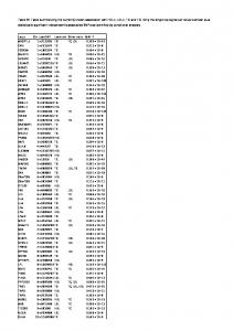
Table 4. Summary of PrnB mutants functional analysis and specificity determination
K m/i (µM) Put4/AdiC residue PrnB allele TMS Binding site L-Proline GABA L-AZC L-Ala Gly %V wt 38 ± 3 n.i 1129 ± 86 >2000 >2000 100 G56P 1 upper G125/G25 n.f n.f n.f n.f n.f n.f T57N 1 upper T126/S26 6 ± 1.9 n.i 610 ± 44 541 ± 53 >2000 48.8 S130C 3 lower C203/A96 20 ± 2.2 >2000 253 ± 18 245 ± 16 >2000 53.8 E138D 3 upper E211/M104 n.f n.f n.f n.f n.f n.f E138Q 3 upper E211/M104 n.f n.f n.f n.f n.f n.f G247A 6 A320/L201 65 ± 4 >2000 895 ± 79 >2000 n.i 86.3 F248W 6 upper - proximal gate F321/W202 21 ± 1.8 >2000 750 ± 67 1013 ± 115 >2000 13.2 F248Y 6 upper - proximal gate F321/W202 33 ± 2.6 >2000 1350 ± 142 >2000 n.i 81.3 F250Y 6 F323/F204 13 ± 1.6 n.i 441 ± 47 563 ± 52 >2000 28.8 F248W/F250W 6 upper 1.9 ± 0.2 >2000 172 ± 14 219 ± 23 >2000 32.9 F252L 6 indirect L325/44 ± 3 >2000 150 ± 16 ~ 2000 >2000 72.9 S253G 6 indirect G326/G206 39 ± 3 >2000 245 ± 19 158 ± 12 ~ 2000 61.3 E255D 6 lower - distal gate E328/E208 2 ± 0.8 n.i 84 ± 6 553 ± 44 >2000 86.9 E255Q 6 lower - distal gate E328/E208 n.f n.f n.f n.f n.f n.f W351F 8 both - middle gate F424/W293 26 ± 3.3 >2000 178 ± 18 403 ± 44 n.i 28.7 W351E 8 both - middle gate F424/W293 27 ± 3 n.i 463 ± 52 >2000 n.i 24.2 W351A 8 both - middle gate F424/W293 n.f n.f n.f n.f n.f n.f T414A 10 lower S487/S357 27 ± 2.9 n.i 461 ± 51 ~ 2000 >2000 8.2 T414S 10 lower S487/S357 31 ± 2.6 >2000 257 ± 28 475 ± 51 529 ± 56 17.3 S130C/T414S 44 ± 4 >2000 576 ± 54 176 ±18 125 ±11 94.3 34.6 5M 29± 2.6 338 ± 31 129 ± 14 52 ± 5 106 ± 10 Put4* 31 110 120 72.5 n.k. n.f., not functional; n.d., not done; # Transport based on the growth tests of figure 9; The rest are as in the legend of Table 3
21
Transport based on growth tests
L-Proline +++ -
++ ++ -
+++ ++ +++ ++ +++ +++ ++
L-Ala#
Gly#
n.d n.d n.d n.d n.d n.d n.d n.d -
n.d n.d n.d n.d n.d n.d n.d n.d n.d n.d -
+
-
n.d n.d
+ +
+ +
-
-
+ + +++ ++
++ -
++
++ ++
Downloaded from http://www.jbc.org/ at ULB on January 9, 2015
Downloaded from http://www.jbc.org/ at ULB on January 9, 2015
Downloaded from http://www.jbc.org/ at ULB on January 9, 2015
Downloaded from http://www.jbc.org/ at ULB on January 9, 2015
Downloaded from http://www.jbc.org/ at ULB on January 9, 2015
Downloaded from http://www.jbc.org/ at ULB on January 9, 2015
Downloaded from http://www.jbc.org/ at ULB on January 9, 2015
Downloaded from http://www.jbc.org/ at ULB on January 9, 2015
Downloaded from http://www.jbc.org/ at ULB on January 9, 2015
Downloaded from http://www.jbc.org/ at ULB on January 9, 2015
Membrane Biology: The Aspergillus nidulans proline permease as a model for understanding the factors determining substrate binding and specificity of fungal amino acid transporters Christos Gournas, Thomas Evangelidis, Alexandros Athanasopoulos, Emmanuel Mikros and Vicky Sophianopoulou J. Biol. Chem. published online January 8, 2015
Find articles, minireviews, Reflections and Classics on similar topics on the JBC Affinity Sites. Alerts: • When this article is cited • When a correction for this article is posted Click here to choose from all of JBC's e-mail alerts This article cites 0 references, 0 of which can be accessed free at http://www.jbc.org/content/early/2015/01/08/jbc.M114.612069.full.html#ref-list-1
Downloaded from http://www.jbc.org/ at ULB on January 9, 2015
Access the most updated version of this article at doi: 10.1074/jbc.M114.612069