1 2 3 4 5 6 7 8 9 10 11 12 13 14 15 16 17 18 19 20 21 22 23 24 25 26
Identification and Characterization of cis- and trans-Acting Elements Involved in Prophage Induction in Streptococcus thermophilus J34
27
Summary
28 29 30 31 32 33 34 35 36 37 38 39 40 41 42 43 44
The genetic switch region of temperate Streptococcus thermophilus phage TP-J34 contains two divergently oriented promoters and several predicted operator sites. It separates lytic cyclepromoting genes from those promoting lysogeny. A polycistronic transcript comprises the genes coding for repressor Crh, metalloproteinase-motif protein Rir, and superinfection exclusion lipoprotein Ltp. Weak promoters effecting monocistronic transcripts were localized for ltp and int (encoding integrase) by Northern blot and 5`-RACE-PCR. These transcripts appeared in lysogenic as well as lytic state. A polycistronic transcript comprising genes coh (encoding Cro homolog), ant (encoding putative antirepressor), orf7, orf8 and orf9 was only detected in the lytic state. Four operator sites, of which three were located in the intergenic regions between crh and coh, and one between coh and ant, were identified by competition electromobility shift assays. Cooperative binding of Crh to two operator sites immediately upstream of coh could be demonstrated. Coh was shown to bind to the operator closest to crh only. Oligomerization was proven by crosslinking Crh by glutaraldehyde. Knock-out of rir revealed a key role in prophage induction. Rir and Crh were shown to form a complex in solution and Rir prevented binding of Crh to its operator sites.
45
Introduction
46 47 48
Temperate phages are latent sources for generation of virulent phages (Mercanti et al., 2011). Certain environmental conditions may activate their induction, leading to the release of new virions (Marcó et al., 2012; Nanda et al., 2015). With their potential to lyse starter cells,
Sabrina Koberg*, Mazhar Desouki Ali Mohamed§, Katharina Faulhaber$, Horst Neve, Knut J. Heller Department of Microbiology and Biotechnology, Max Rubner-Institut (Federal Research Institute of Nutrition and Food), Kiel, Germany *For correspondence: Sabrina Koberg, Department of Microbiology and Biotechnology, Max Rubner-Institut (Federal Research Institute of Nutrition and Food), Hermann-Weigmann Str. 1, D-24103 Kiel, Germany; Phone: +494316092357; Fax: +494316092306; Email:
[email protected] Present addresses: § Microbiology Department, Faculty of Agriculture, Sohag University, Sohag, Egypt $ Chair Organismic Interactions, Interfaculty Institute of Microbiology and Infection Medicine, Tübingen University, Tübingen, Germany
Running title: Lysogeny Module of TP-J34 Keywords: promoter mapping, operator mapping, CI-repressor-homologous protein Crh, Crohomologous protein Coh, repressor-inactivating protein Rir
49 50 51 52 53 54 55 56 57 58 59 60 61 62 63 64 65 66 67 68 69 70 71 72 73 74 75 76 77 78 79 80 81 82 83 84 85 86 87 88 89 90 91 92 93 94 95 96 97 98 99
temperate phages can represent a problem for the dairy and other food fermentation industries, albeit not to the same extent as virulent phages, which may cause not only fermentation delays and alteration of product quality, but also complete loss of product in severe cases (Dupont et al., 2004). On the other hand prophages have been suggested to contribute to the fitness of their hosts through genes coding for lysogenic conversion (Mc Grath and van Sinderen, 2007; Sun et al., 2009a; Sun et al., 2009b). However, the exact role of temperate phages in dairy fermentations still remains unclear ( Sun et al., 2009a; Sun et al., 2009b; Marcó et al., 2012). Lysogeny, i.e. the integration of a prophage, is common among dairy lactococci, while lysogenic Streptococcus thermophilus strains occur only rarely (Josephsen and Neve, 2004). Currently, ca. 10 S. thermophilus strains harboring prophages have been identified and five genomes of corresponding phages (O1205: accession no. U88974 (Stanley et al., 1997), Sfi21: accession no. AF115103 (Desiere et al., 1998), TP-778: accession no. HG917970 (Ali et al., 2014), TP-J34: accession no. HE861935 (Ali et al., 2014), TP-DSM20617: accession no. HG424323) have been published. The TP-J34 host strain S. thermophilus J34 is used as a starter culture for yogurt and cheese production (Neve et al., 2003). All S. thermophilus phages are members of the Siphoviridae type B1, which are characterized by a similar morphology with small isometric heads and long, non-contractile tails (Ackermann, 1992; Brüssow and Desiere, 2001) and are classified in three general groups (Mahony and van Sinderen, 2014). Analysis of minor restriction bands of the phage DNA identified TP-J34 as pac-type phage, using the headful DNA packaging method (Neve et al., 2003). The calculated genome is 45,606 bp, harboring 60 open reading frames (orfs). Upon infection, temperate phages like TP-J34 choose between two possible life-cycles (Madsen et al., 1999; Madsen et al., 2001; Ptashne, 2004; Alsing et al., 2011; Duerkop et al., 2014), i.e., either a lysogenic or a lytic cycle (Madsen et al., 1999). In the lysogenic cycle, the phage DNA integrates as prophage into the host genome and is propagated passively until induction of the lytic cycle occurs spontaneously, or is stimulated by host DNA-damaging environmental signals like mitomycin C or UV light. Thus, the switch of the prophage to lytic mode is initiated by the host SOS response (Kim and Ryu, 2013). Due to a defect in orf48, encoding the putative host-specificity protein (Ali et al., 2014), approximately 95% of released TP-J34 particles are defective and non-infectious, consisting of DNA-filled heads which lack tails (Neve et al., 2003). The lysis vs. lysogeny decision is made by the genetic switch (Ptashne, 2004; EngelbergKulka and Kumar, 2015). This regulatory mechanism is a part of the phage lysogeny module, which shows a common genetic organization of lysogeny-related genes of temperate Siphoviridae stemming from low mol% GC content Gram-positive bacteria (Lucchini et al., 1999). The genetic switch region of TP-J34 harbors two divergently oriented promoters, P1 and P2, separating the potential lytic cycle-promoting genes coh (cro homolog), ant and probably also orf6 through orf9 from the four adjacent lysogeny promoting orfs crh (cI-repressor homolog), orf3 (renamed as rir), ltp and int (Neve et al., 2003) (see Figure 1). The latter genes code for the putative repressor Crh, the putative metalloproteinase Rir, the superinfection exclusion (sie) mediating lipoprotein Ltp (Sun et al., 2006; Bebeacua et al., 2013; Ali et al., 2014), and the putative integrase Int (Neve et al., 2003; Ali et al., 2014). They are encoded on the DNA strand opposite to the one on which all other orfs in the phage genome are orientated, a situation which is also known to occur in other phages (Desiere et al., 2001). Crh and Coh both show significant similarities to the HTH-XRE helix-turn-helix family of proteins, comprising prokaryotic DNA binding proteins belonging to the xenobiotic response element family of transcriptional regulators (Marchler-Bauer et al., 2013). The rir gene product shows significant similarity to domain of unknown function 955 (DUF955) type proteins, which are a family of bacterial and viral proteins with undetermined function. A conserved H-E-X-XH amino acid motif occurring in these proteins is suggestive of a catalytic active site, similar to that found in Zn peptidases (Marchler-Bauer et al., 2013). The X-ray structure of the soluble domain of the Ltp lipoprotein revealed a tandem of three-helix helix-turn-helix (HTH) domains
100 101 102 103 104 105 106 107 108 109 110 111 112 113 114 115 116 117 118 119 120 121 122 123 124 125 126 127 128 129 130 131 132 133 134 135
with a highly negatively charged surface (Bebeacua et al., 2013). Ltp consists of three different functional units: a lipid domain for membrane anchoring, a serine-rich spacer region, and the mentioned repeat domain responsible for sie. LtpTP-J34-like lipoproteins were found in three further temperate S. thermophilus phages:TP-EW, TP-DSM20617 and TP-778 (Ali et al., 2014). The int gene product shows similarity to site-specific recombinase proteins with a Cterminal catalytic domain, which is found in many bacterial and phage integrases, as well as an AP2-like DNA-binding integrase domain (Marchler-Bauer et al., 2013). The ant gene product shows significant similarity to proteins of the cluster of orthologous groups COG3561, which are phage anti-repressor proteins (Marchler-Bauer et al., 2013). The genetic switch is typically regulated by phage-encoded repressor proteins, which block transcription from early divergent promoters by binding to specific operator sites (Ptashne, 2004; Alsing et al., 2011; Biswas et al., 2014). Another mechanism, known for example for temperate S. thermophilus phage Sfi21, is that the Cro-like repressor interacts directly with the CI-like repressor and in this way prevents its binding to the operator sites (Bruttin et al., 2002). This mode of action is typical for antirepressors (Susskind and Botstein, 1975; Mardanov and Ravin, 2007; Fogg et al., 2011). Characterization of the relationship between integrase, excisionase and antirepressor activities associated with a superinfecting Shiga toxin encoding acteriophage. Nucleic Acids Res 39:2116-2129). Prophage genes encoding antirepressors are apparently either transcribed in opposite direction (Bruttin et al., 2002; Alsing et al., 2011) or are located distantly from the repressor genes (Shearwin et al., 1998; Mardanov and Ravin, 2007; Lemire et al., 2011; Kim and Ryu, 2013). The repressor of lytic genes is usually a host RecA-dependent auto-cleavable protein (Ptashne, 2004; Galkin et al., 2009; Pal and Chattopadhyaya, 2009; Kim and Ryu, 2013). Within the intergenic region between crh and coh, three 15 bp operator sites (O1A, O2, O3) showing a more or less dyad symmetry have been predicted (Neve et al., 1998). Between coh and ant, a fourth operator (O1B), revealing the same sequence like O1A, appears to be located. O1A partially overlaps with the lytic promoter P1 and O3 with the lysogenic promoter P2. This investigation provides data on important functions in prophage induction of cis- and transacting elements in the lysogeny module of TP-J34. Our results suggest that Crh represses transcription, which starts immediately upstream of coh, thus stabilizing the lysogenic state. Coh on the other hand appears to interfere with transcription, which starts immediately upstream of crh. The product of the rir gene, transcribed within the same operon as crh, apparently inactivates Crh and thereby induces the lytic development of the phage. It thus acts as an antirepressor. Based on this finding, we propose this gene to be called rir (repressorinactivating regulator).
136
Results
137 138 139 140 141 142 143 144 145
Prophage induction in lysogenic S. thermophilus J34 appears to be a rather slow process. After addition of different concentrations of mitomycin C (MC) to growing cultures at different optical densities, growth continued unaffected for ca. 40 min with the same growth rate as the untreated culture (Figure S1). Thereafter, the growth rate decreased and, after maximal cell densities were reached at ca. 60 through 80 min after induction, lysis started, as observed by the decrease of OD 620 nm values. When probing by RT-PCR for expression of late genes using primers targeting orf43 (encoding the tape measure protein TMP), we found that this RNA started to be produced ca. 40 min after addition of MC, whereas RNA transcribed from the int gene was already detectable without addition of MC (Figure 1).
146
147
Transcripts formed during TP-J34 prophage induction
148 149 150 151 152 153 154 155 156 157 158 159 160 161 162 163 164 165 166 167 168 169 170 171 172 173 174 175 176 177 178 179 180 181 182 183 184 185 186 187 188 189 190 191 192
Based on the DNA sequence of the lysogeny module of TP-J34, some cis- and trans-acting elements were proposed (Neve et al., 1998; Ali et al., 2014) (Figure 2). For Northern blot analyses, therefore hybridization probes targeting the genes int, ltp, rir and ant, were investigated, respectively (Figure 3). For control purposes the amounts of 16S and 23S RNA were documented and thus it could be ascertained that equal amounts of total RNA were applied in all cases. For leftward transcription we could identify three different transcripts. The rir probe identified a transcript of ca. 1200 nt, corresponding to a transcript comprising the three genes crh, rir and ltp. To verify this transcript, we mapped the promoter by 5’-RACE analysis. This confirmed that the transcript started at the consensus promoter sequence P2 upstream of crh (Figure 2). When inspecting the DNA sequence downstream of ltp, a potential terminator structure could be detected (Figure S2), suggesting the exact length of the transcript to be 1322 nt. The transcript was detectable without induction by MC, confirming that it was permanently produced during the lysogenic state. Upon induction, it was enhanced after 20 min and – after a reduction at 40 min – even further enhanced after 60 min (Figure 3). When a probe targeting the ltp gene was applied, two transcripts were detected: a weak one corresponding to the ca. 1200 nt transcript detected also by the rir probe (a heavily overexposed picture (Fig. S3) shows the same pattern of enhanced and reduced transcription as seen in Fig. 3), and a more intensively labeled one of ca. 500 nt. Again, a promoter was mapped by 5’RACE analysis. In this case, a “-10” region sufficiently similar to consensus was detected at an appropriate position upstream of the start of the transcript, while no sequence sufficiently similar to a “-35” region could be detected (Figure S2). However, it should be noted that the transcriptional start point was at the downstream basis of a stem-loop structure. A transcript of 524 nt was thus theoretically calculated. The transcript was also detectable prior to addition of MC. Thereafter, a marginal increase in transcription was observed. At 80 min after induction the same amount of transcript appeared to be present as without induction (Figure 3). A probe targeting int hybridized to RNA of less well defined size (Figure 3), however, maximal intensities were seen for the transcripts of ca. 1800 nt length. Since the intensities seen were rather low, transcription appeared to be weak. It should be noted that in the case of the int transcript not only were smaller transcripts labeled (apparently indicating rather rapid degradation) but that also larger ones were detected. To find out whether this indicated some weak transcription occurring from one of the upstream promoters (identified by ltp and rir probes), we carried out reverse transcription-PCR (RT-PCR) in the leftward transcribed region of the lysogeny module on total RNA isolated from uninduced S. thermophilus J34 cells. Seven different primer pairs were applied and conventional PCR was carried out in addition to control for sizes of RT-PCR products (Figure 4). The results showed that i) crh, rir and ltp are transcribed in one operon, ii) the int transcript extends beyond the 3’-end of int into the chromosomal DNA of the host, and iii) int is transcribed independently of the other three genes. Finally, a probe targeting the ant gene hybridized to a transcript of ca. 1900 nt. The transcript was not visible prior to induction. Transcription was seen at 20 min and became very strong at 60 and 80 min after induction (Figure 3). We did not map the starting point of transcription, since consensus -10 and -35 promoter sequences with 17 bp distance were clearly discernible and were almost 100% identical to the crh promoter. According to its size, the transcript should comprise genes coh, ant, orf7, orf8, and orf9. Downstream of orf9 a large stem-loop structure of ca. 150 bp could be deduced. Termination at the beginning or at the end of this structure would yield transcripts of 1800 and 1950 nt, respectively.
193
Protein over-production and purification
194 195 196 197 198 199 200 201 202 203 204 205 206 207 208
For functional characterization we overproduced and purified the three proteins Crh, Coh and Rir using basically the same method as previously described for Ltp of TP-J34 (Bebeacua et al., 2013). Overproduction involved cloning of the PCR-amplified open reading frames of the genes into pGEX-6P-1 vector, resulting in protein fusions in which the C-terminal end of GST was joined to the N-terminal end of each of the three proteins (Figure S4, showing cloning of crh as an example). The GST-fusion proteins are easily purified from destructed cells by binding to glutathione-columns and discarding the effluent containing contaminating proteins. Either the fusion proteins or the fused protein moieties only were released from the columns by washing with glutathione or by incubation with PreScission Protease® followed by washing with buffer, respectively (Figure 5). All proteins obtained by cleavage with PreScission Protease® contained five amino acids (GPLGS) of the protease cleavage site at their N-terminal ends (Figure S5). To distinguish them from the native proteins in this manuscript, they are all marked by asterisks (Crh*, Coh*, Rir*). It appears that Rir* is less stable than Crh* and Coh* (Figure 5).
209
Crh and Coh are DNA-binding proteins
210 211 212 213 214 215 216 217 218 219 220 221 222 223 224 225 226 227 228 229 230 231 232 233 234 235 236 237 238 239 240 241 242
For mapping potential Crh- and Coh-binding sites by electrophoretic mobility shift assay (EMSA), we used purified Crh* and Coh* derivatives and five PCR-products of 214 bp (DO1A), 309 bp (DO1B), 222 bp (DO1Bcoh), 141 bp (DOcrh) and 87 bp (DOcoh), respectively. The latter were incubated with increasing amounts of Crh* or Coh* (Figure 6). DO1A contained the entire switch region between crh and coh, while DO1B contained most of the coh and part of the ant gene as well as the short coh-ant intergenic region with one predicted operator site. DO1Bcoh and DOcoh were generated by restriction cleavage of DO1B. DOcoh contained sequences internal to coh, whereas DO1Bcoh carried the predicted operator site. Finally, DOcrh contained sequences internal to crh (Figure 6). DO1A and DO1Bcohwere readily shifted by Crh*. In contrast, DOcrh and DOcoh were not shifted at all. DO1Bcoh was shifted into one distinctive larger band at low Crh* concentrations and into an even larger band at increasing concentrations. DO1A was similarly shifted by Crh*, however, no distinctive band was formed at low Crh* concentrations. Coh* did not shift DO1B at any of the concentrations tested. On the other hand, DO1A was readily shifted. In contrast to Crh*, only one shift was observed for Coh* with DO1A: larger, distinctive bands were not observed (Figure 6). It should be noted that even the GST-tag at the N-terminal end of Crh did not prevent, but only impaired binding of GST-Crh to DNA to some extent (Figure S6). To identify the exact sequences of operator sites, we tested short, double-stranded (ds) DNA oligonucleotides for their potential to compete with binding of Crh* and Coh* to operatorcontaining DNA-fragments. The ds-oligonucleotides consisted of 21 bp comprising the 15 bp forming the putative operator sequences, plus three bp each flanking the operators on both sides. Figure 7 shows that the O1A operator sequence was the most effective in inhibiting binding of Crh* to DO1B. Operator sequences O2 and O3 appeared to be equally effective but less than O1A. In contrast, binding of Coh* was inhibited by O3 only. No inhibition was seen with the following two ds-oligonucleotides (only the upper strand is shown): TACTTGAAAATTAGTAAAGTC (O3X) and TATTTGTAATAAATACAACAT (O4) (the 15 bp potential operator sites are shown in bold, complementary bases of inverted repeats are underlined) (see Table 1 for O4). Due to the odd number of nts for the perfect palindromic sequence of O1A, two different, perfectly matching oligo-nts had to be used, since a mismatch in the middle position of the operator apparently completely prevented binding of Crh (not shown). To test for potential cooperativity of operators O1A and O2, we compared the inhibition of binding of Crh to DO1B by the two ds-oligonucleotides O1A and O2 with one dsoligonucleotide (O2+O1) comprising both operator sequences O1A and O2 and the original nt-
243 244 245 246 247 248
sequence between them. Figure 7b shows that O2+O1 already inhibited some binding of Crh to DO1B (as visible by appearance of the unshifted band) at the lowest amount (3.75 pmoles) of ds-oligonucleotide tested, whereas the same effect was seen for both separately added operator sites at amounts 15 pmoles each. We interpret this effect as cooperativity of binding of Crh to operator sites O1A and O2.
249
Function of the rir gene product
250 251 252 253 254 255 256 257 258 259 260 261 262 263 264 265 266 267 268 269 270 271 272 273 274 275
We had previously isolated the S. thermophilus mutant strain J34-12, which carried an apparently intact prophage that was no longer inducible by MC (Neve et al., 2003). To identify the cause of lack of induction, we amplified by PCR and sequenced the part of the lysogeny module comprising genes int through ant. The entire region was identical to the corresponding region of TP-J34 wild type (Ali et al., 2014) with the exception of just one nucleotide, an additional “A” inserted between nt positions 1939 and 1940. The corresponding frame-shift resulted in premature termination of rir after 41 in frame and two frame-shift codons. To confirm that rir was necessary for induction, we constructed a rir knock-out mutant. This was achieved by cloning an internal rir fragment into pG+host9 (Maguin et al., 1996), yielding pKOorf3, and integrating the latter plasmid into the S. thermophilus J34 genome by homologous recombination (Figure S7). Successful integration was demonstrated by Southern hybridization of HindIII-cleaved S. thermophilus J34 DNA and PCR using a primer pair binding to DNA sequences within pG+host9 and outside of pKO-orf3 (not shown). We analysed the resulting strain for prophage induction by MC and found that no induction was visible, in contrast to control strains (Figure 8). To exclude polar effects on int, we cured pKO-orf3 from S. thermophilus J34::pKO-orf3 by growing the strain for more than 100 generations in the absence of erythromycin, followed by plating on agar plates without erythromycin, and subsequently streaking single colonies on both plates with and without erythromycin. From several thousand colonies we isolated 74 erythromycin-sensitive colonies, which were shown by PCR with primer pair ltp.F and pri.CI-R to be cured of the plasmid. Of these 74 cured strains, one (J34cu50) turned out to be non-inducible by MC. Sequencing of rir revealed a transversion (A to C) at nt position 1903 of the TP-J34 wild type genome (Ali et al., 2014), resulting in the amino acid replacement D29A. When S. thermophilus J34cu50 was transformed with pMZorf3, a pMG36e derivative expressing the wild type rir gene (Table 1, cloned as PCR-product generated with primers orf3-C-f and orf3 HindIII. R), induction by MC was restored (Figure 8).
276 277 278 279 280 281 282 283 284 285 286
When blasting the Rir amino acids sequence, the corresponding protein of S. thermophilus phage Sfi21 (which is identical to that of S. thermophilus TP-J34) appeared as “similar to cIlike repressor, metallo-proteinase motif “ (Desiere et al., 1998). However, the isoelectric point of Rir with a calculated pI value of 5.22 does not agree with a potential function as DNA binding protein. Indeed, when testing Rir* by EMSA, no binding to DNA of the genetic switch region (DO1A and DO1B) could be observed (Figure S8). It was then tested, whether Rir* could interfere with the function of Crh* by preventing binding to PCR product DO1B carrying just the operator O1B. EMSA with a fixed amount of Crh* and increasing amounts of Rir*, showed that Rir* was indeed able to inhibit binding of Crh* to DO1B, but only if Crh* and Rir* were simultaneously added to the DNA (Figure 9A). If Rir* was added after formation of the DNACrh* complex, no effect of Rir* was observed (Figure 9B).
287 288
Rir* inhibits dimer formation of Crh*
289 290 291
Typical repressors are known to act as homodimers (Ptashne, 2004; Donner et al., 1997). After incubation in the presence of glutaraldehyde, dimer and tetramer formation in solution was easily detected for Crh* by SDS-PAGE (Figure 10). The crosslinked protein bands were fuzzy
292 293 294 295 296 297 298 299 300 301 302 303 304 305 306 307 308 309 310 311 312 313 314 315 316 317 318 319 320 321 322 323 324
and – compared to standard proteins – appeared somewhat larger than the predicted molecular masses of the Crh* mono-, di- and tetramer would suggest. However, this is most probably due to intra-molecular crosslinking interfering with proper migration in SDS-PAGE. To test for binding of Rir to Crh, the GST-Rir fusion protein was used as commercial anti-GST antibodies were available for detection of GST in Western blots. When analysed by SDS-PAGE after incubation in the presence of glutaraldehyde of GST-Rir either alone or together with either Coh* or Crh*, complex formation, as determined by Western blotting, was only seen for Crh* (Figure 10). The molecular mass of the smallest, dominating complex of ca. 55 kDa suggests that the complex was formed between monomers of GST-Rir (41.56 kDa) and Crh* (13.69 kDa). Under the conditions applied for detecting dimerization, GST, which is known to form homo-dimers itself (Rufer et al., 2005), failed to produce dimers (Figure 10), thus ruling out homo-dimerization of GST-Rir. A BLASTP search for Rir-like sequences was done and was followed by inspection of the genomes of the ca. 15 best hits for potential repressors encoded by the gene upstream of rir. A “FDGKPL” motif appears to be 100% conserved within the species S. thermophilus, but less so with increasing phylogenetic distance: in Lactococcus lactis KF147, for instance, this motif is "FDGRPL" (Siezen et al., 2010) and in Streptococcus mutans SF12 (contig54) it is "FEGKPV" (Cornejo et al., 2013). When comparing the sequences, the highly conserved “FDGKPL” motif was detected close to the C termini of the repressor proteins, which corresponds to the domain necessary for dimerization (Figure 11). As a control, we did a BLASTP search for sequences similar to Crh, followed by selection of some sequences not showing similarities within the C-terminal sequences and definitely lacking the “FDGKPL” motif. From the corresponding genomes, we could see that the genes encoded immediately downstream did not show any similarities to Rir-like proteins, supporting the view that the “FDGKPL” motif indicates interaction with Rir. Furthermore, we noticed that the non“FDGKPL” motif repressor proteins were larger (ca. twice as large) than the “FDGKPL” motif repressor proteins and that their C-terminal regions were characterized by a “S24_LexA-like” domain, that is known to undergo auto-cleavage during SOS-response (Marchler-Bauer et al., 2013). A secondary structure analysis using PSIPRED (Buchan et al., 2013) revealed that the "FDGKPL" motif represents an unstructured region exposed between two α-helices. Such a region may easily be accessible for proteolytic cleavage. A PredictProtein (Yachdav et al., 2014) analysis predicted a protein binding site (Ofran and Rost, 2007) for the two amino acids G and K exactly in the middle of the “FDGKPL” motif.
325
Discussion
326 327 328 329 330 331 332 333 334 335 336 337 338 339 340 341
In this study, we have functionally characterized in temperate phage TP-J34 cis- and transacting elements, which had been suggested to be involved in regulation of lysogeny and lytic propagation (Neve et al., 1998; Ali et al., 2014). Based on the results of this study, the following functional assignments are be proposed: i) The crh gene encodes the repressor that maintains the lysogenic state. This was shown both by demonstrating transcription of the gene during lysogeny, and by binding of a purified Crh-derivative to appropriately located, short inverted repeat sequences within the genetic switch region. ii) The coh gene encodes the regulator Coh, involved in establishing the lytic cycle by antagonizing Crh. This was shown by demonstrating a) transcription of the coh gene upon induction of lytic propagation of the phage, and b) binding of a purified Coh-derivative to a short inverted repeat sequences overlapping with the crh promoter P2. iii) The rir gene encodes a regulator essential for induction of lytic propagation of the phage. This was shown by demonstrating lack of induction in rir knock-out mutants of the prophage. iv) The ltp gene had previously been shown to be involved in superinfection exclusion (Sun et al., 2006; Ali et al., 2014; Bebeacua et al., 2013). v) The int gene encodes the integrase of the phage. No further investigations were necessary, since both the very high homology to other well characterized integrases (Bruttin et al., 1997) and its topological
342 343 344 345 346 347 348 349 350 351 352 353 354 355 356 357 358 359 360 361 362 363 364 365 366 367 368 369 370 371 372 373 374 375 376 377 378 379 380 381 382 383 384 385 386 387 388 389 390 391 392
position in close vicinity to the attP site (Bruttin et al., 1997) are sufficient proof for its function as integrase. Presently, however, it is not yet clear, whether the integrase also functions as excisionase. Transcription was shown to increase during induction. Breüner et al., (1999) found that the integrase of lactococcal phage TP901-1 was also active in excision, albeit with low activity. Activity increased considerably by binding of the excisionase, a small basic protein encoded by the third gene of the first lytic operon. However, no gene homologous to an excisionase was detected in the genome of TP-J34 (Ali et al., 2014) nor could a gene encoding a small basic protein be found within the first lytic operon. These findings together with the increased transcription upon induction may argue for a dual function of the int gene product as integrase and excisionase. The four genes of the TP-J34 lysogeny module with leftward transcription (crh, rir, ltp, int) were transcribed during lysogenic state without induction. This had already been known for ltp, since its gene product had been detected by Western blotting in non-induced, lysogenic S. thermophilus J34 cells (Sun et al., 2006). For the putative repressor gene crh, transcription without induction is a prerequisite for maintaining the lysogenic state. Upon induction, this transcription should first be increased, due to RecA-mediated inactivation of Crh and relief of self-repression. Thereafter, derepression of coh transcription should result in Coh-mediated repression of crh transcription. This was observed during the first 40 min after induction (Figure 3). However, at 60 min after induction transcription of crh increased considerably. A similar behavior has already been described for temperate S. thermophilus phage Sfi21, however under conditions of infection of a non-lysogenic S. thermophilus strain with phage Sfi21 (Ventura et al., 2002). We assume that accumulation of intracellular phage DNA, which appears to peak between 45 and 60 min (as shown during infection of non-lysogenic S. thermophilus J34-6f with TP-J34 (Sun, 2002), provides increasing numbers of potential Coh binding-sites competing for binding of Coh and thus relieving Coh-mediated repression. Two transcripts were found for ltp, one polycistronic comprising in addition crh and rir, and the other monocistronic comprising just ltp. The latter appears to be unaffected by induction with mitomycin C. The polycistronic transcript starts at the crh promoter P2, the sequence of which is 92% identical to consensus. The promoter for the monocistronic ltp transcript was mapped in the rir-ltp intergenic region, where it overlaps with a stem-loop structure. The promoter sequence, although exhibiting relatively poor similarity to consensus (67% identity in both -10 and -35 regions) may be responsible for transcription in the lysogenic state by unaltered host RNA polymerase. The promoter of int was mapped in the intergenic region between ltp and int, where it overlapped with a potential terminator sequence. Transcription of int appears to start right within the 3'-strand of the terminator stem. We could demonstrate by RT-PCR presence of a terminator, since there was no contiguous transcript spanning the intergenic region. The -35 (AAGCCC) and -10 (GACAAT) regions of the int promoter show poor similarities to consensus. This may explain its weak level of transcription, however, it also raises the question, whether the int transcript might be produced by RNase cleavage of a larger precursor comprising one or more of the genes located upstream of int. Although this would explain why the int transcript was inducible by mitomycin C (which would be the case if transcription started at the crh promoter), the facts that i) no contiguous transcript was detectable, ii) no polycistronic crh-ltp transcript larger than 1.2 knt was detected, and iii) int transcripts larger than 1.8 knt were shown to arise from run-off transcription into the host genome downstream of int, argue against RNase processing. However, it still remains unclear how int transcription is induced by mitomycin C, since no operator sites for Crh are detectable in or near the int promoter region. Mapping of potential operator sites was done by inhibition of Crh-induced electromobility changes of TP-J34 DNA fragments comprising different parts of the genetic switch regionFor competition assays, the method of Steinmetzer et al. (2002) was applied, who used synthetic
393 394 395 396 397 398 399 400 401 402 403 404 405 406 407 408 409 410 411 412 413 414 415 416 417 418 419 420 421 422 423 424 425 426 427 428 429 430 431 432 433 434 435 436 437 438 439 440 441 442 443
oligonucleotides, each containing a postulated binding site, as competitors for the binding sites on DNA fragments. However, deviating from this protocol, unlabeled oligonucleotides were used and the shifted DNA was detected after staining with SYBR® Safe. The sensitivity of our assays, applying DNA and protein in concentrations of 15-30 nM and ca. 1 µM, respectively, appears to confirm the appropriateness of our assay. The genetic switch region of TP-J34 shows some similarities to that between the genes encoding CI and Cro of coliphage lambda, the prototype of temperate phages (Ptashne, 2004). Three repressor binding sites are located between genes crh encoding the repressor maintaining the lysogenic state, and coh encoding the regulator blocking transcription of the repressor gene. Similar as with lambda, the TP-J34 Crh repressor binds i) to all three sites, ii) with highest affinity to the site overlapping with the coh promoter P1, and iii) cooperatively to the two sites immediately upstream of coh. However, in contrast to phage lambda, the Coh regulator binds to the site overlapping with the crh promoter P2 only. When comparing the different operator sites with sites to which no binding of Crh occurred (Table 1), it appears that a mismatch in the third position of the operator site is incompatible with binding of Crh as is a T in position 4, and that an A in position 2 discriminates for binding of Coh and against binding of Crh. In our study, we used proteins obtained by proteolytic cleavage (by PreScission Protease ®) from a GST-tag, which in turn was used for protein purification. Thus, the purified proteins retained five amino acids of the PreScission Protease ® cleavage site at their N-terminal ends. Apparently, our data showed that these derivatives functioned appropriately with comparable specificities and affinities as their wild type counterparts as they at least exhibited the same qualitative properties: (i) binding to TP-J34 DNA was specific for those fragments apparently carrying operator sites (Figure 6), (ii) binding to such DNA could be seen even with derivatives still containing the comparatively large GST-tag (Figure S6), indicating that even a large Nterminal extension was non-fatal, (iii) the specificities of binding to the operator sites are as predicted. Crh should bind at least to the two sites overlapping with the coh promoter and the crh promoter, respectively. The latter is necessary for self-regulation during lysogeny. Coh, on the other hand, only needs to bind to the operator overlapping with the crh promoter, since Coh is not active at all during lysogeny. Self-regulation is not necessary during lytic propagation, as lytic propagation results in death of the cell, and (iv) with regard to affinities of the applied protein derivatives, we think that they are also at least close to those of the wild type proteins: When taking into account the number of molecules e.g. for Crh* that show successful EMSA and calculate how many molecules this would be per volume of a cell, a number of several hundred Crh per one cell was the result. This appears to be a slightly high but nevertheless reasonable number (Ptashne, 2004). A rather surprising finding of our study was that the product of the rir gene, which is transcribed in one operon together with crh, is essential for induction of lytic propagation. This was unequivocally demonstrated by i) knocking out rir and demonstrating failure of induction by mitomycin C, and by ii) complementation of an inactive rir gene by a cloned copy of rir expressed from a plasmid. We could not demonstrate any DNA-binding activity for Rir (Figure S7), instead, we could show binding to free, monomeric Crh. Binding to Crh already associated with DNA could not be demonstrated. These results indicated that Rir acts by directly binding to Crh, interfering with dimer formation and, as a consequence, with binding of Crh to its operator sites. Inactivation of the repressor is the mode of action typical for prophage-encoded antirepressors (Susskind and Botstein, 1975; Mardanov and Ravin, 2007). However, cotranscription of repressor and antirepressor in one polycistronic message is an unusual situation, which to our knowledge has not been described so far. Usually, repressor and antirepressor genes are either transcribed in opposite direction from the genetic switch region (Bruttin et al., 2002; Alsing et al., 2011; Fogg et al., 2011), or they are located in distant regions
444 445 446 447 448 449 450 451 452 453 454 455 456 457 458 459 460 461 462 463 464 465 466 467 468 469 470 471
of the prophage genome where their transcription is differently regulated (Shearwin et al., 1998; Mardanov and Ravin, 2007; Lemire et al., 2011; Kim and Ryu, 2013). The peculiar arrangement of the Rir encoding gene in one operon with the repressor encoding gene, suggests that in the lysogenic state of TP-J34, just binding of Rir to Crh may not be efficient enough to result in induction. Upon induction, an additional component may become involved: RecA, which is induced by mitomycin C in S. thermophilus (Giliberti et al., 2006) and which is known to stimulate cleavage of HdiR, the repressor replacing LexA in Lactococcus lactis (Boutry et al., 2013) and to some extent in S. thermophilus (Savijoki et al., 2003). It may be speculated that a trimeric complex involving Crh, Rir and RecA is necessary for induction, and that the metallo-proteinase motif of Rir may be involved in this process. We were not able to show any in vitro degradation of Crh* through Rir*, so perhaps RecA would be needed to induce the proteolytic activity of Rir. This would be a converse effect as has been shown for the Hex protein of phage 434, the gene of which is located downstream of the phage repressor gene and which prevents RecA-mediated repressor autocleavage (Shkilnyj et al., 2013). Another possibility could be that Rir, which appears to be intrinsically unstable (as is suggested from Figure 5), is stabilized by a component of the SOS response. Whatever the mechanism is, it has to make sure that Rir, despite of the fact that its gene is transcribed during lysogenic state, is only active upon induction, and not during lysogenic state.
472
Experimental Procedures
473
Bacterial strains, plasmids and oligonucleotides
474 475 476 477 478 479 480 481 482 483 484 485 486 487 488
The bacterial strains, plasmids and oligonucleotides used in this study are listed in Table S1 of Supplemental Material. E. coli strains were grown aerobically at 37°C in LB-medium (Sambrook and Russell, 2010). Gram-positive strains were cultivated in M17 medium (Terzaghi and Sandine, 1975) supplemented with 8% lactose for S. thermophilus (thLM17, (Krusch et al., 1987) or 5% glucose for L. lactis (GM17), respectively. S. thermophilus grew at 40°C except for pG+host9 harboring strains which were cultivated at 30°C to avoid plasmid integration. L. lactis was cultured at 30°C. Plasmid harboring strains were selected with antibiotics as follows: 100 µg ml-1 ampicillin (Ap) for E. coli, 100 µg ml-1 erythromycin (Em) for E. coli and 5 µg ml-1 for S. thermophilus and L. lactis. Prophages of S. thermophilus J34 and its derivatives were induced with 0.3, 0.6 and 0.9 µg/ml mitomycin C (MC), if not stated otherwise. MC was added to the culture at an optical density (OD620nm) of 0.08, 0.15 or 0.2 and the growth behavior was monitored until turbidity reached the lowest level. Protein overproduction in E. coli was induced with 0.1 mM isopropyl-ß-D-thiogalactopyranoside (IPTG) at OD585nm 0.6 -1.0.
Based on the data presented in this study, we propose that Rir together with RecA plays an important and essential role in prophage induction in TP-J34, while the role of Coh may be more supportive and less essential. To prove this aspect, coh should be knocked out in the prophage. So far, we have not been able to construct such a knock-out mutant. However, we think that Coh and Ant (for which we do not have any functional data at all) are more important for the process of establishing lysogeny after infection of a host bacterium. Under these conditions RecA is not activated and thus Rir should be only marginally effective.
489
Basic DNA methods
490 491 492 493 494 495 496 497 498 499 500 501 502 503 504 505
Isolation of chromosomal DNA was performed as described previously (Ali et al., 2014). Plasmid DNA of S. thermophilus and L. lactis was isolated as described by Anderson and McKay, (1983). For plasmid isolation from E. coli the “NucleoSpin Plasmid” kit (MachereyNagel Düren, Germany) was applied. PCRs were performed as described previously (Bebeacua et al., 2013). Restriction analysis of DNA and DNA treatment with modifying enzymes were done according to the protocols provided by the manufactures (Thermo Scientific, New England Biolabs). For DNA purification the “DNA Clean & ConcentratorTM-5” kit (Zymo Research, Irvine, USA) was applied and the extraction of DNA out of agarose gel was performed with the “NucleoSpin Gel and PCR clean-up” kit (Macherey-Nagel, Düren, Germany). Agarose gel electrophoresis and Northern blot analysis were carried out as described by Sambrook and Russell (2010). E. coli was transformed by heatshock or electroporation (Sambrook and Russell, 2010). For the transformation of DNA into L. lactis the protocol of Holo and Nes (1989) was used. Competent S. thermophilus cells were electroporated as described previously (Sun et al., 2006). Sequencing of plasmids and DNA fragments was done by Eurofins MWG Operon.
506
Transcriptional analysis of TP-J34 lysogeny module
507 508 509 510 511 512 513 514 515 516 517 518 519 520 521 522 523 524 525 526 527 528 529 530
Isolation of RNA. All works with RNA were done under RNase-free conditions. Total RNA of S. thermophilus cells was isolated with RNeasy® Mini Kit (Qiagen, Hilden, Germany) according to protocol 5 (Enzymatic Lysis, Proteinase K Digestion, and Mechanical Disruption of Bacteria; RNAprotect ® Bacteria Reagent Handbook) with some modifications. 1 ml (1 volume) of either uninduced or induced S. thermophilus cells at OD620 0.6 was mixed with 2 volumes RNAprotect ® Bacteria Reagent by vortexing for 10 s. After incubation at room temperature for 5 min, the cells were collected by centrifugation (5 000 x g for 10 min) and pellets were stored at -20°C. For mechanical disruption the pellet was resuspended in 100 µl TE buffer (Sambrook and Russell, 2010) containing 0.1 mg lysozyme and 1.5 mg proteinase K. The suspension was incubated at room temperature for 20 min while vortexing every 2 min for 10 s. After adding buffer RLT the cell mixture was transferred into a tube with 50 mg hydrochloric acid-washed glass beads (150-600 µm) and disrupted (10 times for 30 s at 4 m/s, FP120 Fastprep Cell Disrupter, Bio 101 Savant Instruments, Inc. USA). Cell debris was sedimented and the supernatant carefully transferred into a safe-lock-tube. 600 µl ethanol (80%) was added and the mixture was purified according to the protocol of the provider. The integrity and purity of RNA was analysed by 1% agarose formaldehyde gel (1 g agarose melted in 72 ml DEPC-H2O and 10 ml 10x MOPS (Ambion, Austin, USA), cooled down to 60-70°C and supplemented with 18 ml formaldehyde (37%)). Samples were mixed with equal volumes of 2x RNA loading dye (Thermo Scientific, Braunschweig, Germany) and heated at 70°C for 10 min. The loaded gel was run at 74 V for 90 min using 1x MOPS as running buffer. RNA detection was obtained as described before for DNA using RiboRulerTM RNA Ladder, High Range (Thermo Scientific, Braunschweig, Germany) for quantification. Contaminating genomic DNA was removed using the RNase-free DNase set (Qiagen, Hilden, Germany).
531 532 533 534 535 536 537 538 539 540 541 542 543 544 545 546 547 548
Northern blot hybridization. For detection of specific RNA transcripts, DIG-labeled PCR fragments were used, which were generated with primer pairs MZ ant.F and MZ ant.R (Δant 646 bp), int. F and int. R (Δint 543 bp) and ltp. F and ltp. R (Δltp 400 bp). Labeling was performed with the DIG DNA labeling mix (Roche Diagnostics GmbH, Mannheim, Germany). Deviating from the provider`s protocol, 1 µg denatured DNA was mixed with 10 µl 10x Hexanucleotide Mix, 10 µl DIG DNA labeling mix and 5 µl Klenow enzyme filled up with dH20 to 100 µl. After overnight incubation, DNA was precipitated with pure ice-cold ethanol and washed with 70% ethanol. The pellet was dissolved in 50 µl TE buffer. The efficiency of probe labeling was determined by comparison with a DIG-labeled control DNA. RNA was separated on an agarose formaldehyde gel as described above. Before detection, the gel was washed 3 times for 20 min in DEPC-H2O. Blotting was performed in 20x SSC for 4 h using the downward transfer assembly (Chomczynski, 1992). The membrane was rinsed 1 min in 2x SSC and fixed by baking at 120°C for 30 min in a hybridization oven (OV 3, Biometra, Göttingen, Germany). For hybridization and detection with CSPD, the protocols of the provider (Roche Diagnostics GmbH, Mannheim, Germany) were used. The membrane was exposed to an X-ray film (Fuji photo film, Düsseldorf, Germany) in the dark for 30 min or more. Bands were visualized with a photo developing solution (Tetenal, Norderstadt, Germany) and stabilized with fixing solution (Adefo-Chemie, Nürnberg, Germany).
549 550 551 552 553 554 555 556
Reverse transcription PCR (RT-PCR). For RT-PCR, a one-step RT-PCR kit (Qiagen, Hilden, Germany) and appropriate primer pairs were used (see Table 1). RT-PCR programs were performed in a thermocycler (Mastercycler 5333, Eppendorf, Hamburg, Germany), which was preheated to 50°C before placing samples. Reactions contained 15 pmoles of each primer and were adjusted to 25 µl with RNase-free water. RT-PCR started with a denaturation stept at 95°C for 15 min. Then the following program was applied: 1. initial denaturation (94°C, 1 min); 2. primer binding (5°C below Tm of primers, 1 min); 3. Elongation (72°C, 1 min); 4. final elongation (72°C, 10 min); 5. holding (4°C). Steps 2, 3, and 4 were repeated 30 times.
557
Identification of transcriptional start sites
558 559 560 561 562 563 564 565 566
For localization of postulated promoters P2, Pltp and Pint the 5`/3`RACE Kit, 2nd Generation (Roche, Mannheim, Germany) was used. The procedure was according to the protocol of the provider. Specific primers are shown in Table 1. For localization of promoter P2 primers GSP1_P2 and GSP2_P2 were used. Primers GSP1_ltp and 3-O were applied to obtain the ltp promoter region and primers GSP1-int and MZR2PstI to obtain the int promoter region. Total RNA of S. thermophilus J34f-2 was isolated as described above. 5’-RACE PCR products were examined by agarose gel electrophoresis and fragments with accurate sizes were sequenced by Eurofins MWG Operon (Ebersberg, Germany).
567
Knock out of TP-J34 rir
568 569 570 571 572 573 574 575 576 577 578 579 580 581 582 583 584 585
For knocking out rir in S. thermophilus J34f-2 and J34-12 a construct based on the thermosensitive plasmid pG+host9 was created. An internal 255 bp rir-fragment was generated by PCR using primer pair EcoRI-ko and ko-EcoRI. This fragment was digested with EcoRI, purified and ligated overnight at 16°C by T4 ligase (Thermo Scientific, Braunschweig, Germany) with EcoRI cleaved pG+host9, yielding pKO-orf3. After transformation of pKO-orf3 into S. thermophilus J34f-2 and J34-12, the cells were kept at a permissive temperature of 30°C. Integration of the plasmid into the genome by homologous recombination was initiated as follows. 5 ml thLM17 containing 2 µg/ml erythromycin was inoculated with 10% overnight culture (grown at 30°C) and incubated for 6 h at 40°C. Then the culture was mixed with thLM17 topagar and plated onto thLM17 plates with erythromycin. After overnight incubation at 40°C, colonies were analysed for plasmid integration via PCR. To exclude that integration of pKOorf3 caused a polar effect on other genes in the operon, a mutation in rir was created by "imprecise" excision of the plasmid. The integrant S. thermophilus J34f-2 KO-orf3 was cultured for 40 passages in thLM17 without erythromycin. Thereafter, appropriate amounts were plated on thLM17 agar plates without erythromycin. Single colonies were picked and plated on thLM17 in the presence and absence of erythromycin. Those, which could grow only in the absence of erythromycin, had apparently lost the plasmid and were further analysed.
586
Overexpression and purification of TP-J34 proteins Crh, Rir and Coh
587 588 589 590 591 592 593 594 595
The genes crh, rir and coh were amplified by PCR using TP-J34 DNA and defined primer pairs (Table 1). For amplification of the crh DNA fragment (385 bp) primer pair crh_for_BamHI and crh_rev_NotI was used. The rir fragment (387 bp) was generated using primers orf3BamHI-rev and orf3-NotI-fwd, and the coh fragment (222 bp) was obtained by using cro_BamHI_for and cro_EcoRI_rev. Fragments generated were cloned downstream of the glutathione S-transferase (GST) gene of vector pGEX-6P-1 (GE Healthcare, Freiburg, Germany) resulting in constructs pSK6, pGEX-6P-1orf3 and pBSKcro, respectively. After the establishment of these constructs in E. coli DH5α, three flasks with 500 ml LB broth supplemented with 100 µg ml-1 of ampicillin were inoculated 1:100 with saturated, overnight
596 597 598 599 600 601 602 603 604 605 606 607 608 609 610 611 612 613 614 615 616 617 618 619 620 621 622 623 624 625 626 627 628
cultures of E. coli DH5α (pSK6), (pGEX-6P-1orf3) and (pBSKcro), respectively. Overexpression was induced at mid-log phase (OD585 0.6-1.0) with a final concentration of 0.1 mM IPTG. Cultures were incubated at 37°C for further 4-5 h until the cells were harvested by centrifugation (11 000 g, 15 min, 4°C). Cells were resuspended in 5 ml PBS buffer (140 mM NaCl, 10 mM Na2HPO4, 2.7 mM KCl, 2 mM KH2PO4), and mechanically disrupted with glass beads (FastPrep 120-230; 10x 30 s at 4 m/s). Cellular debris and glass beads were removed by centrifugation (13 000 g, 15 min, 4°C). GST-tagged proteins were purified from whole cell lysates according to the protocol “batch/gravity-flow purification of GST-tagged proteins” (Macherey-Nagel, Düren, Germany). Polypropylene chromatography columns (Bio-Rad, München, Germany) were filled with 1.333 ml Protino Glutathione Sepharose 4B (MachereyNagel, Düren, Germany) and equilibrated with 10 ml PBS. Each column was loaded with the supernatant fraction of one culture and washed 3x with 10 ml PBS. GST-tagged proteins were eluted with 4x 0.5 ml Tris-HCl-buffer containing glutathione (50 mM Tris base (pH 8.0), 10 mM glutathione). For cleaving proteins from GST-tags, fusion proteins were treated with PreScission Protease® (GE Healthcare, Freiburg, Germany) (10 µl in 1 ml protease buffer (50 mM Tris-HCl, 150 mM NaCl, 1 mM EDTA, 1 mM dithiothreitol (DTT), pH 7.0)) at 4°C overnight, before they were eluted in fractions of 6x 0.5 ml 1xPBS from the columns. Overexpression levels and purification steps were monitored by SDS-PAGE stained with Coomassie blue as described before (Bebeacua et al., 2013). Samples were mixed 1:6 with SDS loading buffer (Sambrook and Russell, 2010), boiled at 95°C for 3 min and run at 200 V for 1.5 h (Biometra, Göttingen, Germany). Prestained Protein Marker VI (10-245 kDa) (AppliChem, Darmstadt, Germany) was used as molecular weight marker. To specifically detect GST fusion proteins, they were electro-transferred to a polyvinylidene difluoride (PVDF) membrane by semidry blot (Sun et al., 2006) using 10 V for 40 min. The membrane was incubated 1 h in blocking solution (5% skim milk, 0.1% Tween20 in 1x PBS) followed by incubation with rabbit-anti-GST (Thermo Scientific, Braunschweig, Germany) diluted 1:500 in blocking solution for 2 h under gentle agitation. After being washed three times (0.5% skim milk, 0.1% Tween20 in 1x PBS), the membrane was incubated 1 h with goat-anti-rabbit (Life Technologies, Darmstadt, Germany) diluted 1:5000 in washing solution. After four washing steps in PBS-Tween20 (10% 10x PBS, 0.1% Tween20) detection was performed with NBT and BCIP (Promega, Mannheim, Germany) in detection buffer (100 mM Tris-HCl, pH 9.0, 150 mM NaCl, 1 mM MgCl 2).
629
Chemical crosslinking of proteins
630 631 632 633 634 635
For chemical crosslinking, 0.005-0.05% (v/v) glutaraldehyde was added to protein in 1x PBS in reactions of 15 µl total volume. Incubation was for 5 min at room temperature and reaction was stopped by adding 10% (v/v) 1 M Tris (pH 8.0). Crosslinked samples were analysed by SDS-PAGE as described above. For visualization of protein bands the Roti®-Black P Kit (Roth, Karlsruhe, Germany) based on silver staining was utilized.
636
Electrophoretic mobility shift assay (EMSA)
637 638 639 640 641 642 643 644
DNA fragments comprising predicted operator sites were amplified by PCR. The 214 bp DO1A fragment was generated with primers crh_O1A and coh_O1A, the 309 bp DO1B fragment was obtained using primers coh_O1B and ant_O1B, the two 222 bp DO1Bcoh and 87 bp DOcoh were obtained by RsaI hydrolysis of DO1B, and the 141 bp DOcrh by pri. CI-F and pri. CI-R. Each binding reaction of 10 µl contained 30 ng DNA fragment and variable/increasing amounts of purified protein (2.4-142 pmoles) in binding buffer (0.1 M Na-hepes, 0.1 M K-glutamate, 20 mM Mg-aspartate, 0.0022% IGEPAL®CA-630, 1 mg/ml BSA). After incubation at room temperature for 20 min, the reaction mixtures were mixed with 6x gel-loading buffer (0.25%
645 646 647 648 649 650 651 652 653 654 655 656 657 658 659 660
bromphenol blue, 0.25% xylene cyanol FF, 30% glycerol in water (Sambrook and Russell, 2010)) and separated on 6% non-denaturing polyacrylamide gels at 200 V for 50 min. In a control sample only DNA was applied. To visualize the small DNA amounts, SYBR® Safe DNA gel stain (Life Technologies, Darmstadt, Germany) was used (1x concentrated in 0.5x TBE (Sambrook and Russell, 2010)) for 30 min in the dark. Competition assays were performed as described above with the exception that 30 ng DNA (150-300 fmoles) were combined with constant protein amounts (50 pmoles of Crh*, 150 pmoles of Coh*) and increasing amounts of synthetic oligonucleotides (5`→3` sequences are listed in Table 1) (Steinmetzer et al., 2002). The assays with Crh* contained a 47-, 94-, 219-, 344- and 469-fold molar excess of competitors ds-O1A, ds-O2 and ds-O3 over the DO1B fragment, while the assays with Coh* contained a 250-, 500-, 1000-, 2000- and 3000-fold molar excess of these competitors over the DO1A fragment. Complementary oligonucleotide pairs were incubated 1 h at their specific melting temperatures to allow double strand formation. DNA and oligonucleotides were mixed before protein was added to the reaction. Control samples contained only DNA (DO1A, DO1B fragment) or DNA with protein, respectively.
661
Analysis of protein interactions
662 663 664 665 666 667 668 669 670 671 672 673 674 675 676
Interaction of Crh* and Rir* was analysed by competition assays and crosslinking experiments. Competition assays were performed as described before, except for using purified Rir* protein as competitor instead of synthetic oligonucleotides. Either both proteins were incubated simultaneously with the DNA or Rir* was applied after Crh* had been incubated with DNA for 20 min. 0.16 pmoles of the DO1B fragment and constantly 16 pmoles Crh* were combined with a 3.75-, 7.5-, 15-, 30-, and 45-fold molar excess of Rir*. Crosslinking experiments with the proteins Crh*, Rir*, Coh* and the GST-tagged derivatives were performed as described above, incubating for 5 min at room temperature.
677
References
678 679
Ackermann, H.-W. (1992) Frequency of morphological phage descriptions. Archives of Virology 124: 201-209.
680 681 682
Ali, Y., Koberg, S., Heßner, S., Sun, X., Rabe, B., Back, A. et al. (2014) Temperate Streptococcus thermophilus phages expressing superinfection exclusion proteins of the Ltp type. Front Microbiol 5: 98.
683 684
Alsing, A., Pedersen, M., Sneppen, K., and Hammer, K. (2011) Key players in the genetic switch of bacteriophage TP901-1. Biophys J 100:313–321
685 686
Anderson, D. G., and McKay, L. L. (1983) Simple and rapid method for isolating large plasmid DNA from lactic streptococci. Appl Environ Microbiol 46: 549–552.
687 688 689
Bebeacua, C., Lorenzo Fajardo, Juan Carlos, Blangy, S., Spinelli, S., Bollmann, S., Neve, H. et al. (2013) X-ray structure of a superinfection exclusion lipoprotein from phage TP-J34 and identification of the tape measure protein as its target. Mol Microbiol 89: 152–165.
Acknowledgements We thank Imke Bokelmann for constructing a rir-knock-out mutant and Charles M.A.P. Franz for critically reading the manuscript. None of the authors has any conflict of interest to declare.
690 691 692
Biswas, A., Mandal, S., and Sau, S. (2014) The N-terminal domain of the repressor of Staphylococcus aureus phage Φ11 possesses an unusual dimerization ability and DNA binding affinity. PloS one 9: e95012.
693 694 695
Boutry, C., Delplace, B., Clippe, A., Fontaine, L., and Hols, P. (2013) SOS response activation and competence development are antagonistic mechanisms in Streptococcus thermophilus. J Bacteriol 195: 696–707.
696 697 698
Breüner, A., Brøndsted, L., and Hammer, K. (1999) Novel organization of genes involved in prophage excision identified in the temperate lactococcal bacteriophage TP901-1. J Bacteriol 181: 7291–7297.
699 700
Bruttin, A., Foley, S., and Brüssow, H. (1997) The site-specific integration system of the temperate Streptococcus thermophilus bacteriophage phiSfi21. Virology 237: 148–158.
701 702
Bruttin, A., Foley, S., and Brüssow, H. (2002) DNA-binding activity of the Streptococcus thermophilus phage Sfi21 repressor. Virology 303: 100–109.
703 704
Brüssow, H., and Desiere, F. (2001) Comparative phage genomics and the evolution of Siphoviridae: insights from dairy phages. Mol Microbiol 39: 213–222.
705 706
Buchan, D. W., Minneci, F., Nugent, T. C., Bryson, K., and Jones, D. T. (2013) Scalable web services for the PSIPRED Protein Analysis Workbench. Nucleic Acids Res 41: W349-57.
707 708
Chomczynski, P. (1992) Solubilization in formamide protects RNA from degradation. Nucleic Acids Res 20: 3791–3792.
709 710 711
Cornejo, O. E., Lefebure, T., Bitar, Paulina D Pavinski, Lang, P., Richards, V. P., Eilertson, K. et al. (2013) Evolutionary and population genomics of the cavity causing bacteria Streptococcus mutans. Mol Biol Evol 30: 881–893.
712 713 714
Desiere, F., Lucchini, S., and Brüssow, H. (1998) Evolution of Streptococcus thermophilus bacteriophage genomes by modular exchanges followed by point mutations and small deletions and insertions. Virology 241: 345–356.
715 716 717 718
Desiere, F., McShan, W. M., van Sinderen, D., Ferretti, J. J., and Brüssow, H. (2001) Comparative genomics reveals close genetic relationships between phages from dairy bacteria and pathogenic Streptococci: evolutionary implications for prophage-host interactions. Virology 288: 325–341.
719 720
Donner, A. L., Carlson, P. A., and Koudelka, G. B. (1997) Dimerization specificity of P22 and 434 repressors is determined by multiple polypeptide segments. J Bacteriol 179: 1253–1261.
721 722
Duerkop, B. A., Palmer, K. L., and Horsburgh, M. J. Enterococcal bacteriophages and genome defense. Enterococci: from commensals to leading causes of drug resistant infection.
723 724 725
Dupont, K., Vogensen, F. K., Neve, H., Bresciani, J., and Josephsen, J. (2004) Identification of the receptor-binding protein in 936-species lactococcal bacteriophages. Appl Environ Microbiol 70: 5818–5824.
726 727 728
Engelberg-Kulka, H., and Kumar, S. (2015) Yet another way that phage λ manipulates its Escherichia coli host: λrexB is involved in the lysogenic-lytic switch. Mol Microbiol 96: 689– 693
729 730 731 732
Fogg, P.C., Rigden, D.J., Saunders, J.R., McCarthy, A.J., and Allison, H.E. 2011. Characterization of the relationship between integrase, excisionase and antirepressor activities associated with a superinfecting Shiga toxin encoding acteriophage. Nucleic Acids Res 39: 2116-2129.
733 734 735
Galkin, V.E., Yu, X., Bielnicki, J., Ndjonka, D., Bell, C.E., and Egelman, E.H. (2009) Cleavage of bacteriophage lambda cI repressor involves the RecA C-terminal domain. J Mol Biol 385: 779–787.
736 737
Giliberti, G., Baccigalupi, L., Cordone, A., Ricca, E., and Felice, M. de (2006) Transcriptional analysis of the recA gene of Streptococcus thermophilus. Microb Cell Fact 5: 29.
738 739 740
van de Guchte, M., van der Vossen, J. M., Kok, J., and Venema, G. (1989) Construction of a lactococcal expression vector: expression of hen egg white lysozyme in Lactococcus lactis subsp. lactis. Appl Environ Microbiol 55: 224–228.
741 742 743
Holo, H., and Nes, I. F. (1989) High-frequency transformation, by electroporation, of Lactococcus lactis subsp. cremoris grown with glycine in osmotically stabilized media. Appl Environ Microbiol 55: 3119–3123.
744 745 746
Josephsen, J., and Neve, H. (2004) Bacteriophage and antiphage mechanisms of lactic acid bacteria. Lactic Acid Bacteria Microbiology and Functional Aspects, Marcel Dekker, Inc., New York,: pp. 295–350.
747 748
Kim, M., and Ryu, S. (2013) Antirepression system associated with the life cycle switch in the temperate podoviridae phage SPC32H. J Virol 87: 11775–11786.
749 750 751
Krusch, U., Neve, H., Luschel, B., and Teuber, M. (1987) Characterization of virulent bacteriophages of Streptococcus salivarius subsp. thermophilus by host specificity and electromicroscopy. Kieler Milchwirtsch. Forschungsber. 39: pp.155-167.
752 753
Lemire, S., Figueroa-Bossi, N., and Bossi, L. (2011) Bacteriophage crosstalk: coordination of prophage induction by trans-acting antirepressors. PLoS Genet 7: e1002149.
754 755 756
Lucchini, S., Desiere, F., and Brüssow, H. (1999) Similarly Organized Lysogeny Modules in Temperate Siphoviridae from Low GC Content Gram-Positive Bacteria. Virology 263: 427– 435.
757 758
McGrath, Stephen; van Sinderen, Douwe (2007) Bacteriophage. Genetics and molecular biology. Norfolk, UK.Caister Academic Press.
759 760 761
Madsen, P. L., Johansen, A. H., Hammer, K., and Brondsted, L. (1999) The genetic switch regulating activity of early promoters of the temperate lactococcal bacteriophage TP901-1. J Bacteriol 181: 7430–7438.
762 763 764
Madsen, S.M., Mills, D., Djordjevic, G., Israelsen, H., and Klaenhammer, T.R. (2001) Analysis of the genetic switch and replication region of a P335-type bacteriophage with an obligate lytic lifestyle on Lactococcus lactis. Appl Environ Microbiol 67: 1128–1139.
765 766
Maguin, E., Prévost, H., Ehrlich, S. D., and Gruss, A. (1996) Efficient insertional mutagenesis in lactococci and other gram-positive bacteria. J Bacteriol 178: 931–935.
767 768
Mahony, J., and van Sinderen, D. (2014) Current taxonomy of phages infecting lactic acid bacteria. Front Microbiol 5: 7.
769 770 771
Marchler-Bauer, A., Zheng, C., Chitsaz, F., Derbyshire, M. K., Geer, L. Y., Geer, R. C. et al. (2013) CDD: conserved domains and protein three-dimensional structure. Nucleic Acids Res 41: D348-52.
772 773
Marcó, M. B., Moineau, S., and Quiberoni, A. (2012) Bacteriophages and dairy fermentations. Bacteriophage 2: 149–158.
774 775
Mardanov, A.V., and Ravin, N.V. (2007) The Antirepressor Needed for Induction of Linear Plasmid-Prophage N15 Belongs to the SOS Regulon. J Bacteriol 189: 6333-6338.
776 777 778
Mercanti, D. J., Carminati, D., Reinheimer, J. A., and Quiberoni, A. (2011) Widely distributed lysogeny in probiotic lactobacilli represents a potentially high risk for the fermentative dairy industry. Int J Food Microbiol 144: 503–510.
779 780 781
Nanda, A. M., Thormann, K., and Frunzke, J. (2015) Impact of spontaneous prophage induction on the fitness of bacterial populations and host-microbe interactions. J. Bacteriol. 197: 410– 419.
782 783
Neve, H., Geis, A., and Teuber, M. (1984) Conjugal transfer and characterization of bacteriocin plasmids in group N (lactic acid) streptococci. J Bacteriol 157: 833–838.
784 785 786
Neve, H., Zenz, K. I., Desiere, F., Koch, A., Heller, K. J., and Brüssow, H. (1998) Comparison of the lysogeny modules from the temperate Streptococcus thermophilus bacteriophages TPJ34 and Sfi21: implications for the modular theory of phage evolution. Virology 241: 61–72.
787 788 789
Neve, H., Freudenberg, W., Diestel-Feddersen, F., Ehlert, R., and Heller, K. J. (2003) Biology of the temperate Streptococcus thermophilus bacteriophage TP-J34 and physical characterization of the phage genome. Virology 315: 184–194.
790 791
Ofran, Y., and Rost, B. (2007) ISIS: interaction sites identified from sequence. Bioinformatics (Oxford, England) 23: e13-6.
792 793 794
Pal, A., and Chattopadhyaya, R. (2009) RecA-mediated cleavage of lambda cI repressor accepts repressor dimers: probable role of prolyl cis-trans isomerization and catalytic involvement of H163, K177, and K232 of RecA. J Biomol Struct Dyn 27: 221–233.
795 796
Ptashne, Mark (2004) A genetic switch. Phage lambda revisited. 3rd ed. Cold Spring Harbor, N.Y.Cold Spring Harbor Laboratory Press.
797 798 799
Rufer, A. C., Thiebach, L., Baer, K., Klein, H. W., and Hennig, M. (2005) X-ray structure of glutathione S-transferase from Schistosoma japonicum in a new crystal form reveals flexibility of the substrate-binding site. Acta Crystallogr Sect F Struct Biol Cryst Commun 61: 263–265.
800 801
Sambrook, Joseph; Russell, David W. (2010) Molecular cloning. A laboratory manual. 3rd ed. Cold Spring Harbor, N.Y.Cold Spring Harbor Laboratory Press.
802 803 804
Savijoki, K., Ingmer, H., Frees, D., Vogensen, F. K., Palva, A., and Varmanen, P. (2003) Heat and DNA damage induction of the LexA-like regulator HdiR from Lactococcus lactis is mediated by RecA and ClpP. Mol. Microbiol. 50: 609–621.
805 806
Shearwin, K.E., Brumby, A.M., and Egan, J.B. (1998) The Tum protein of coliphage 186 is an antirepressor. J Biol Chem 273: 5708–5715.
807 808
Shkilnyj, P., Colon, M. P., and Koudelka, G. B. (2013) Bacteriophage 434 Hex protein prevents RecA-mediated repressor autocleavage. Viruses 5: 111–126.
809 810 811
Siezen, R. J., Bayjanov, J., Renckens, B., Wels, M., van Hijum, Sacha A F T, Molenaar, D., and van Hylckama Vlieg, Johan E T (2010) Complete genome sequence of Lactococcus lactis subsp. lactis KF147, a plant-associated lactic acid bacterium. J. Bacteriol. 192: 2649–2650.
812 813 814
Stanley, E., Fitzgerald, G. F., Le Marrec, C., Fayard, B., and van Sinderen, D. (1997) Sequence analysis and characterization of phi O1205, a temperate bacteriophage infecting Streptococcus thermophilus CNRZ1205. Microbiology 143: 3417–3429.
815 816 817
Steinmetzer, K., Behlke, J., Brantl, S., and Lorenz, M. (2002) CopR binds and bends its target DNA: a footprinting and fluorescence resonance energy transfer study. Nucleic Acids Res 30: 2052–2060.
818 819 820
Sun, X. (2002) Molecular and functional characterization of a temperate Streptococcus thermophilus phage TP-J34 gene (ltp) encoding a putative lipoprotein. Dissertation. ChristianAlbrechts-University, Kiel. Max Rubner-Institut.
821 822 823
Sun, X., Göhler, A., Heller, K. J., and Neve, H. (2006) The ltp gene of temperate Streptococcus thermophilus phage TP-J34 confers superinfection exclusion to Streptococcus thermophilus and Lactococcus lactis. Virology 350: 146–157.
824 825 826 827
Sun, X., Neve, H., and Heller, K. J. (2009a) Fighting fire by fire – Applying temperate phage for preventing phage infection in food fermentations. In: Adams, H.T. (Ed.) Contemporary Trends in Bacteriophage Research, Nova Science Publishers, Inc., Hauppauge, New York: pp. 3-8.
828 829 830
Sun, X., van Sinderen, D., Moineau, S., and Heller, K. J. (2009b) Impact of lysogeny on bacteria with a focus on lactic acid bacteria. In: Adams, H.T. (Ed.) Contemporary Trends in Bacteriophage Research, Nova Science Publishers, Inc., Hauppauge, New York: pp. 309-336.
831 832
Sun, Z., Chen, X., Wang, J., Zhao, W., Shao, Y., Wu, L. et al. (2011) Complete genome sequence of Streptococcus thermophilus strain ND03. J Bacteriol 193: 793–794.
833 834
Susskind, S.S., and Botstein, D. (1975) Mechanism of action of Salmonella phage P22 antirepressor. J Mol Biol 98: 413-42.
835 836
Terzaghi, B. E., and Sandine, W. E. (1975) Improved medium for lactic streptococci and their bacteriophages. Appl Microbiol 29: 807–813.
837 838
Ventura, M., Bruttin, A., Canchaya, C., and Brüssow, H. (2002) Transcription analysis of Streptococcus thermophilus phages in the lysogenic state. Virology 302: 21–32.
839 840 841
Yachdav, G., Kloppmann, E., Kajan, L., Hecht, M., Goldberg, T., Hamp, T. et al. (2014) PredictProtein--an open resource for online prediction of protein structural and functional features. Nucleic Acids Res 42: W337-43.
842 843 844 845
Tables
846
Table 1: Operator and non-operator sequences within the genetic switch region of TP-J34. Binding2 of
Operator
847 848 849
1
Name
Sequence1
Genome position3
Crh
Coh
O1A / O1B
TTGAATTTAATTCAA
2546..2560 / 2826..2840
+
-
O2
TTGAAAAAAAATCAA
2517..2531
+
-
O3
TAGTAAAGTCTACTA
2467..2481
+
+
O3X
TTTACTAATTTCAA
2475..2489
-
nt
O4
TTGTAATAAATACAA
2760..2774
-
nt
Complementary nts in inverted repeats are underlined. + (binding), - (no binding), nt (not tested). ³See Ali et al., 2014. 2
850 851 852 853
Legends
854 855 856 857 858 859 860 861 862 863 864 865 866
Fig. 1: Time-course of transcription of int and late gene orf43. Total RNA was isolated at different time points (0, 20, 40, 60 and 80 min) after induction by mitomycin C (0.3 µg/ml), purified and adjusted for identical RNA concentrations. A) Aliquots were applied to an agarose formaldehyde gel to control for identical RNA concentrations, as indicated by the amounts of 16 and 23S RNA. B) RT-PCR was carried out using primer pairs tail-F/tail-R targeting orf43 between nt position 23883 and 25239 and yielding a product of 1.35 kb (Ali et al., 2014) and primer pair primer 4/int.R targeting the chromosomal gene encoding 50S ribosomal protein L19 (Bruttin et al., 1997) and the int gene at nt position 793 (Ali et al., 2014), respectively. The size of the latter product is ca. 1 kb, as corresponding to the S. thermophilus NDO3 genome sequence (Sun et al., 2011).
867 868 869 870 871
Fig. 2: Genetic map of the lysogeny module of TP-J34 (modified from Neve et al., 1998). Below the genes of the lysogeny module, the sequence of the switch region is shown with potential promoters, transcription start points and operator sites indicated.
872 873 874 875 876 877 878
Fig. 3: Mapping of transcripts by Northern blotting. Potential transcripts are shown below the genetic map. For each Northern blot, the total RNA isolated is shown in addition to control for correct amounts of RNA added to the gels. RNA markers are indicated by (M) and the sizes (nt) of marker bands are shown in the left or right margin, respectively. The time points after addition (+MC) or without addition (-MC) are indicated below the figures.
879 880 881 882 883 884
Fig. 4: RT-PCR analyses of transcripts in the leftward transcribed region of the lysogeny module. Theoretical products generated by either PCR or RT-PCR are shown below the genetic map of the genes analysed. Numbers of the products correspond to the numbers inserted in the agarose gel pictures below. The sizes of marker (M) bands are shown in the left margin.
885 886 887 888 889 890 891 892
Fig. 5: Overproduction of GST-Crh, GST-Rir and GST-Coh and purification of Crh*, Rir* and Coh*. SDS-polyacrylamide gels stained with Coomassie blue are shown. The sizes of bands of protein markers (M) are shown in the margins alongside the gel pictures. Lanes (+) and (-) show cell extracts with and without induction of GST-tagged fusion proteins, respectively. The arrows point to the fusion proteins. Lane () shows purified protein derivatives released from Glutathione Sepharose 4B columns by cleavage with PreScission® Protease.
893 894 895 896 897 898 899 900 901 902 903
Fig. 6: Confirmation by EMSA of binding of Crh* and Coh* to DNA bearing putative operator sequences. EMSAs were carried out using different PCR-generated DNA products (as shown in the upper part below the genetic map, with potential operator sites indicated in boxes) and increasing concentrations of Crh* (left) or Coh* (right), respectively (upper pictures). Lane (-): DNA without protein; lane (+): DNA with protein. DNA was visualized by SYBR® Safe DNA gel stain.
904 905
906 907 908 909 910 911 912 913 914 915 916 917 918
Fig. 7 a): Inhibiton of Crh*- and Coh*-mediated EMSA by short ds-oligonucleotides bearing potential operator sites. Competition assays are shown using constant concentrations of Crh* (50 pmoles, left) or Coh* (150 pmoles, right) and increasing concentrations of competitor oligonucleotides (ds-O1A, ds-O2, ds-O3, ds-O4), respectively (lower pictures). b): Competition assays investigating cooperative binding of Crh* to operators O2 and O1A. Constant concentrations of Crh* (60 pmoles) and DO1B fragment (200 fmoles) were combined with increasing concentrations of competitor oligonucleotides ds-O1A and ds-O2 (left) or dsO1A-O2 (right), respectively. Sequences of competitor oligonucleotides are shown with arrows indicating the inverted repeat sequences.
1,6
1,4
1,2
OD620nm
1,0 J34f-2 J34-6f J34f-2::pKOorf3 J34f-2-cu50 J34f-2-cu50[pMZorf3]
0,8
0,6
0,4
0,2
0,0 0
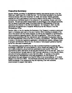
919 920 921 922 923 924 925 926
50
100
150
200
time [min] Fig. 8: Prophage induction with 0.2 g mitomycin C/ml in S. thermophilus strains J34f-2, J346f, J34f-2::pKOint, J34f-2cu50, and J34f-2cu50[pMZorf3] growing in thLM17-medium at 40°C. When reaching OD620nm of ca. 0.2, growing cultures were diluted with the same volume of pre-warmed thLM17-medium containing 0.4 g mitomycin C/ml. Growth was controlled by measuring OD620.
927 928 929 930 931 932
933 934 935 936 937 938 939 940 941 942 943 944 945 946
Fig. 9: Effect of Rir* on binding of Crh* to DNA. DO1B fragment (15 nM) was combined with constant concentrations of Crh* (1.57 µM) and increasing concentrations of Rir*. (A) Crh* and Rir* were added simultaneously to O1B; (B) Crh* was incubated with O1B prior to addition of Rir*. DNA was visualized by SYBR® Safe DNA gel stain.
Fig. 10: Protein-crosslinking of repressor Crh* using glutaraldehyde as analysed by SDSPAGE. a) Crosslinking of 2 μM of purified repressor Crh*: lane (-), Crh* without glutaraldehyde; lane (+), Crh* incubated with 0.005% glutaraldehyde for 5 min; lane (M), protein marker VI, molecular masses of proteins shown in the left margin. 10%SDSpolyacrylamide gel was used and protein was visualized by silver staining. b) Protein-protein interaction between GST-Rir and GST-Rir, Coh* and Crh*, respectively. Addition of proteins and glutaraldehyde is shown in the upper part of the figure. Crosslinked proteins, as detected after electrophoretic separation in 15% SDS-polyacrylamide gels by Western blotting using anti-GST rabbit antibodies is shown below. Lane (M): protein marker VI, molecular masses of proteins shown below the bands.
947 948 949 950 951
Sth Sth Ssa Sth Sth Sth Sag Sag Sor Spy Spy Spy Sth Ssu Smu
phage TP-J34 phage TP-778 phage YMC-2011 phage Sfi21 phage tp20617 phage 5093 strain GB00206 strain Gottschalk 998A phage PH10 phage 315.5 strain SSI-1 strain GA19702 phage O1205 phage phi20c strain NLML9
---------------------------------------MDLNKQRGSRIESLRASKGIS ---------------------------------------MDLNKQRGSRIESLRASKGIS -------------------------------------------MQAEERIKELIIAKYGN ----------------------------MFILLNKGKKCMRNNDEIISLIKSYLDNSSMS ---------------------------------------MRNNDEIISLIKSYLDNSSMS ----------------------------------------------------------MS ------------------------------------------MSVLIDRIRELSNKKGMS ------------------------------------------MSVLIDRIRELSNKKGMS ------------------------------------------MFSTLEKIKELALKRGIS -----------------------------------MR----TNDEIIDLMDNIRKEKRIS -----------------------------------MR----TNDEIIDLMDNIRKEKRIS -----------------------------------MR----TNDEIIDLMDNIRKEKNLS -----------------------------------MKGDVIMKENTAQRLAQIMKERNLR 12 ...HFFDFFSSFFGICTKSLDIKSLFTYNIPMNREVKIMADDKIKYPEVGQRIRELREMRGFE -------------------------------------MSLGNKEIMSKNIQYYMHKKGVD
Sth Sth Ssa Sth Sth Sth Sag Sag Sor Spy Spy Spy Sth Ssu Smu
phage TP-J34 phage TP-778 phage YMC-2011 phage Sfi21 phage tp20617 phage 5093 strain GB00206 strain Gottschalk 998A phage PH10 phage 315.5 strain SSI-1 strain GA19702 phage O1205 phage phi20c strain NLML9
QLELAKMLG--------YKSDSTISKWESGASIPTGTKIVKLA-----QALGTTTDYILF QLELAKMLG--------YKSDSTISKWESGASIPTGTKIVKLA-----QALGTTTDYILF VRAFATESG--------ISYTTVRSILERGIMNAKAENVFKIC-----HLLGISPD--TV MSELANKAG--------IS-KSTLSRYLSGSRVFPLNKADDFA-----RALGLTTE---MSELANKAG--------VS-KSTLSRYLSGSRVFPLNKADDFA-----RALGLTTE---MSELANKAG--------VS-KSTLSRYLSGSRVFPLNKADDFA-----RALGLTTE---LNDLEDTLG--------YSRNSLYSLNENS-KMGKPKEIAQYFNVSLDYLLGLTDN---P LNDLEDTLG--------YSRNSLYSLNENS-KMGKPKEIAQYFNVSLDYLLGLTDN---P LQKVAEDLG--------YSINYLYTLKEKTPKSDRLQEIADYFNVSTDYLLGRTDN---P ISELSRK---------VGMSKSGVSLYFNKARKFPLDRANLFA-----KALGTTPEH-II ISELSRK---------VGMSKSGVSLYFNKARKFPLDRANLFA-----KALGTTPEH-II ISELSRK---------VGMSKSGVSLYFNKARKFPLDRANLFA-----KALGTTPEH-II QVDILRQSEKFQKELGIKLGKSALSQYVSGKSIPDQDKLVLLS-----KTLGVSEAW-LM QLDIANELG--------YKSQSTISKWESGVNLPTGKKLILLA-----EMLDTSTDY-IL RNKIVSDLG---------VKYTTLTDWINGVTYPRIDKIELLA-----NYFGIEKSD--. : . :.
Sth Sth Ssa Sth Sth Sth Sag Sag Sor Spy Spy Spy Sth Ssu Smu
phage TP-J34 phage TP-778 phage YMC-2011 phage Sfi21 phage tp20617 phage 5093 strain GB00206 strain Gottschalk 998A phage PH10 phage 315.5 strain SSI-1 strain GA19702 phage O1205 phage phi20c strain NLML9
GDNSENNDGQQIISTDIDEIIQNAMMFDGKPLTDDDKRAIRGIIAGYMNSKEK--GDNSENNDGQQIISTDIDEIIENAMMFDGKPLTDDDKRAIRGIIAGYMNSKEK--ADWGISNE-QQPSSHDIDNIIDNAMMFDGKPLTDDDKRAIRGIIAGYMSSKEK--QVLNVKPSPKDISSTDIDDIIENAMMFDGKPLTDDDKRAIRGIIAGYMNSKEK--QFLNVKPSPKDTSPTDIDEIIANAMMFDGKPLTDDDKRAIRGIIAGYMSSKEK--QFLNVKPSPKDTSSTDIDEIIENAMMFDGKPLTDEDKRAIRGIIAGYMNSKEK--RIASDETAIIDGQVVDLREAAAHTMLFDGKPLDEDDIDFITAVLSAHFKNKQKD-RIASDETAIIDGQVVDLREAAAHTMLFDGKPLDEDDIDFITAVLSAHFKNKQKD-AIATNGDASAP---LDLRDIAAQSMLFDGKPLTEEDIDFITAVLEAHLKNK----GVTPKNSP---TQTIDLSNLRERVVMFDGKPLSDDDVKKIEAIIKLSLGVGNGEDR GVTPKNSP---TQTIDLSNLRERVVMFDGKPLSDDDVKKIEAIIKLSLGVGNGEDR GVTPKNSP---TQTIDLSNLRERVVMFDGKPLSDTDVEKIEAIIKLSLGVGNGEDK GYDTNESTDVKKESIDLSNLREKVVMFDGKPLSDDDVKKIEQIIRLSIEVTGDEDHGKANDISPLPTPTIDLSNLRERVVLFDGKPLSDEDVEKITKIIELSLEVSASEDR LVEEHKFVPLNDYAIDIENIPETVMLFDGKPLTDKDKIAIKNLIEIYLGGNKDD-*: : ::****** : * * :: :
21 21 17 32 21 2 18 18 18 21 21 21 25 60 23 68 68 62 74 63 44 66 66 67 66 66 66 79 106 66
121 121 114 127 116 97 120 120 115 119 119 119 134 162 120
Fig. 11: Alignment of amino acid sequences of TP-J34 Crh and putative repressor proteins of streptococcal phages. All repressors shown are associated with a topological Rir homolog.