www.nature.com/scientificreports
OPEN
received: 08 July 2016 accepted: 30 September 2016 Published: 24 October 2016
Supernova: A Versatile Vector System for Single-Cell Labeling and Gene Function Studies in vivo Wenshu Luo1,2,*, Hidenobu Mizuno1,2,*, Ryohei Iwata1,2, Shingo Nakazawa1,2, Kosuke Yasuda3, Shigeyoshi Itohara3 & Takuji Iwasato1,2 Here we describe “Supernova” series of vector systems that enable single-cell labeling and labeled cell-specific gene manipulation, when introduced by in utero electroporation (IUE) or adeno-associated virus (AAV)-mediated gene delivery. In Supernova, sparse labeling relies on low TRE leakage. In a small population of cells with over-threshold leakage, initial tTA-independent weak expression is enhanced by tTA/TRE-positive feedback along with a site-specific recombination system (e.g., Cre/loxP, Flpe/ FRT). Sparse and bright labeling by Supernova with little background enables the visualization of the morphological details of individual neurons in densely packed brain areas such as the cortex and hippocampus, both during development and in adulthood. Sparseness levels are adjustable. Labeled cell-specific gene knockout was accomplished by introducing Cre/loxP-based Supernova vectors into floxed mice. Furthermore, by combining with RNAi, TALEN, and CRISPR/Cas9 technologies, IUE-based Supernova achieved labeled cell-specific gene knockdown and editing/knockout without requiring genetically altered mice. Thus, Supernova system is highly extensible and widely applicable for singlecell analyses in complex organs, such as the mammalian brain. The mammalian brain, a complex organ, comprises numerous cells (neurons) densely packed and interconnected with each other to form intricate neural circuits responsible for higher brain function. To understand the precise cellular and molecular mechanisms of the neural circuit development and function, single-cell analyses that dissect connectivity of individual cells and molecular machinery operating in these cells are indispensable. For this purpose, two transgenic/knock-in mouse-based genetic systems, MADM1,2 and SLICK3, have been reported and have received much attention as promising tools4–6. However, unfortunately the use of each system was hampered by its intrinsic weakness (See Discussion). Moreover, systems that solely rely on mouse genetics, such as MADM and SLICK, have common weaknesses, including extensive cost and space requirements for mouse breeding and slow experimental turnover time, making these systems inflexible and hampering their application. Currently, as alternatives to transgenic/knock-in mouse approaches, in utero electroporation (IUE)-based and virus-mediated gene delivery techniques are widely used for cell labeling and gene manipulation in vivo7–16. These methods present extremely rapid experimental turnover, and are applicable for virtually all brain areas and neuron types. Thus, IUE and virus-mediated approaches have many advantages over pure mouse genetic approaches. However, because IUE and virus-mediated systems generally label cells too densely and/or manipulate genes in too many cells in transfected brain areas7,9,10,16–18, using IUE or virus systems for single-cell analysis (i.e., sparse cell labeling and labeled cell-specific gene manipulation) has been a challenge requiring methodological innovation. For the purpose of single-cell labeling, some IUE and virus-based methods are recently reported19–25. Nevertheless, further innovation is still necessary for more effective and wider applications (See Discussion). Moreover, for the purpose of achieving sparse and bright cell labeling and labeled cell-specific gene manipulation simultaneously, to date, only the initial primitive version of IUE-based “Supernova” system was briefly described18, and further innovation is awaited. Here we improved and expanded the initial Supernova substantially to develop the Supernova series of vector systems, and characterized these systems in detail to enable wider applications. When Supernova vectors are delivered to a specific brain area using IUE, they achieve sparse cell labeling with extremely high fluorescent 1
Division of Neurogenetics, National Institute of Genetics, Mishima, 411-8540, Japan. 2Department of Genetics, SOKENDAI (The Graduate University for Advanced Studies), Mishima, 411-8540, Japan. 3Laboratory for Behavioral Genetics, RIKEN Brain Science Institute, Wako, 351-0198, Japan. *These authors contributed equally to this work. Correspondence and requests for materials should be addressed to T.I. (email:
[email protected]) Scientific Reports | 6:35747 | DOI: 10.1038/srep35747
1
www.nature.com/scientificreports/ intensities and labeled cell-specific gene knockout in vivo. Background labeling (ratio of darkly labeled cells) is drastically reduced from the original version. By detailed and systematic characterizations of the systems, we have shown following: (1) The sparseness and brightness of Supernova labeling are constant at different developmental stages and in adulthood. (2) Labeling sparseness is adjustable without affecting the brightness. (3) Supernova enables simultaneous multiple gene expression in a cell. (4) Particularly, the incorporation of RNAi26,27, transcription activator-like effector nuclease (TALEN)28,29, and CRISPR/Cas916,30 technologies into Supernova is a critical leap. These sets of Supernova achieved single-cell gene knockout/knock-down even without requiring a genetically altered mouse. We further developed adeno-associated virus (AAV)-based Supernova system and demonstrated that it also enables both bright and sparse cell labeling and labeled-cell-specific gene knockout. Thus, Supernova, a promising tool, is simple, rapid, and widely applicable for single-cell analyses of complex organs, including the mammalian brain.
Results
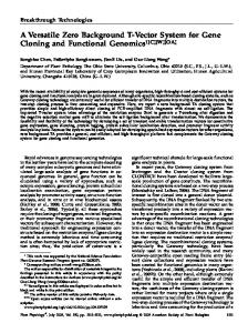
The Supernova series of vector systems. To allow sparse and bright cell labeling and labeled cell-specific gene manipulation in the mammalian brain, we developed Supernova series of vector systems that use IUE for vector delivery. The elementary composition of IUE-based Supernova includes a set of two vectors: TRE-SSRWPRE-pA [TRE-SSR; TRE: tetracycline response element; SSR: site-specific recombinase, such as Cre, Flpe31 and Dre32] and CAG-RT-stop-RT-XFP-ires-tTA-WPRE-pA (CAG-RT-stop-RT-XFP-tTA; RT: recombination target site, such as loxP, FRT, and rox; XFP: fluorescent proteins, such as GFP and RFP; tTA: tetracycline transactivator) (Fig. 1a). The initial Supernova is composed of TRE-Cre and CAG-loxP-stop-loxP-RFP-tTA vectors18. For wider applications, we re-designed the CAG-RT-stop-RT-XFP-tTA vector backbone such that one XFP type can be easily replaced with another XFP type or with any gene of interest (Supplementary Fig. 1 and Supplementary Table 1). When cells are transfected with a Supernova vector set, in a very small population among these cells, TRE leakage drives above-threshold but weak SSR expression, followed by tTA weak expressions. Then, only in these sparse cells, tTA binds with TRE, which further facilitates XFP expression through positive feedback cycles (Fig. 1b). IUE was employed to transfect Supernova vectors into cells in objective brain regions, including each cortical layer and the hippocampus (Supplementary Fig. 2). The significance of tTA/TRE enhancement in the system was clearly demonstrated (See Supplementary Fig. 3 and its legend). IUE-based Supernova enables single-cell labeling with high fluorescence intensity in vivo.
To enable wider applications of the Supernova system, we systematically and comprehensively evaluated Supernova labeling properties. To estimate sparseness and brightness extents of Supernova labeling, we delivered the Supernova GFP vector set (SnGFP) based on Flpe/FRT31 [Flpe-SnGFP: TRE-Flpe and CAG-FRT-stop-FRT-GFP-tTA (CAG-FSF-GFP-tTA)] into L2/3 cortical neurons via IUE at embryonic day 15.5 (E15.5). The CAG-RFP vector was co-electroporated to label the transfected neurons. In brains at postnatal day 22 (P22), a small and sparse population (1.2% ± 0.2%, n = 5 mice) of transfected neurons was GFP-positive (Fig. 1c). Most SnGFP-labeled cells were so bright that morphological details, including dendritic spines and axonal boutons, were clearly visible (Fig. 1d–g). By combining an optical clearing method33 and two-photon microscopy, we could image and reconstruct a single L2/3 callosal projection neuron having a long axon, in an intact fixed brain (Supplementary Fig. 4). Furthermore, by introducing Flpe-SnRFP into cortical L4 neurons using IUE at E14.5 and observing at P5 using two-photon microscopy, we could visualize the morphological details of dendrites and axons even in vivo (Fig. 1h). Note that in vivo imaging of single neurons located in deep cortical layers, such as L4, requires excellent sparseness and brightness. These results indicate that Supernova labeling (Flpe-based version) is extremely sparse and bright. Next, we evaluated the background level of Supernova labeling by delivering Flpe-SnRFP into L2/3 cortical neurons using IUE at E15.5. Notably, almost all (26/28 cells, four mice) Flpe-SnRFP-labeled cells were so bright that visualizing the whole dendritic morphologies of these cells was possible at P6. Only a few (2/28 cells) RFP-positive cells were defined as dark cells, which failed to label some of the basal dendrites to their tips. Thus, Flpe-Supernova achieved high intensity fluorescent neuronal labeling with little background.
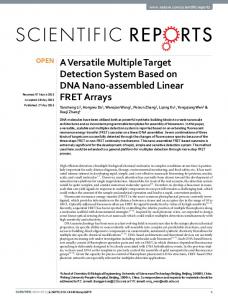
IUE-based Supernova is applicable for several developmental stages and in adulthood. We quantitatively examined the sparseness of Supernova labeling at different developmental stages and in adulthood by transfecting Flpe-SnGFP and CAG-RFP (control) together. We dissected the brains at P8, P22, 2 months (2 M), 4 M and 8 M (Fig. 2a) and evaluated sparseness as the ratio of Flpe-SnGFP-positive to RFP-positive neurons. The ratios (Fig. 2b) and brightness (Fig. 2a) were similar at all ages examined. Our results imply that the sparseness and brightness of Supernova labeling are constant at different developmental stages and in adulthood. Labeling sparseness is adjustable in Supernova system without affecting labeling brightness. We speculate that Supernova-labeling sparseness could be achieved by copy number variability of the TRE-SSR vector among transfected cells. Only cells in which TRE-SSR vector copy numbers are higher than a certain level might drive initial above-threshold SSR expression and finally achieve extremely bright cell labeling by tTA/TRE positive feedback (Fig. 1b). If this scenario is the case, it raises the possibility of adjusting labeling sparseness by changing the TRE-SSR vector concentration in the Supernova vector mixture. To test this attractive possibility, we prepared a series of Flpe-SnGFP vector mixture, containing different concentrations of the TRE-Flpe vector (5, 50, and 500 ng/μl), and introduced each mixture into L2/3 cortical neurons using IUE at E15.5 (Fig. 2c). The CAG-RFP was co-electroporated to label transfected neurons. The ratio of GFP/RFP-positive cell numbers was quantified at P8 (Fig. 2d). We found that, when 5 ng/μl (standard concentration) was used, only a very small population of RFP-positive neurons (1.4% ± 0.1%, n = 5 mice) was labeled by
Scientific Reports | 6:35747 | DOI: 10.1038/srep35747
2
www.nature.com/scientificreports/
Figure 1. IUE-based Supernova enables sparse and bright cell labeling with little background. (a) Schematics showing the elementary components of IUE-based Supernova: TRE-SSR-WPRE-pA (TRE-SSR) and CAG-RT-stop-RT-XFP-ires-tTA-WPRE-pA (CAG-RT-stop-RT-XFP-tTA). TRE: tetracycline response element; tTA: tetracycline transactivator; SSR: site specific recombinase (e.g. Cre, Flpe); RT: recombination target site (e.g. loxP, FRT); XFP: fluorescent proteins (e.g. GFP, RFP); WPRE: WHP post-trascriptional response element; ires: internal ribosome entry site. (b) Schematics showing how Supernova works: (1) Initially, only in a sparse population among many cells that are transfected with both vectors, the leakage of TRE drives abovethreshold but weak SSR expression. (2) This low level of SSR excises the RT-stop-RT cassette in a few copies of CAG-RT-stop-RT-XFP-tTA vector, initiating the transcription of XFP and tTA, albeit weakly. (3) Through binding with TRE, tTA facilitates expression of SSR. (4) Then RT-stop-RT cassette is excised from many copies of CAG-RT-stop-RT-XFP-tTA vector, and expression of XFP and tTA is increased. This positive loop of tTA/ TRE enhancement (See Supplementary Fig. 3) leads to extremely high levels of expression of both SSR and XFP, only in a small population of transfected cells. (c–g) Supernova labeling is sparse and bright enough to visualize the detailed morphology of single neurons including dendritic spines (f) and axonal branches and boutons (g). An Flpe-based Supernova GFP (Flpe-SnGFP) vector set (TRE-Flpe and CAG-FRT-stop-FRT-GFP-tTA) was introduced into layer 2/3 (L2/3) cortical neurons by in utero electroporation (IUE). The CAG-RFP vector was co-electroporated to mark the transfected neurons. Higher-power images of the rectangles in c were shown in (d). The square in (d) were further magnified in (e). (f,g) Show higher-magnification images of the rectangles in (e). (h) Two-photon in vivo imaging of L4 cortical neurons labeled by Flpe-based Supernova RFP (Flpe-SnRFP) in P5 mouse. The traces of imaged cortical neurons were shown in right panel. Black lines indicate the dendrites of labeled neurons. The axons of these neurons are represented by red and blue lines, separately. Scale bars, 250 μm (c); 100 μm (d); 50 μm (e,h); 4 μm (f); 10 μm (g).
Scientific Reports | 6:35747 | DOI: 10.1038/srep35747
3
www.nature.com/scientificreports/
Figure 2. The sparseness of Supernova labeling is stable and adjustable. (a,b) The sparseness and brightness of Supernova labeling were constant from early postnatal stages to adulthood. Images of L2/3 cortical neurons labeled by Flpe-SnGFP, in which concentration of the TRE-Flpe vector was 5 ng/μl (standard concentration), were shown (a). CAG-RFP was co-electroporated to label all the transfected cells. Coronal sections were made from P8, P22, 2 month-old (2M), 4M and 8M brains. The ratio of Flpe-SnGFP-labeled cell number to transfected cell number at each age was shown under the image in (a) and as diagram in (b) [mean ± SEM; n = 5 mice at P8 (30/2162 cells), P22 (8/654 cells), 2M (8/562 cells), n = 3 mice at 4M (9/529 cells), n = 2 mice at 8M (7/555 cells)]. Scale bars, 100 μm. (c,d) The degree of sparseness of Supernova-labeling is adjustable. Concentrations of TRE-Flpe vector used for IUE were 5 ng/μl, 50 ng/μl and 500 ng/μl [n = 5 mice for each group; 30/2162 cells (5 ng/μl), 890/1880 cells (50 ng/μl), 2507/2539 cells (500 ng/μl)]. L2/3 cortical neurons were labeled by Flpe-SnGFP together with CAG-RFP. Data obtained using 5 ng/μl are common in panels (a,c). Scale bars, 100 μm.
SnGFP. When 50 ng/μl was used, approximately half of the RFP-positive neurons (48.0% ± 5.4%, n = 5 mice) were SnGFP-positive. When the TRE-Flpe vector concentration was increased to 500 ng/μl, almost all RFP-positive neurons (98.7% ± 2.5%, n = 5 mice) were SnGFP-positive. These data indicate that the sparseness of Supernova
Scientific Reports | 6:35747 | DOI: 10.1038/srep35747
4
www.nature.com/scientificreports/ labeling is adjustable by simply changing the TRE-SSR vector concentration in the DNA solution for IUE. It is also important that labeling brightness was not altered by changing labeling sparseness. The initial version of Supernova, which is Cre-based SnRFP, is developed to meet a requirement for in vivo imaging of single L4 neurons in the neonatal mouse cortex, and therefore characterization of the system is restricted to the specific research purpose18. In the present study, we characterized Cre-based SnXFPs in various brain areas and ages and showed that they achieve sparse and bright cell labeling (Supplementary Figs 1b,c, 2a and 5) similar to that achieved by Flpe-based Supernova. However, we also noticed a few differences between Flpe-based and Cre-based versions (See Supplementary Fig. 6 and the legend for details). First, compared with the fluorescence signals expressed by Flpe-based Supernova, those expressed by Cre-based Supernova became visible more quickly after IUE (Supplementary Fig. 6a–h). Second, when the same TRE-SSR vector concentration was used, the sparseness of Flpe-Supernova labeling was superior to that of Cre-Supernova labeling (Supplementary Fig. 6i). Third, although the background level of Cre-Supernova labeling was low, that of Flpe-Supernova labeling was much lower (superior) (Supplementary Fig. 6c,d,g,h). In conclusion, although Flpe- and Cre-based versions show some differences, both Supernova versions are useful for visualizing the morphological details of individual neurons in densely packed brain areas, such as the cortex, by achieving sparse and bright cell labeling with low background.
Supernova enables Cre-independent single-cell labeling in Cre-expressing transgenic mice. To date, many Cre-expressing mouse lines have been reported and have contributed in advancing our understanding of the brain by generating region- and cell type-specific knockout mice34–37. To examine whether Supernova can be used in Cre-expressing mice, we introduced Flpe-SnGFP into the cortical neurons of an Emx1-Cre knock-in mouse, which shows Cre-mediated recombination in all excitatory neurons of the cortex34,38,39. As a recombination indicator, CAG-loxP-stop-loxP-RFP-WPRE (CAG-LSL-RFP) vector was co-expressed (Supplementary Fig. 7a). CAG-LSL-RFP labeling was overcrowded in the cortex, confirming ubiquitous Cre expression in the cortical excitatory neurons of Emx1-Cre mice. In contrast, Flpe-SnGFP labeled only a very small neuron population, even in Cre-expressing cortex; therefore, the morphological details of labeled neurons could be clearly observed (Supplementary Fig. 7b). We also developed Dre/rox-based Supernova, which gave similar results (Supplementary Fig. 8). Thus, Supernova labeling (Flpe- and Dre-versions) is suitable for Cre-independent sparse labeling in Cre-expressing brain areas. Supernova enables simultaneous multiple gene expression in a cell. To examine the possibility of simultaneous multiple gene expressions in a sparse cell population, we introduced SnGFP and SnRFP together (i.e., TRE-Flpe, CAG-FSF-GFP-tTA, and CAG-FSF-RFP-tTA) into cortical neurons using IUE at E15.5. We found that at P8, most labeled neurons expressed both GFP and RFP (RFP+/GFP+ = 51/53 cells, GFP+/RFP+ = 51/51 cells, n = 5 mice) (Fig. 3a). Using the simultaneous multiple gene expressions in single cells, we visualized the cell and nucleus together through the co-transfection of SnGFP and Supernova nuclear localization signal (nls)-RFP (SnnlsRFP) into cortical neurons (Fig. 3b). We also simultaneously transfected SnRFP and Supernova postsynaptic density protein (PSD) 95-fused GFP (SnPSD95-GFP) into L4 cortical neurons using IUE, and visualized both whole dendritic morphology and individual spines in single cells with RFP and GFP, respectively (Fig. 3c). Supernova system enables sparsely labeled cell-specific gene knockout in floxed mice.
Cre-based Supernova system design predicts high Cre expression levels in Supernova-labeled cells, whereas Cre expression absence in non-labeled cells (Fig. 1b); thus, a floxed gene in the genome is deleted only in Supernova-labeled cells (Fig. 4a). Here, we systematically and quantitatively analyzed the efficiency and specificities of Cre-based Supernova for knocking out an endogenous floxed gene in the mouse. We chose α2-chimaerin (α2-Chn) as a target gene because α2-chimaerin is ubiquitously expressed in the hippocampal CA1 pyramidal neurons at a high level40,41 and an anti-α2-chimaerin antibody that is applicable for immunohistochemistry is available42. We introduced Cre-SnRFP into the hippocampal neurons of α2-Chnflox/flox mice using IUE at E14.5 (Fig. 4b). The brains were dissected at P14, when the α2-chimaerin signals are strongest in the CA1 region40,41. α2-Chimaerin is ubiquitously expressed in CA1, but specifically lacks in SnRFP-labeled neurons (Fig. 4b). We quantified the ratios of α2-chimaerin-positive cells in RFP-negative (control) and RFP-positive (SnRFP-labeled) cells. In the hippocampal CA1 region, 97.7% ± 0.1% of RFP-negative cells expressed α2-chimaerin, whereas only 5.9% ± 3.0% of RFP-positive neurons showed α2-chimaerin signals (n = 3 mice, Fig. 4c). Furthermore, even in these SnRFP-labeled α2-chimaerin-positive neurons (n = 14 cells, three mice), α2-chimaerin expression levels were much lower than those in RFP-negative control cells (n = 14 cells, three mice) (Fig. 4d). These results demonstrate extremely high specificity and efficiency of the Supernova-induced gene knockout, and provide compelling evidence that Supernova is suitable for conditional gene knockout using a floxed mouse.
Supernova-mediated RNAi enables sparsely labeled cell-specific gene knockdown without using genetically altered mice. To achieve sparsely labeled cell-specific manipulation of endogenous
gene expression without using a floxed mouse, we modified the Supernova system by combining it with RNA interference (RNAi) technology26,27. Small hairpin RNA (shRNA) can be transfected into cortical neurons by the IUE-mediated transfection of CAG promotor/microRNA30-based RNAi vector (CAG-LSL-mir30)14. By IUE of Supernova-mediated expression vectors carrying shRNA against target genes (details see Methods and Supplementary Table 2), we efficiently reduced their expression level in sparsely labeled cortical neurons (See Fig. 5 and its legend). Scientific Reports | 6:35747 | DOI: 10.1038/srep35747
5
www.nature.com/scientificreports/
Figure 3. Simultaneous visualization of multiple proteins in a single cell using the Supernova. (a) Using the Supernova, RFP and GFP were expressed in sparsely labeled neurons with high co-expression efficiency (RFP+/GFP+ = 51/53 cells, GFP+/RFP+ = 51/51 cells, n = 5 mice). Flpe-based Supernova vector sets (TRE-Flpe, CAG-FSF-RFP-tTA and CAG-FSF-GFP-tTA) were introduced by IUE. (b) RFP fused with nuclear localization signal (nlsRFP) and GFP was co-expressed in a small population of cortical neurons by introducing Supernova vectors (TRE-Cre, CAG-LSL-GFP-tTA and CAG-LSL-nlsRFP-tTA). (c) Simultaneous visualization of RFP and PSD95-GFP in the same single neuron. Upper panel shows low-power images of a tangential section through the somatosensory cortex L4 transfected with SnRFP and Supernova PSD95-GFP (SnPSD95-GFP) vectors (TRE-Cre, CAG-LSL-RFP-tTA and CAG-LSL-PSD95-GFP-tTA). Bottom shows higher-magnification images of the rectangles in the upper panel. Scale bars, 100 μm (a,b); 25 μm (c).
Supernova-mediated TALEN enables sparsely labeled cell-specific gene knockout/editing in vivo. To achieve more effective gene suppression, we adapted TALEN-based genome editing technology28,29
to the Supernova system (Flpe-based version, Fig. 6). To examine the efficiency of Supernova-mediated TALEN, we constructed a pair of TALEN vectors targeting the endogenous α2-Chn gene and co-transfected them into the hippocampal CA1 neurons with TRE-Flpe and CAG-FSF-RFP-tTA vectors (Fig. 6a). We used immunohistochemistry to detect α2-chimaerin expression. DAPI-staining was performed to visualize cell body location (Fig. 6b). We categorized DAPI-stained cells in the hippocampal CA1 region into three groups depending on the intensity of anti-α2-chimaerin signals: α2-chimaerinhigh, α2-chimaerinnlow, and α2-chimaerinnegative (See Materials and Methods and Fig. 6 legend for details). We found that 76.0% ± 6.0% (n = 3 mice) of SnRFP-labeled cells were α2-chimaerinnegative cells. The rest were α2-chimaerinlow cells, whereas there were no α2-chimaerinhigh cells. In contrast, α2-chimaerinhigh and α2-chimaerinlow cells were 95.1% ± 0.5% (n = 3 mice) and 3.2% ± 0.1% (n = 3 mice), respectively, of RFP-negative neurons (Fig. 6c), i.e., in cells that express α2-Chn-targeting TALENs,
Scientific Reports | 6:35747 | DOI: 10.1038/srep35747
6
www.nature.com/scientificreports/
Figure 4. Labeled cell-specific gene knockout via Cre-based Supernova in floxed mice. (a) Schematic for Supernova-mediated single-cell knockout (KO) of endogenous gene of interest (GOI) flanked by two loxP sites. (b) α2-Chimaerin protein is expressed ubiquitously in the hippocampal CA1, while it is undetected specifically in SnRFP-labeled neurons (arrows), indicating that Cre-based Supernova-mediated gene knockout is highly specific to the labeled cells. Cre-SnRFP vectors were introduced into α2-chimaerin (α2-Chn)flox/flox mouse CA1 by IUE. α2-chimaerin immunohistochemistry and DAPI-staining were performed. Upper panels show the hippocampus of α2-Chnflox/flox mouse. A set of enlarged example images is shown in the bottom. Note that because extremely high intensity signal of Supernova labeling hinders detection of α2-chimaerin signal, we partially photobleached SnRFP signal in this experiment. (c,d) Quantification of Supernova-dependent α2-Chn knockout efficiency and specificity. (c) α2-Chimaerin expression was detected in almost all of SnRFP-negative CA1 hippocampal cells (97.7% ± 0.1%, n = 3 mice; 785 cells/804 cells, 464 cells/474 cells, 1011 cells/1036 cells), while only in 5.9% ± 3.0% (n = 3 mice; 3 cells/31 cells, 0 cell/18 cells, 3 cells/37 cells) of SnRFP-positive CA1 neurons, indicating labeled cell-specific knockout. All values represent as mean ± SEM; (**): 0.001