SUPPORTING SOIL REMEDIATION AT FERNALD BY ELECTRON BEAM METHODS. E. C. Buck, N. R. Brown, N. L. Dietz, and J. C. Cunnane. Chemical ...
r SUPPORTING SOIL REMEDIATION AT FERKALD BY ELECTRON BEAM METHODS
E. C. Buck, N. R. Brown, N. L. Dietz, and J. C. Cunnane ARGONNE NATIONAL LABORATORY Chemical Technology Division 9700 South Cass Avenue Argonne, IL 60439-4837
The submitted manuicnpt has been authored by a contractor of the U. S. Govtrnrrutnt under contract No. W-3M09-£NG-3SAccordingly, tha U. S. Government nuins i nonexclusive, royalty-free license to publish or reproduce the published form of thit contribution, or allow others to do so, for U. S. Government purposes.
Submitted to Waste Management '94 Tucson, Arizona February 27-March 3, 1994
Work supported by the U.S. Department of Energy, Office of Technology Development, as part of the Uranium in Soils Integrated Demonstration Program, under contract W-31-109-ENG-38.
SUPPORTING SOIL REMEDIATION AT FERNALD BY ELECTRON BEAM METHODS E. C. Buck, N. R. Brown, N. L. Dietz, and J. C. Cunnane Chemical Technology Division, Argonne National Laboratory ABSTRACT Electron beam techniques have been used to characterize uraniumcontaminated soils at the Fernald Site, Ohio. The major uranium phases have been identified by analytical electron microscopy (AEM) as uranyl phosphate (autunite), uranium oxide (uraninite), and uranium phosphite [U(PO3)4]. Luminescence and X-ray absorption spectrosccpy incorrectly identified uranium oxide hydrate (schoepite) as the major phase in Fernald soils. The solubilities of schoepite and autunite are very different, so a solubility-dependent remediation method selected for schoepite will not be effective for removing autunite. AEM is the only technique capable of precisely identifying unknown submicron phases. The uranium phosphite has been found predominantly at the incinerator site at Fernald. This phase has not been removed successfully by any of the chemical remediation technologies. We suggest that an alternative physical extraction procedure be applied to remove this phase. INTRODUCTION The remediation of radioactively contaminated sites in the United States has become an important issue for the U.S. Department of Energy (DOE) in recent years. Many technologies are available for decontamination, but if the nature of the contamination is not understood (i.e., its physical and chemical characteristics), it becomes impossible to select the most suitable method for cleaning a particular site. At the Fernald DOE facility, which is located northwest of Cincinnati, Ohio, detailed characterization of the site is being provided to the remediation groups, which helps them optimize their treatment methods. At Argonne National Laboratory (ANL), we have used electron beam methods of analysis to determine the nature of the uranium in contaminated soils from the Fernald site. We have proved that the technique of analytical electron microscopy (AEM) can provide important characterization information that allows the remediation effort to be directed on a sound scientific basis [1]. AEM has, in fact, provided the most useful and reliable characterization data compared to many other techniques which are currently being used in the Uranium in Soils Integrated Demonstration (USID). Its advantage is that it is not a "fingerprint" method: that is, total unknowns can be examined, sometimes without the need to consult a data base. Soil fractions containing the highest uranium contents have been characterized by a variety of techniques, including X-ray diffraction (XRD), inductively coupled plasma-atomic emission spectroscopy (ICP-AES), and alpha spectroscopy [2]. X-ray absorption spectroscopy (XAS) has also been used to determine the uranium oxidation state of the bulk soil samples. It was estimated that at least 80% of the soil uranium was in the [U(VD] state [3]. However, this was determined by using the position of the uranium LI1Z X-ray absorption edge, which may be misleading in a complex sample. Recently, a more sensitive method of obtaining the radial distribution function from the extended fine structure has been adopted. However, XAS and ultraviolet luminescence spectroscopy (both fingerprinting techniques) cannot identify phases
with confidence, and misinformation from these techniques has led to the selection of remediation methods which are inappropriate. The number of uranium minerals is large, so techniques that try to match spectral features ("fingerprints") cannot easily identify phases. In addition, if a phase is present that has not been suspected, identification is impossible. Electron beam methods, such as those used at ANL to analyze Fernald soils, allow identification of unique uranium-bearing phases. AEM involves a combination of methods that provide compositional and structural data, allowing identification completely unknown phases [1,4]. This paper describes an electron beam analysis of uranium-contaminated soils from the Fernald site that was performed in support of remediation groups. EXPERIMENTAL Fernald soil samples were infiltrated with a water-soluble melamine resin, and uranium-rich particles were located by scanning electron microscopy (SEM) combined with backscattered electron (BSE) imaging. These particles were isolated and prepared as transmission electron microscopy (TEM) thin sections by ultramicrotomy [3,4]. This method of sample preparation allowed direct comparison between SEM and TEM images, which means that the characterization of TEM samples is representative of the bulk sample. The samples were analyzed in a JEOL 2000 FXII TEM operated at 200 kV and equipped with X-ray energy dispersive spectrometers (EDS). Phases were identified by a combination of EDS and selected area electron diffraction. RESULTS In an earlier paper, the utility of AEM for identifying uraniumbearing phases in contaminated soils was demonstrated [1]. In this follow-up paper, we present examples of how AEM characterization could improved soil decontamination processes. SEM investigations of untreated soils have shown that uranium is contained within particles that are typically 1 |im to 100 nm in diameter. Further analysis with AEM has shown that these uranium-rich regions are made up of discrete uranium-bearing phases. The distribution of these uranium phases was found to be inhomogeneous at the microscopic level. Many phases have been found [4], including uranium adsorbed onto iron oxides, uranium silicates, uranium phosphates (autunites), uranium oxides (UO,), and uranium contained within a calcium fluorite phase. These results suggest that the majority of these phases contained uranium in the [(VI)] oxidation state; however, particles of uranium [(IV)] phases have also been identified, including uranium silicide (USi2) , uranium oxides (UO2) , and uranium phosphite (which is of special concern in this paper). Work is underway to find suitable remediation technologies for cleaning up the site. Recent investigations have concentrated on uranium-contaminated soils around the incinerator stack at Fernald-termed "A-soils". The other contaminated sites at Fernald have been labeled SP2-3, SP4, and SP5 [4]. Most of the phases at SP4 were uranium in a very fine form (uranium oxide particles -20 nm in diameter) and uranium adsorbed onto iron oxides. Soil samples from SP4 have been found to be among the easiest to clean up, and we believe that this is because most of
the uranium is absorbed onto other soil particles. However, samples from the incinerator site (A-soils) and other regions of the plant (such as SP2-3) have been found to be difficult to clean up, even after repeated application of extraction procedures. Much of the contamination in these regions is particulate in nature. Citrate Carbonate Extraction Carbonate and citrate complexing agents are being used by Francis et al. to remove [U(VI)] phases [5]. XAS synchrotron studies indicated that >80% of the uranium was in the [U(VD] state in all soils. However, only 50% of the total uranium contamination was removed during carbonate/citrate washing. Contaminated A14 soils had around 500 ppm uranium, whereas the citrate- and carbonate-treated soils had 180 ppm-300 ppm uranium. Electron beam analysis of citrateand carbonate-treated A14 soil samples revealed micron-sized uraniumbearing phases, identified as uranium oxide and uranium phosphite [U(PO3)4]. Some particles of autunite remained, but this process seemed to be effective at removing the uranyl phosphate. By comparing SEM and AEM information, one can begin to develop a database that allows uranium phases to be identified with greater efficiency. This improvement in identification allows representative characterization and semiquantitative estimates of the relative amounts of uranium phases present in the treated soil samples. The majority of the uranium observed in the treated soils (60-7 5%) was in uranium phosphites, about 20% was in uranium oxide [UO2] , and the remainder was in uranyl phosphate. Therefore, repeated extractions may reduce the uranium concentration by a fraction, but new processes must be developed to remove the uranium phosphite phases completely. Dithionite/Tiron Extraction AEM has shown why the use of the complexing agent Tiron has been ineffective at Fernald. For the USID at Fernald, Brainard et al. selected a soil washing process that involves the use of Tiron, a synthetic analog of a microbially produced complexing agent [5]. In this method of soil remediation, a catalyst, dithonite, is used to improve complexing. Dithonite reduces the [U(VD] species (uranyl) to [U(IV)], then these reduced species are removed from solution through complexation with Tiron. On the basis of characterization data supplied by Morris et al. in which the "fingerprint" technique of luminescence indicated that a major phase present in Fernald soils was a uranium oxide hydrate (schoepite), the remediation method should have been successful. However, AEM showed that this characterization was incorrect: the major [U(VD] phase in Fernald A-soils and SP2 soils is not schoepite but uranyl phosphate (autunite) [3,4]. Soil washing with Tiron is not considered to be effective for removing uranyl phosphates, which accounts for the problems with the technique in this instance. It appears that this method of treatment is not selectively removing any particular species (Table I). However, when one compares the ratios of autunite to uranium phosphite in the treated and untreated soils, it is evident that Tiron has affected these phases. The first two columns show the results of SEM analyses of untreated A14 samples received from the aqueous biphasic extraction group and Brainard et al., respectively. The data indicate that the distribution of
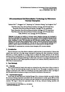
uranium phases differed between the two samples (i.e., the A14 samples were inhomogeneous). This inhomogeneity was found by XAS. The sample from Brainard et al. had a much lower concentration of autunite relative to uranium phosphite. From the treated sample results, it appears that dithionite use has led to the removal of uranium oxide phases. In both dithionite-only and dithionite/Tiron treatments, the concentration of uranium oxide has been substantially reduced. Uranium/iron phases observed in the untreated soils were absent in the treated samples. Bacterial Acid Digestion Delwiche et al. are are investigating a process for treating uranium contamination in which Fe3+ is used to oxidize U(IV) to U(VI) and then bacteria are used to oxidize the reduced iron [6]. The uranium is removed by a highly acidic solution (pH = 2 ) , which may have damaging effects on the soil structure. It is unclear whether this process is controlled by bacteria or whether it is simply acid digestion. When the results of chemical analysis of Fernald soils by ICP-AES are compared with the results from the more representative neutron activation analysis of bulk uranium content, the ICP-AES values are consistently lower than the neutron activation values [5]. This is perhaps due to the presence of insoluble uranium phases in the soil. Uranium phosphite phases have been found to be still present in the treated A-soils which have undergone this treatment. Identification of Uranium Phosphite Phase During the AEM characterization of uranium contamination in Fernald A-soils, an electron-beam-stable, ceramic-like phase was found, which was identified as uranium phosphite (also known, less accurately, as uranium metaphosphate). This phosphite phase did not possess the fine needle-like morphology characteristic of many uranium minerals, including uranyl phosphates (autunite) [4]. Figure 1 shows an example of the ceramic-like phase in a treated soil sample (A14). PLACE FIG. 1 HERE The diffraction patterns were very stable even under a condensed beam, which tends to amorphize natural uranium minerals rapidly. The uranium phosphite phase is unlikely to be a weathering product, but instead probably originated from the incinerator, where it formed at temperatures over 400°C and then was deposited in the surrounding area. After investigating these phases in A-soil samples, we have found that none of the USID remediation processes have successfully removed these phases. Identification of uranium phosphorus phases (autunite), uranium oxide, and uranium phosphite (see Tables II and III), observed in untreated, dithionite-treated, and dithionite/Tiron-treated samples, has been confirmed by TEM analysis. The structure of uranium phosphite [U(PO3)4] has not been completely elucidated; however, there are several varieties, all of which require high temperatures and fairly harsh reaction conditions to form [7]. DISCUSSION Effective removal of uranium from the Fernald soils can be enhanced by detailed knowledge of the chemical and physical characteristics of the
waste and its environment. We have found that unless the characterization technique can determine the exact nature of the phase, incorrect interpretations of data may lead to the selection of inappropriate remediation methods. The belief that schoepite was a major phase in Fernald soils suggested that remediation of Fernald soils would be easily achievable. It was not until AEM determined that the major phase was a uranium phosphate (autunite) that the problems with remediation experienced at Fernald were explained. The characterization methods described above, in combination with other methods under development [1,2], will allow remediation technology groups to find a more direct and efficient way of removing the contamination. Characterization of contaminated sites following the EPA protocol [8,9] will allow the most efficient remediation technology to be developed. These techniques can be transferred for implementation at contaminated sites operated by the DOE and private sector. Electron beam techniques for the preparation of soil samples have already been used with uranium- and thorium-contaminated soils from Ecotek, Inc. [10]. They are also being used to study plutonium contamination at Johnston Atoll [11,12]. There, a conveyor-belt technique developed by Thermo-Analytical is being used in an attempt to isolate contaminated soil and reduce its volume. Detailed soil analysis is required to determine whether this particular technique is feasible or whether another technique should be adopted. Evidence to date suggests that due to the redistribution of plutonium within calcite particles, the technique will be unable to effectively reduce the volume of contaminated soils [12]. CONCLUSION An unexpected uranium-bearing phase (uranium phosphite) has been found in soils from the Fernald incinerator site. This phase is a ceramic and not a natural uranium mineral; it may have formed while the incinerator was in use. The phase does not break down easily, and, therefore, we recommend that a physical extraction procedure be employed. The nature of the uranium phosphite phase would have proved extremely difficult for a "fingerprint" technique to identify. Because AEM involves a combination of structural and compositional techniques, it was able to characterise the phase positively. Based on the AEM data, the carbonate/citrate remediation technique has been effective on many of the autunite particles, but it has failed to remove the uranium oxides and uranium phosphite phases. The dithionite/Tiron method does not appear to work as predicted. The technique is somewhat successful, but it also does not remove the uranium phosphite particles. Acid leaching has also failed to remove this ceramic phase; in addition, this method causes substantial damage to the soil structure. Further studies of core and processed samples will continue to investigate the effectiveness of separation processes by characterizing the uranium-bearing phases isolated from the soil. At Fernald, electron beam methods have proved to be the most effective and reliable method of characterization. Similar techniques should be utilized at other DOE contaminated sites. We have shown that characterization can supply information which will allow the selection of appropriate technologies for remediating a contaminated site. ACKNOWLEDGMENTS
This work was supported by the U.S. Department of Energy, Office of Technology Development, as part of the Uranium in Soils Integrated Demonstration Program, under contract W-31-109-ENG-38. REFERENCES 1.
E. C. BUCK, J. C. CUNNANE, J. K. BATES, and N. L. DIETZ, "Analytical Electron Microscopy of Uranium Contaminated Soils at Fernald," Waste Management '93, Tucson, AZ, March 2-5, 1993.
2.
S. Y. LEE and J. D. MARSH, "Characterization Studies," ORNL/TM11980, Oak Ridge National Laboratory (1992).
3.
D. E. MORRIS, S.D. CONRADSON, C. DREW-TAIT, C. J. CHISHOLMBRAUSE, J. BERG, and J. MUSGRAVE, "Uranium Speciation in Fernald Soils," Progress Report (May 1992).
4.
E. C. BUCK, N. R. BROWN, and N. L. DIETZ, "The Distribution of Uranium Phases in Fernald Soils," Scientific Basis of Nuclear Waste Management, Boston, MA, November 29-December 3, 1993.
5.
C. W. FRANCIS et al., Soil Decon Task Group, "Removal of Uranium from Uranium Contaminated Soils Phase I: Bench-scale Testing," ORNL-6762, Oak Ridge National Laboratory (1993).
6.
M. E. DELWICHE, J. E. WEY, A. E. TORMA, D. T. MAIERS, R. G. REDDY, and J. C. CUNNANE, "Uranium Extraction from Contaminated Soils by Bioprocessing," Waste Management '94, Tucson, AZ, February 27-March 3, 1994.
7.
GMELIN Handbook of Inorganic Chemistry, Supplement Vol. C14, "Uranium Compounds with P, As, Sb, Bi, and Ge," Springer-Verlag, Berlin (1981).
8.
U.S. EPA, "Characterization Protocol for Radioactive Contaminated Soils," U.S. EPA office of Solid Waste and Emergency Response, Washington, D.C., Publication 9380.1-10FS (May 1992).
9.
J. NEIHIESEL, "Petrographic Methods in Characterization of Radioactive and Mixed Waste," Hazardous Materials Control Resources Institute, Superfund '92, Monitoring and Sampling, op. 192-195 (1992).
10.
N. R. BROWN, B. CARLSON, E. C. BUCK, N. L. DIETZ, and J. K. BATES, "Characterization of U and Th Contaminated Soils from a Nuclear Fuel Facility," Waste Management '94, Tucson, AZ, February 27-March 3, 1994.
11.
E. T. BRAMLITT, "Plutonium Mining for Clean-Up at Johnston Atoll," Health Physics 55, 451 (1988).
12.
S. F. WOLF, J. K. BATES, E. T. BRAMLITT, N. R. BROWN, E. C. BUCK, AND M. GONG, to be presented at Spectrum '94, Atlanta, GA, August 14-18, 1994.
Table I. Distribution (%) of uranium phases in soil samples treated with dithionite/Tiron. This table reflects relative concentrations in samples and not absolute amounts of the uranium phases. A14 (Chaiko)
A14 (Brainard)
Dithionite/ Tiron
Dithionite
Tiron
Autunite
80
55
68
76
11
U(PO 3 ) 4
10
34
27
20
69
U Oxide
8
5
3
0
11
Other U
2
5
2
4
10
Table II. Electron diffraction data from uranium phosphite [U(PO,)4] d spacings (nm) (Experimental)
d spacings (nm) (JCPDS 20-1348)
0.664
0.627
0.513
0.513
0.440
0.441
0.3825
0.385
0.365
0.366
0.2715
0.270
0.317
0.314
0.296
0.295
0.221
0.220
0.180 0.215
0.216
0.202
0.202
Table III.
Crystallographxc data for a- andft-IUtPO,)41 (taken from the GMSLIN Har.dhook. Symmetry
a/nm
b/nm
c/nm
1.495
0.639
a
P
P-tU(PO,)4]
Orchorhomic
0.895
a-[u(po,)j
Orthorhomic
0.6913
1.4967
0.8986
P-[U(PO314)
Orthorhomic
0.6907
1.4947
0.8986
1.4940
0.8987
P-[t3(PO,)4]
Orthorhomic
1.3821
P-[O(POj)4]
Orthorhomic
1.380
P-[U(PO3)4]
Orthorhomic
1.3826
a-[U(PO3)4j
Monoclinic
2.342
O-[U(P03)4]
Triclinic
(X-[U(PO3)4J
Triclinic
1.492
y
0.900 2.9933
0.8986
1.302
2.300
90.0
1.543
0.8147
0.8734
117.64
112.59
1.543
0.8147
1.547
115.15
89.08
89.30
Fig. 1.
A E M identification of uranium phosphite phase found in citrate-treated A14: (a) SEM/BSE micrograph of phase, where bright contrast is related to atomic number; !b} TEM image of phase; (c) selected area electron diffraction pattern of phase; and id) E D S analysis of phase.
* 1• 1 •
m
•
0.5um
•j
1 •
1
p
u Cu ! i 0
4
3 12 Energy (keV)
16
20