Hindawi Publishing Corporation Journal of Spectroscopy Volume 2013, Article ID 702310, 7 pages http://dx.doi.org/10.1155/2013/702310
Research Article Synthesis, Characterization, and Photochemical Properties of a New Square Mn(I)-Ru(II) Complex Using Pyrazine as Bridge Ligand Inara de Aguiar,1 Simone D. Inglez,2 Antonio Claudio Tedesco,3 and Rose Maria Carlos1 1
Departamento de Química, Universidade Federal de São Carlos, CP 676, 13565-905 São Carlos, SP, Brazil Faculdade de Ciências Exatas e Tecnologia, Universidade Federal da Grande Dourados, CP 533, 79804-970 Dourados, MS, Brazil 3 Departamento de Ciências Farmacêuticas, Faculdade de Filoso�a, Ciências e �etras de Ribeirão Preto, Universidade de São Paulo, 14040-901 Ribeirão Preto, SP, Brazil 2
Correspondence should be addressed to Rose Maria Carlos;
[email protected] Received 1 July 2012; Revised 19 September 2012; Accepted 8 October 2012 Academic Editor: R. P. S. Chakradhar Copyright © 2013 Inara de Aguiar et al. is is an open access article distributed under the Creative Commons Attribution License, which permits unrestricted use, distribution, and reproduction in any medium, provided the original work is properly cited. e photochemical properties of the complexes cis,fac-[Ru(phen)2 (pz)2 -Mn(CO)3 Br]2 4+ (I), cis-[Ru(phen)2(pz)2 ]2+ (II), and fac-Mn(CO)3 (pz)2 Br (III) where phen is phenanthroline and pz is pyrazine in acetonitrile solution are reported. e three complexes were characterized using 1 H NMR, UV-vis and FTIR spectroscopy and electrochemical (cyclic voltammetry and spectroelectrochemical) techniques. e complexes show intense absorption in the visible region assigned to the population of MLCT excited states. e absorption spectrum of I is the sum of the spectra of the mononuclear species II and III, and the two oxidation potentials at +1.10 and +1.56 V versus Ag/AGCl observed in I are ascribed to the different coordination environments of metal centers. e photolysis in the acetonitrile solution resulted in the pz dissociation to give the monoacetonitrile complexes for I, II, and III, respectively.
1. Introduction e investigation of spectroscopic, electrochemical, and photochemical properties of manganese compounds has attracted much attention due to the potential application of these compounds in the development of the supramolecular system which may work photochemically for clean energy sources in renewable solar fuels [1, 2]. Most of current research is being focused on the covalent coupling of a photoactive Ru(II) polypyridinic complex to a high-valence mono- and/or multinuclear 𝜇𝜇-oxo bridged manganese complexes. Herein, we wish to report a novel, square pyrazine donor-acceptor complex, composed of a triscarbonyl manganese complex linked to two ruthenium(II) phenanthroline complexes via two pyrazine bridge ligands. Our approach, introducing three good 𝜋𝜋 acceptor (CO) and one good 𝜋𝜋 donor (Br− ) ligand, was chosen because of the abilities of these ligands to accept and donate electronic density to
metal centers and in this way to stabilize the high oxidation states that the manganese center may acquire during the photoinduced electron transfer reaction. For this reason, in this work the photochemical stability of the complexes was studied.
2. Experimental 2.1. General. All synthesis and electrochemical and spectroscopic experiments were carried out under puri�ed N2 atmosphere, using Schlenk techniques. RuCl3 ⋅xH2 O, 1,10′ -phenanthroline (phen), pyrazine (pz), and lithium chloride were from Aldrich; tetrabutylammonium hexa�uorophosphate (TBAPF6 ) and bromide pentacarbonyl manganese from Strem. HPLC grade acetonitrile and dichloromethane were distilled prior to use. e solutions were carefully handled in the dark before the experiments were
2
Journal of Spectroscopy
6 7 7
3
N 88
15000
2 1 α
β
N
I
N
N
(mol− 1 L cm − 1 )
4
5
Ru N
N
N
N
6 5
4
3
10000
2
II
5000 III 0 300
10.5
10
9.5
9
8.5 (ppm)
8
7.5
7
6.5
400
500 Wavelength (nm)
600
700
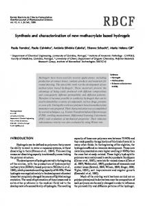
F 2: Electronic spectra in CH3 CN solution of (I) cis,fac[Ru(phen)2 (pz)2 -Mn(CO)3 Br]2 , (II) cis-[Ru(phen)2 (pz)2 ], (III) fac[Mn(CO)3 (pz)2 Br].
(a)
10.5
10
9.5
9
8.5 (ppm)
III
8
7.5
7
6.5 I
(b)
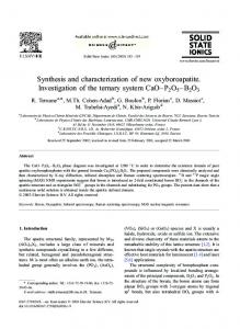
F 1: 1 H NMR spectra in CD3 CN of the aromatic region to the complex II (a) and complex I (b). 2200
performed. e complexes cis-[Ru(phen)2 Cl2 ]⋅2H2 O [3], cis[Ru(phen)2 CO3 ]⋅2H2 O [4], cis-[Ru(phen)2 (H2 O)2 ](PF6 )2 [5], and [Mn(CO)3 (pz)2 Br] [6] were prepared by the literature routes. FTIR spectra were measured in CaF2 windows in CH2 Cl2 solution on a Bomem-Michelson 102 spectrometer in the 4000–1000 cm−1 region. UV-visible spectra were recorded on an HP-8453 A (Diode array) spectrophotometer. NMR spectra were recorded using a Bruker DRX400 spectrometer. All chemical shis (𝛿𝛿) are given in ppm units with reference to the hydrogen signal of the methyl group of tetramethylsilane (TMS) as internal standard. Monochromatic irradiations at 350 nm and 420 nm were generated using an RMR-600 model Rayonet Photochemical reactor using RMR-3500 and RMR-4200 lamps, respectively. e continuous photolysis experiments were followed by UV-vis. Time-resolved optical spectra were obtained using a laser �ash-photolysis apparatus containing a Continuum Q-switched Nd:YAG laser (Continuum, Santa Clara, CA) with excitation provided by the third harmonic at 𝜆𝜆 355 nm. Cyclic voltammetry was performed using an 𝜇𝜇 Autolab Type III potentiostat. Voltammograms were obtained in CH3 CN (1 mM TBPF6 ) at 22∘ C in a lightprotected voltammetric cell with a platinum cylinder disc for both the working and the auxiliary electrodes. A silver wire
2150
2100
2050
2000
1950
1900
1850
1800
Wavenumber (cm − 1 )
F 3: FTIR spectra of complexes III (black line) and I (red line) in CH2 Cl2 at room temperature, with CaF2 window.
coated with silver chloride was used as reference electrode, connected to the bulk of the solution by a Luggin capillary �lled with the same solvent and electrolyte. Solutions were deoxygenated with a stream of N2 and maintained under a positive pressure of N2 during the measurements. e concentration of the complexes was kept always at 1 mM. 2.2. Synthesis 2.2.1. cis,fac-[Ru(phen)2 (pz)2 -Mn(CO)3 Br]2 (PF6 )4 (I). Mn(CO)5 Br (74 mg, 0.27 mmol) was dissolved in degassed acetone (50 mL), and the complex cis-[Ru(phen)2 (pz)2 ] (250 mg, 0.27 mmol) was added. It was stirred under dark for 12 hours at room temperature. e orange precipitate formed was �ltrated and dried under vacuum. Yield � 80%. 2.2.2. cis-[Ru(phen)2 (pz)2 ](PF6 )2 (II). [Ru(phen)2 Cl2 ]⋅2H2 O (200 mg, 0.35 mmol) and pyrazine (282 mg, 3.52 mmol) were
Journal of Spectroscopy
3
0.6 /( A)
0.3 0
I
− 0.3 0
0.6
1.2
1.8
0.6 /( A)
0.3 0
III
− 0.3 0
0.6
1.2
1.8
0.6 /( A)
0.3 0
II
− 0.3 0
0.6
1.2
1.8
(V)
F 4: Cyclic voltammograms of complexes I, II and III (1 mM), TBPF6 (0.1 M) versus Ag/AgCl, scan rate 100 mV/s, 25∘ C, in CH3 CN. 1.8
1.8
1.6
1.6 II
1.4
1.4
1.2
1.2
Absorbance
Absorbance
I
1
1
0.8
0.8
0.6
0.6
0.4
0.4
200
300
400
500
600
700
Wavelength (nm)
(a)
200
300
400
500
600
700
Wavelength (nm) (b)
F 5: Spectroelectrochemical of complex I (1.5 mM in CH3 CN; 0.1 M TBAPF6 ) and complex II (1.5 mM in CH3 CN; 0.1 M TBAPF6 ) in an OTTLE cell, oxidation 𝐸𝐸applied = 1.5 V versus Ag/AgCl.
re�uxed in a 1 : 1 EtOH/H2 O solution for 6 hours. Upon cooling to room temperature, a saturated solution of ammonium hexa�uorophosphate was added to the solution to precipitate an orange product, which was isolated by �ltration. e orange solid was recrystallized using acetone/ether. Yield: 70%. 2.2.3. fac-[Mn(CO)3 (pz)2 Br](III). Mn(CO)5 Br (200 mg, 0.73 mmol) was dissolved in degassed CH2 Cl2 (50 mL) and pyrazine (117 mg, 1.45 mmol). It was stirred under dark for 12 hours at room temperature. e yellow precipitate formed was �ltrate and dried under vacuum. Yield: �0%.
3. Results and Discussion e square complex was synthesized in a manner similar to a previously published procedure [1], starting from cis-[Ru(phen)2 (pz)2 ]2+ and considering the complex Mn(CO)5 Br as a ligand (Scheme 1). e 1 H NMR spectral data for the ligands and complexes in CD3 CN are listed in Table 1 using the numbering scheme as represented for complex II as follow. e signals of the complex I were assigned by comparison and analysis of the precursor Ru(phen)2 Cl2 and the free pyrazine. In the complex I, the phenanthroline protons appeared as eight
4
Journal of Spectroscopy
Abs
− 0.02 0 0.02
/mol L cm
20000 15000 10000 5000 0 − 5000 300
350
400
450
500
550
600
650
Wavelength (nm)
F 6: Transient (upper) and experimental (low) absorption spectra of complex I in acetonitrile.
N N N
Ru N
N
Br
N +
N
CO CO
N
Br Mn CO
CO
Ru
C3 H6 O
N
Br OC
N
N Mn
CO
Mn
N
N
CO
CO CO CO
N N
N
Ru
CO
S 1: Synthesis of square complex I.
T 1: 1 H NMR spectral data of complexes I and II in CD3 CN. 𝛿𝛿H (ppm) Proton type H 1 H 1′ H2 H2′ H3 H3′ H4 H4′ H5 H5′ H6 H6′ H7 H7′ H8 H8′ H𝛼𝛼 H𝛼𝛼′ H𝛽𝛽 H𝛽𝛽′
Complex II
Complex I
9.35 (2H, d) 8.25 (4H, m) 8.84 (2H, d) 8.25 (4H, m) 8.14 (2H, d) 8.50 (2H, d) 7.57 (2H, m) 7.98 (2H, dd) 8.49 (4H, d) 8.45 (4H, d)
9.35 (4H) 8.26 (8H) 8.84 (4H) 8.26 (8H) 8.15 (4H) 8.50 (4H) 7.58 (4H) 7.98 (4H) 8.50 (8H) 8.50 (8H)
signals instead of four signals to the free ligand, as expected for the cis isomer. e two pyrazine 𝛼𝛼 and 𝛽𝛽 protons were observed at 8.49 (4H, d) and 8.45 (4H, d). On coordination of Ru(II) to Mn(I) (Figure 2, Table 1), the linewidth of all signals were broadened due to the presence of the bromide ion coordinated at the manganese center. e absence of new signals in the whole spectrum, as expected to a triangle complex, suggests the formation of the square complex, which was further con�rmed by signals integration [7]. In the
T 2: Electrochemistry properties of complexes I, II, and III in CH3 CN.
Complex I
𝐸𝐸ox /𝐸𝐸red , 𝑉𝑉 1.10 0.79/0.73
𝜈𝜈CO cm−1
𝜆𝜆máx , nm (𝜀𝜀) 394 (11500); 442 (10400)
2032, 1938
Complex II
1.56/1.49
—
394 (9100) 425 (8000)
Complex III
1.10 0.87/0.89
2041, 1953, 1932
381 (2900)
square complex, all the assignments were done using the same numbering �gure as complex II, Figure 1. 3.1. Absorption Properties. e absorption spectra of complexes I, II, and III in CH3 CN solution are shown in Figure 2. e absorption maximum of 380 nm (𝜀𝜀max = 1400 mol−1 L cm−1 ) for III appears as a shoulder on the strong 𝜋𝜋-𝜋𝜋∗ absorption bands of the ligands, while the complex II exhibits the intense and broad MLCT absorption (𝜆𝜆max = 397 nm; 𝜀𝜀max = 9200 mol−1 L cm−1 ) typical of RuII polypyridine complexes [8]. e absorption spectrum of I (𝜆𝜆máx = 397 nm; 𝜀𝜀máx = 11230 mol−1 L cm−1 ) is consistent with the
Journal of Spectroscopy
5
1
1 III
Absorbance
Absorbance
II
0.5
0 300
400
500 Wavelength (nm)
600
0.5
0 300
700
400
(a)
500 Wavelength (nm)
600
700
(b) 1
Absorbance
I
0.5
0 300
400
500 Wavelength (nm)
600
700
(c)
F 7: Photochemistry of complex II (1.5 mM in CH3 CN, 𝜆𝜆irr = 420 nm), complex III (0.95 mM in CH3 CN, 𝜆𝜆irr = 350 nm), and complex I (1.5 mM in CH3 CN, 𝜆𝜆irr = 420 nm).
superposition of bands characteristics of the corresponding mononuclear complexes. e data for complex I show that the energy of the charge transfer band RuII → L (394 e 442 nm) remains unchanged upon the introduction of Mn ion into the Ru complex, whereas it is slightly shied to shorter wavelength compared to the tris-(phenanthroline) complexes (422 and 446 nm) [9], which is characteristic of ligands serving as better 𝜋𝜋-acceptors ligands than phen. e FTIR spectrum of III, shown in Figure 3, exhibitsthree intense v(CO) absorptions at 2041, 1953, and 1932 cm−1 consistent with the facial arrangement of the three COs in the coordination sphere. For complex I, the v(CO) stretching frequency appears as weak and broad bands around 2032 and 1940 cm−1 , and the two lower energy bands are overlapped suggesting the attachment of Mn(CO)3 unit into the RuII complex.
3.2. Electrochemistry. e voltammetric data are summarized in Table 2. Figure 4 shows a cyclic voltammogram (scan rate 100 mVs−1 ) for a 1 mM solution of the complexes I, II, and III over the range 0–1.8 V (versus AG/AGCl) in acetonitrile (TBPF6 1 mM).
Complex II exhibits a redox couple at 𝐸𝐸1/2 = 1.52 V (𝐸𝐸ox = 1.56 V and 𝐸𝐸red = 1.49 V versus Ag/AgCl) of RuII/III which is more positive than those found to [Ru(phen)3 ]2+ [10]. e complex III, on the other hand, displays a shoulder at 0.80 V corresponding to MnI/II oxidation followed by an oxidation peak at 1.10 V attributed to the oxidation of MnII to MnIII , which is paired with a nonreversible reductive wave at 0.90 due to MnIII/II reduction. e Mn(CO)3 Br coordination on the Ru(phen)2 (pz)2 did not change the redox potential values of the Ru(II) metal center. e cyclic voltam ogram of complex I exhibits one irreversible oxidation at 1.10 V and two redox couple 𝐸𝐸1/2 (1) = 0.76 V (𝐸𝐸ox = 0.79 V and 𝐸𝐸red = 0.73 V versus Ag/AgCl) and 𝐸𝐸1/2 (2) = 1.52 V (𝐸𝐸ox = 1.56 V and 𝐸𝐸red = 1.49 V versus Ag/AgCl).
3.3. Spectroelectrochemistry. Insights into the bonding characteristics of the Ru(II) complexes for complexes I and II were obtained by spectroelectrochemical experiments. For the Ru(II) complexes, a constant potential 1.5 V (determined from cyclic voltammetry) was applied and the extent of oxidation to Ru(III) was monitored by UV-vis spectroscopy
6
Journal of Spectroscopy complexes I and II, exhaustive photolysis leads to the same �nal stable spectrum assigned to the monosolvated complex.
1
Absorbance
0.8 0.6 0.4 0.2 0 300
400
500
600
700
800
Wavelength (nm)
F 8: Spectral changes accompanying the consume of reduced methyl viologen (MV•+ ), from the thermal reaction MV•+ → MV2+ in H2 O, (black line—spectrum of the mixture: complex III and MV2+ before irradiation).
(Figure 5). e spectrum shows the disappearance of the broad absorption band at 400 nm. Aer 30 min of oxidative electrolysis, the spectral changes were completed. e oxidative spectroelectrochemistry at 1.5 V leads to disappearance of the MLCT band indicating that oxidation was a RuII/III process which is irreversible. 3.4. Transient Absorption Spectra. Figure 6 shows the excited state absorption spectrum for complex I in CH3 CN solution aer excitation with an 8 ns pulse at 355 nm irradiation. ere is a bleach of absorption band at 400 nm and new structured absorption with maxima centered at 350 nm and 600 nm consistent with formation of an MLCT (Ru → phen) excited state [11].
3.5. Photochemistry. e complexes are stable in deaerated solutions in the absence of light. When solutions of complex II were subject to continuous photolysis, the resulting optical spectral changes were consistent with the substitution of only one pyrazine molecule by a solvent molecule (1) and (2) 2+
𝑐𝑐𝑐𝑐𝑐𝑐-Ruphen2 pz2
+ CH3 CN
⟶ 𝑐𝑐𝑐𝑐𝑐𝑐-Ruphen2 pz CH3 CN
𝑐𝑐𝑐𝑐𝑐𝑐-Ruphen2 pz CH3 CN
2+
2+
+ pz
+ CH3 CN
⟶ 𝑐𝑐𝑐𝑐𝑐𝑐-Ruphen2 CH3 CN2
2+
(1)
(2)
For example, Figure 7 illustrates the spectral changes seen when an acetonitrile solution of complex I (0.22 mM) was irradiated at 𝜆𝜆irr = 420 nm, 𝐈𝐈0 = 1.27 × 10⁻⁸ einstein s−1 . e spectra show a progressive depletion of the characteristic absorption band at 400 nm concomitant with formation of two broad shoulders at 385 and 422 nm, in accordance with the formation of complex Ru(phen)2 (CH3 CN)2 [12]. For
3.6. Photoinduced Electron Transfer Reactions. Figure 8 shows the UV-vis spectral changes of the thermal reaction (MV•+ → MV2+ ) of a solution containing complex III and MV2+ (methyl viologen) in pure water immediately aer 10 s continuous irradiation at 355 nm light. Before irradiation, the absorption spectrum of the mixture shows the characteristic absorption of starting complex (𝜆𝜆max = 380 nm). A broad absorption band with maximum near 605 nm and a peak at 394 nm appeared just aer irradiation. ese new absorptions match the methyl viologen radical (MV•+ ) absorption spectrum [13]. e photoinduced electron transfer reactions for complexes I and II did not occur, since in the UV-vis spectra was not observed any MV•+ characteristic band even aer exhaustive photolysis. ese results show that the intermolecular photoinduced electron transfer reaction is activated in water only in certain conditions. e presence of Ru(II) unit inhibits the MV2+ reduction.
References [1] I. De Aguiar, S. D. Inglez, F. C. A. Lima et al., “Photochemical reactions of fac-[Mn(CO)3 (phen)imidazole]+ : evidence for long-lived radical species intermediates,” Inorganic Chemistry, vol. 47, no. 24, pp. 11519–11526, 2008. [2] V. C. Nogueira, C. Longo, A. F. Nogueira, M. A. Soto-Oviedo, and M. A. D. Paoli, “Solid-state dye-sensitized solar cell: improved performance and stability using a plasticized polymer electrolyte,” Journal of Photochemistry and Photobiology A, vol. 181, no. 2-3, pp. 226–232, 2006. [3] B. P. Sullivan, D. J. Salmon, and T. J. Meyer, “Mixed phosphine 2,2′ -bipyridine complexes of ruthenium,” Inorganic Chemistry, vol. 17, no. 12, pp. 3334–3341, 1978. [4] E. C. Johnson, B. P. Sullivan, D. J. Salmon, S. A. Adeyemi, and T. J. Meyer, “Synthesis and properties of the chloro-bridged dimer [(bpy)2 RuCl]2+ 2 and its transient 3+ mixed-valence ion,” Inorganic Chemistry, vol. 17, no. 8, pp. 2211–2215, 1978. [5] P. Bonneson, J. L. Walsh, W. T. Pennington, A. W. Cordes, and B. Durham, “Six-coordinate complexes with 1,10-phenanthroline ligands in the trans con�guration. Preparation of trans-bis(1,10-phenanthroline)ruthenium(II) complexes and crystal structure of trans-bis(1,10-phenanthroline)bis(pyridine)ruthenium(II) hexa�uorophosphate,” Inorganic Chemistry, vol. 22, no. 12, pp. 1761–1765, 1983. [6] R. M. Carlos, I. Ap. Carlos, B. S. Lima Neto, and M. G. Neumann, “Spectroscopic and electrochemical properties of [Mn(phen)(CO)3 (imidazole)](SO3 CF3 ) complexes,” Inorganica Chimica Acta, vol. 299, no. 2, pp. 231–237, 2000. [7] L. A. Berben, M. C. Faia, N. R. M. Crawford, and J. R. Long, “Angle-dependent electronic effects in 4,4′ -bipyridine-bridged Ru3 triangle and Ru4 square complexes,” Inorganic Chemistry, vol. 45, no. 16, pp. 6378–6386, 2006. [8] K. Kalyanasundaram, Photochemistry of Polypiridyne and Porphyrin Complexes, Academic Press, London, UK, 1992. [9] C.-T. Lin, W. Böttcher, M. Chou, C. Creutz, and N. Sutin, “Mechanism of the quenching of the emission of substituted polypyridineruthenium(II) complexes by iron(III),
Journal of Spectroscopy
[10]
[11]
[12]
[13]
chromium(III), and europium(III) ions,” Journal of the American Chemical Society, vol. 98, no. 21, pp. 6536–6544, 1976. F. Barigelletti, A. Juris, V. Balzani, P. Belser, and A. Von �elewsky, “In�uence of the ligand structure on the electrochemical and spectroscopic properties of ruthenium(II)polypyridine complexes,” Inorganic Chemistry, vol. 26, no. 24, pp. 4115–4119, 1987. C. Creutz, M. Chou, T. L. Netzel, M. Okumura, and N. Sutin, “Lifetimes, spectra, and quenching of the excited states of polypyridine complexes of iron(II), ruthenium(II), and osmium(II),” Journal of the American Chemical Society, vol. 102, no. 4, pp. 1309–1319, 1980. P. Bonneson, J. L. Walsh, W. T. Pennington, A. W. Cordes, and B. Durham, “Six-coordinate complexes with 1,10-phenanthroline ligands in the trans con�guration. Preparation of trans-bis(1,10-phenanthroline)ruthenium(II) complexes and crystal structure of trans-bis(1,10-phenanthroline)bis(pyridine)ruthenium(II) hexa�uorophosphate,” Inorganic Chemistry, vol. 22, no. 12, pp. 1761–1765, 1983. T. Watanabe and K. Honda, “Measurement of the extinction coefficient of the methyl viologen cation radical and the efficiency of its formation by semiconductor photocatalysis,” Journal of Physical Chemistry, vol. 86, no. 14, pp. 2617–2619, 1982.
7
International Journal of
Medicinal Chemistry Hindawi Publishing Corporation http://www.hindawi.com
Volume 2014
Photoenergy International Journal of
Organic Chemistry International Hindawi Publishing Corporation http://www.hindawi.com
Volume 2014
Hindawi Publishing Corporation http://www.hindawi.com
Volume 2014
International Journal of
Analytical Chemistry Hindawi Publishing Corporation http://www.hindawi.com
Volume 2014
Advances in
Physical Chemistry Hindawi Publishing Corporation http://www.hindawi.com
Volume 2014
International Journal of
Carbohydrate Chemistry Hindawi Publishing Corporation http://www.hindawi.com
Journal of
Quantum Chemistry Hindawi Publishing Corporation http://www.hindawi.com
Volume 2014
Volume 2014
Submit your manuscripts at http://www.hindawi.com Journal of
The Scientific World Journal Hindawi Publishing Corporation http://www.hindawi.com
Journal of
International Journal of
Inorganic Chemistry Volume 2014
Journal of
Theoretical Chemistry
Hindawi Publishing Corporation http://www.hindawi.com
Hindawi Publishing Corporation http://www.hindawi.com
Volume 2014
Spectroscopy Hindawi Publishing Corporation http://www.hindawi.com
Analytical Methods in Chemistry
Volume 2014
Hindawi Publishing Corporation http://www.hindawi.com
Volume 2014
Chromatography Research International Hindawi Publishing Corporation http://www.hindawi.com
Volume 2014
International Journal of
Electrochemistry Hindawi Publishing Corporation http://www.hindawi.com
Volume 2014
Journal of
Hindawi Publishing Corporation http://www.hindawi.com
Volume 2014
Journal of
Catalysts Hindawi Publishing Corporation http://www.hindawi.com
Journal of
Applied Chemistry
Hindawi Publishing Corporation http://www.hindawi.com
Bioinorganic Chemistry and Applications Hindawi Publishing Corporation http://www.hindawi.com
Volume 2014
International Journal of
Chemistry Volume 2014
Volume 2014
Spectroscopy Volume 2014
Hindawi Publishing Corporation http://www.hindawi.com
Volume 2014