expression during fracture repair is regulated temporally, with elevated levels seen as early as PF day 3 and ... The data presented in this study complement previous re- ... diographs were taken to ensure a clean, simple transverse ... max film overnight at room temperature. .... entire soft and hard callus (i.e., PF day 10, Fig.
JOURNAL OF BONE AND MINERAL RESEARCH Volume 15, Number 6, 2000 © 2000 American Society for Bone and Mineral Research
Temporal Expression of the Chondrogenic and Angiogenic Growth Factor CYR61 During Fracture Repair MICHAEL HADJIARGYROU,1,2 WILLIAM AHRENS,2 and CLINTON T. RUBIN1,2
ABSTRACT The repair of a fractured bone is a complex biological event that essentially recapitulates embryonic development and requires the activity of a number of different cell types undergoing proliferation, migration, adhesion, and differentiation, while at the same time expressing a host of different genes. To identify such genes, we employed differential display and compared messenger RNA (mRNA) populations isolated from postfracture (PF) day 5 calluses to those of intact rat femurs. One such gene in which expression was up-regulated at PF day 5 is identified as CYR61, a member of the CCN family of secreted regulatory proteins. CYR61 is a growth factor that stimulates chondrogenesis and angiogenesis. We show that its mRNA expression during fracture repair is regulated temporally, with elevated levels seen as early as PF day 3 and day 5, rising dramatically at PF day 7 and day 10, and finally declining at PF day 14 and day 21. At the highest peak of expression (PF day 7 and day 10, which correlates with chondrogenesis), CYR61 mRNA levels are approximately 10-fold higher than those detected in intact femurs. Similarly, high protein levels are detected throughout the reparative phase of the callus, particularly in fibrous tissue and periosteum, and in proliferating chondrocytes, osteoblasts, and immature osteocytes. The secreted form of CYR61 also was detected within the newly made osteoid. No labeling was detected in hypertrophic chondrocytes or in mature cortical osteocytes. These results suggest that CYR61 plays a significant role in cartilage and bone formation and may serve as an important regulator of fracture healing. (J Bone Miner Res 2000;15:1014 –1023) Key words:
CYR61, fracture, healing, mRNA, expression
INTRODUCTION ESPITE A plethora of information on the expression of a number of extracellular matrix (ECM) proteins and growth factors during fracture healing (reviewed in Ref. 1), very little is known about the role of numerous other genes in the repair process. Considering that fracture healing is essentially a replay of embryonic development that involves many different cell types (i.e., endothelial, fibroblasts, chondroblasts, osteoblasts, osteoclasts, etc.) and cellular processes (i.e., adhesion, proliferation, migration, differentiation, etc.), it is highly probable that a host of other genes, some known and some novel, also will be expressed. To
D
identify such genes, we have utilized differential display(2) to compare directly messenger RNA (mRNA) populations isolated from intact femurs to those of postfracture (PF) day 5 calluses. One such gene identified by this approach and in which relative temporal and spatial expression is reported here is CYR61. CYR61 belongs to the CCN family (a term coined by Bork(3) referring to the original members, CYR61, CTGF, and NOV) of secreted extracellular proteins that are characterized by having all of their 38 conserved cysteine residues present in their secreted portions (reviewed in Ref. 4). Initially, it was identified as an immediate early gene that is activated transcriptionally by serum or purified growth fac-
1
Program in Biomedical Engineering, State University of New York at Stony Brook, Stony Brook, New York, U.S.A. Department of Orthopaedics, State University of New York at Stony Brook, Stony Brook, New York, U.S.A.
2
1014
CYR61 EXPRESSION DURING FRACTURE REPAIR
tors.(5) Biological assays showed that CYR61 is a secreted protein that associates with the ECM and cell surface,(6) and promotes proliferation, migration, and adhesion of endothelial cells, suggesting that its likely function is one of an ECM signaling molecule.(7) During mouse embryonic development, CYR61 expression correlates with chondrogenesis. More specifically, CYR61 is expressed in mesenchymal cells of both mesodermal and ectodermal origins in developing limbs, ribs, and prevertebrae, as well as Meckel’s cartilage.(8) More recently, Wong et al.(9) showed that the CYR61 protein promoted chondrogenesis in micromass cultures of limb bud mesenchymal cells in vitro, as well as enhanced cell-cell aggregation, an initial step in chondrogenesis. Furthermore, CYR61 was identified by differential mRNA display to be expressed by human fetal osteoblasts in response to 1␣,25-dihydroxyvitamin D3 and its expression to be regulated by cytokines, growth factors, and serum,(10) as well as induced by bone morphogenetic proteins (BMPs) in embryonic stem cells.(11) Taken together, these studies suggest that CYR61 may play an important role during chondrogenesis (the hallmark of endochondral ossification), a process that occurs during fracture healing of long bones, as well as osteoblast function and differentiation, events that critically contribute to the development of the mammalian skeleton. Here, we report the identification and differential expression of rat CYR61 during fracture healing and show the presence of elevated levels of CYR61 mRNA in fracture calluses from various PF days. In addition, we show that the CYR61 protein is expressed in intact bone as well as various cells of the healing callus. Robust protein staining also is detected within the newly made osteoid matrix of the callus. The data presented in this study complement previous reports that link CYR61 expression to chondrogenesis and further suggest that CYR61 plays an important role during endochondral ossification leading to the regeneration of the adult mammalian skeleton.
MATERIALS AND METHODS Fracture model All methods and animal procedures were reviewed and approved by the University’s Lab Animal Users Committee and met all guidelines for the humane use of animals in research. The rat femur fracture model was based on that of Bonnarens and Einhorn(12) and previously used in our studies.(13–16) Briefly, Sprague-Dawley adult male rats were anesthetized before surgery and their right hind leg was prepared for aseptic surgery. The knee was flexed, and a-1 cm incision was made just medial to the patellar groove of the femoral condyle. The patella was retracted out of the groove to rest lateral to the condyle. This procedure permitted visualization of the entire articular surface of the distal femur. Gently rotating an 18-gauge syringe needle, a small midline hole was made through the grooves of the articular cartilage to access the femoral intramedullary (IM) canal. A 0.45 Kirschner wire (K-wire) was passed through this hole and diaphysis and was engaged with the trabecular bone in the proximal femoral metaphysis. The wire was then drawn
1015
back 2 mm, cut, and anchored back into place. With this procedure, the distal, cut surface of the wire lay beneath the femoral groove, allowing the patella to glide unhindered back into place. At this point the wound was sutured and stapled closed in two layers. After completion of the IM nailings, an impact device(12) was used to create a controlled closed transverse diaphyseal femur fracture. Immediately after fracture production, radiographs were taken to ensure a clean, simple transverse fracture, which was well immobilized by the wire and remained reduced. A total of 27 animals were prepared in such a manner. One set of three animals was killed 3 days PF, six animals at each time point of 5, 7, and 10 days, and three animals at both 14 days and 21 days. After death by CO2 inhalation, the contralateral control femur from each animal, as well as the fracture calluses from 22 animals were dissected free and processed for RNA extraction. For the remaining five animals (one from each time point of 5, 7, 10, 14, and 21 days), the fractured femur was removed and processed for histochemistry.
RNA purification Total RNA was isolated from the fracture calluses and intact bones (which included bone marrow and articular and normal growth plate cartilage) using the ToTALLY RNA kit (Ambion, Inc., Austin, TX, U.S.A.) based on the method of Chomczynski and Sacchi.(17) The intact femurs initially were pulverized in liquid nitrogen using a pestle and mortar and then added to the denaturing solution. In contrast, each callus was added directly to the denaturing solution. Each sample was homogenized with a polytron (Brinkmann Instruments, Inc., Westbury, NY, U.S.A.) and extracted once with phenol-chloroform-isoamyl alcohol using centrifugation (10,000g). The aqueous phase was then transferred to a fresh tube and a 1/10 aqueous phase volume of sodium acetate solution was added. The sample was again extracted with acid-phenol-chloroform. The aqueous phase was transferred to a fresh tube, mixed with an equal volume of isopropanol, and incubated at ⫺20°C for at least 1 h to precipitate the RNA. Finally, the RNA was pelleted by centrifugation (10,000g), washed with 70% ethanol, airdried, and dissolved in RNase-free water/0.1 mM EDTA. The concentration of each RNA sample was determined spectrophotometrically and the integrity of all RNA samples was monitored on agarose gels.
Differential mRNA display The differential mRNA display method was used as described by Liang and Pardee(2) using the RNAimage kit (GenHunter Corp., Nashville, TN, U.S.A.). Before reverse transcription, RNA was treated with DNase I to ensure that no traces of DNA were present. The DNA-free total RNA was then mixed with 1 M of each of the degenerate oligo-dT-primers (H-T11A, H-T11C, and H-T11G, in which H is the HindIII site sequence), 1⫻ reverse transcription buffer, and 20 M deoxynucleoside triphosphate (dNTPs). The solution was heated for 5 minutes at 65°C and then cooled to 37°C for 10 minutes followed by the addition of 200 U of reverse transcriptase (RT). After incubation at
1016
37°C for 1 h, the mixture was heated for 5 minutes at 95°C followed by cooling and storage at ⫺20°C. Polymerase chain reaction (PCR) was performed in thin-walled tubes containing 0.2 vol of RT reaction, 1⫻ PCR buffer (10 mM Tris-Cl, pH8.4, 50 mM KCl, 1.5 mM MgCl2, and 0.001% gelatin), 2 M dNTPs, 33P-deoxyadenosine triphosphate (dATP) (0.25 l of 1200 Ci/mmol), 1 M of each of the degenerate oligo-dT-primers, 0.2 M arbitrary primer, and 10 U of AmpliTaq DNA polymerase. Light mineral oil was overlaid in each tube and the PCR reactions were performed as follows: denaturing at 94°C for 30 s, annealing at 40°C for 2 minutes, and extending at 72°C for 30 s for 40 cycles followed by 1 cycle of extension at 72°C for 5 minutes. DNA sequencing loading buffer was added to an aliquot of each reaction and incubated at 80°C for 2 minutes. Each sample was then loaded onto a 6% denaturing DNA sequencing gel and electrophoresed at 1700 V. The gel was dried without fixation and exposed directly to Kodak Biomax film overnight at room temperature. The specific band of interest was excised from the gel, placed in 100 l water for 10 minutes, and boiled for 15 minutes. After a 2-minute spin, the supernatant was transferred to a new tube and 10 l of 3 M sodium acetate, 5 l of glycogen (10 mg/ml), and 450 l of 100% ethanol were added. After a 30-minute incubation at ⫺80°C, the sample was centrifuged for 10 minutes at 4°C to pellet the DNA. The pellet was washed with 85% ethanol, air-dried, and dissolved in 10 l water. Four microliters of the sample was used for reamplification using the corresponding primer set (arbitrary primer ⫽ AAGCTTGACCTTT; oligo-dT-primer ⫽ AAGCTTTTTTTTTTTA; AAGCTT in both primers represents a HindIII site). After reamplification the complementary DNA (cDNA) fragment was checked on an agarose gel for size consistency and subcloned into the PCR-tartrateresistant acid phosphatase (TRAP) vector (GenHunter). The cDNA fragment was subsequently sequenced using the Sequenase version 2.0 DNA sequencing kit (Amersham, Uppsala, Sweden).
Northern blot analysis Total RNA (20 g) from multiple samples was prepared, fractionated on a 1% formaldehyde/agarose gel, transferred to a nylon membrane (Nytran), and UV cross-linked according to standard procedures. cDNA probes were randomlabeled with 32P-deoxycytosine triphosphate (dCTP) and hybridized to the membrane at 65°C overnight in a solution containing 15% formamide, 200 mM NaPO4 (pH 7.2), 1 mM EDTA, 7% sodium dodecyl sulfate (SDS), and 1% bovine serum albumin (BSA). After hybridization, the blot was washed in a solution of 2⫻ SSC/1% SDS at 50°C for 30 minutes, 0.2⫻ SSC/1% SDS at 50°C for 30 minutes, and 0.2⫻ SSC/0.1% SDS at 65°C for 30 minutes. Finally, the blot was exposed to Kodak Biomax film at ⫺80°C. The amount of bound probe was quantitated by scanning the X-ray film and measuring the integrated optical density (IOD) of each band using Image-Pro Plus software (Media Cybernetics, Inc., Silver Spring, MD, U.S.A.). The values of bound CYR61 were normalized to the corresponding levels of 18S ribosomal RNA (rRNA; on membrane), and plotted as the ratio of CYR61 to 18S rRNA in arbitrary units.
HADJIARGYROU ET AL.
Histochemistry and immunocytochemistry After anesthesia, PF femurs (day 5, 7, 10, 14, and 21) and intact bones were removed carefully from each animal, cleared of soft tissue, fixed in 10% buffered formalin, decalcified in 5% formic acid, and embedded in paraffin (PolyFin; Polysciences, Inc., Niles, IL U.S.A.). Serial longitudinal sections (10 m) were cut from each bone and either stained with safranin O–fast green for the presence of cartilage (using standard histochemical procedures) or labeled with an anti-mouse CYR61 rabbit polyclonal antibody (Munin Corp., Chicago, IL, U.S.A.). For antibody labeling, all sections were deparaffinized using xylene and graded alcohol washes before the addition of the antibody. Briefly, all sections were blocked with diluted normal horse serum (1.5%) for 30 minutes at RT followed by a phosphatebuffered saline (PBS) wash for 5 minutes at RT. The primary antibody was added at a dilution of 1:1000 in PBS and incubated overnight at 4°C. After washing the sections 3⫻ with PBS (5 minutes/wash), a secondary goat anti-rabbit biotinylated antibody was added at a dilution of 1:200 and the sections were incubated further at RT for 45– 60 minutes. The sections were washed again 3⫻ with PBS followed by the addition of the ABC reagent (Vectastain; Vector Laboratories, Inc., Burlingame, CA, U.S.A.). After a 30-minute RT incubation, the sections were washed with PBS and incubated with the peroxidase-substrate solution (DAB; Vector Laboratories, Inc.). Finally, the sections were washed with tap water, mounted, viewed with a Zeiss Ultraphot microscope, and photographed using a Princeton cooled-CCD digital camera. (SDR Clinical Technology, Middle Cove, Australia)
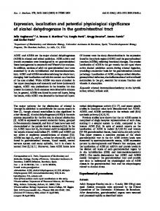
RESULTS Differential mRNA display To identify genes that are expressed differentially during the early stages of fracture repair, initially we compared mRNA populations isolated from PF day 5, 7, and 10 rat femoral calluses, and we were surprised to identify only a very small number of genes in which expression changed as a function of time. Based on these results, we hypothesized that comparing mRNA from a fracture callus, which involves a wider array of cell types, to mRNA from an intact bone would yield a broader spectrum of information about the families of genes involved in the repair process. To this end, we decided to examine mRNA isolated from a PF day 5 callus to that of intact femur (unfractured and included bone marrow and articular and normal growth plate cartilage). Using specific combinations of primers, we identified a large number of cDNA fragments that appeared upregulated in the PF day 5 callus in each of the two different animals examined.(16) One such fragment is shown in Fig. 1, lanes 1 and 2 (arrow). This cDNA was isolated from the gel, purified, reamplified using the corresponding primer pair, and subcloned. The cloned cDNA was then sequenced and found to be 284 nucleotides (nt) long (Fig. 2A). A nucleotide sequence search of GenBank revealed a 99% homol-
CYR61 EXPRESSION DURING FRACTURE REPAIR
1017
Analysis of CYR61 and collagen type II mRNA expression
FIG. 1. Differential display of mRNA populations isolated from PF day 5 calluses and intact femurs. Lanes 1 and 2 represent cDNA fragments amplified from RNA isolated from two different PF day 5 calluses. Lanes 3 and 4 represent cDNA fragments amplified from RNA isolated from two different intact femurs. The candidate clone (CYR61) showing differential mRNA expression is indicated by the arrow.
ogy with the 3⬘-untranslated region of rat CYR61 (gb AB015877; Fig. 2B). Similar to the 3⬘-most region of murine CYR61 mRNA,(17) this rat 284 nt cDNA also contains a number of the ATTTA-like sequence motifs that are thought to be important for rapid RNA degradation(19) (Fig. 2, bold), as well as the classic polyadenylation site AATAAA (Fig. 2, small case letters).
To confirm the results obtained from the differential mRNA display, we used the subcloned cDNA as a probe for Northern analysis. We prepared a nylon membrane with total RNA samples isolated from six different PF day 5 calluses and six different intact femurs (included bone marrow and articular and normal growth plate cartilage). As shown in Fig. 3, the 2.4-kilobase (kb) CYR61 mRNA transcript is expressed at low levels in the intact femurs (lanes 1– 6), whereas it is highly expressed in the PF day 5 calluses (lanes 7–12), confirming the differential display results. In addition to the major 2.4-kb transcript, a second but minor transcript of approximately 4 kb also was detected (Fig. 3, arrowhead). This larger transcript has been previously reported and is thought to occur as a result of differential splicing or from a different polyadenylation signal.(10,20) We further investigated CYR61 mRNA expression during the development of the healing callus, starting with PF day 3 and progressing to PF day 5, 7, 10, 14, and 21. These days include the various cellular events that occur during fracture healing, soft callus formation (cartilage tissue), hard callus formation (osteoid formation under periosteum), endochondral ossification, and osteoclast remodeling of new bone.(21) CYR61 mRNA expression increases as early as PF day 3, gradually rises to higher levels at PF day 5, peaking at PF day 7 and day 10, and declines gradually at PF day 14 and day 21 (Fig. 4A). To investigate whether CYR61 expression corresponds with active chondrogenesis, we examined the expression of collagen type II [Coll(␣1)II] during the same PF time points. Similar to CYR61 expression levels, the highest level of Coll(␣1)II mRNA was detected at PF day 7 and day 10 indicating active cartilage
FIG. 2. Identification of CYR61. (A) The nucleotide sequence of the cDNA clone isolated and subcloned from Fig. 1. The nucleotide sequence of the primers used in differential mRNA display are underlined. The ATTTA-like sequence motifs are shown in bold and the classic polyadenylation site (aataaa) appears in lowercase letters and bold. (B) Results from a BLAST search indicating the homology between the nucleotide sequence of the cDNA clone shown in A to that of rat CYR61 (gb AB015877).
1018
HADJIARGYROU ET AL.
FIG. 3. Northern analysis of rat CYR61 cDNA. Total RNA (20 g) isolated from six different PF 5-day calluses and six different intact femurs was fractionated on a 1% formaldehyde/agarose gel and Northern blotting was carried out as described in the Materials and Methods sectopm using a random labeled rat CYR61 probe (284 bp cDNA fragment from Fig. 2). Lanes 1– 6 represent RNA from six intact femurs, whereas lanes 7–12 represent RNA from the six different PF day 5 calluses. Arrowhead shows the 4 kb CYR61 mRNA transcript. Arrow indicates the major 2.4-kb CYR61 mRNA transcript. The membrane containing the ethidium bromide–stained RNA used in this experiment is shown below indicating the integrity and amounts of RNA loaded per lane.
formation (Fig. 4A). The high level of Coll(␣1)II expression in intact bone reflects the presence of mRNA derived from both articular and normal growth plate cartilage present in our RNA sample. The relative levels of CYR61 mRNA during the development of the fracture callus were determined by integrated optical density measurements (normalized to that of 18S rRNA) and are shown in Fig. 4B. When compared with intact bone, CYR61 mRNA expression in the callus begins with an approximate 2-fold increase by PF day 3, rises to 4.5-fold by PF day 5, and dramatically reaches its highest levels, 10.2- and 9-fold, by PF day 7 and day 10, respectively. It then declines to lower levels, at 2- and 2.5-fold higher than those seen with intact bone by PF day 14 and day 21, respectively (Fig. 4B).
Analysis of CYR61 protein expression To examine the cellular location of CYR61 within the fracture callus, we decided to analyze its protein expression via immunocytochemistry. Areas of cartilage formation within the various fracture calluses were first confirmed by safranin O–fast green staining, as shown in a PF day 10 femur shown at low magnification (Fig. 5A). An adjacent section stained with the anti-CYR61 antibody is shown in Fig. 5B for comparison. In the numerous immunostained sections used from each time point (PF day 5, 7, 10, 14, and 21), we observed strong labeling for CYR61 throughout the entire soft and hard callus (i.e., PF day 10, Fig. 5B) at all time points (see below). Specifically, the CYR61 protein was strongly detected in fibrous tissue adjacent to the fracture site (PF day 5, Figs. 6A and 6G), in the cytoplasm of active proliferating chon-
FIG. 4. Temporal expression of CYR61 mRNA during fracture healing. (A) Total RNA from an intact femur (IF) and different PF day calluses (3, 5, 7, 10, 14, and 21 days) was fractionated on a 1% formaldehyde/agarose gel and Northern analysis was carried out as described in the Materials and Methods sectopn using random labeled probes [rat CYR61 and Coll(␣1)II]. The membrane containing the ethidium bromide–stained RNA used in this experiment is shown below indicating the integrity and amounts of RNA loaded per lane. (B) Plot indicating the relative ratio (in arbitrary units) of CYR61 mRNA to that of 18S rRNA versus intact and different PF day calluses based on IOD measurements of bands shown in panel A (CYR61 blot and photo of RNA-containing membrane). drocytes found in regions of new cartilage formation (PF day 7 and day 10, Figs. 6B and 6C, respectively), in osteoblasts present in regions of newly made trabecular bone during intramembranous ossification (PF day 10, Figs. 6D and 6H, arrows), in the fibrous periosteum (Fig. 6D), and in immature osteocytes of the newly made trabecular bone (PF day 21 and day 10, Figs. 6F and 6H, arrowheads, respectively). Strong labeling also was detected within the osteoid matrix, indicating the presence of the secreted form of CYR61 (Fig. 6C, arrows). An unexpected finding was the spatial decline in CYR61 staining as the proliferating chon-
CYR61 EXPRESSION DURING FRACTURE REPAIR
1019
FIG. 5. Histology of a PF day 10 callus. (A) Section was stained with Safranin O–fast green. (B) Adjacent section immunostained with an anti-CYR61 polyclonal antibody at 1:1000 dilution. Arrows indicate the fracture site. Specific regions are labeled as follows: Bm, bone marrow; Ca, cartilage; Cb, cortical bone; Tb, trabecular bone.
drocytes differentiated into hypertrophic chondrocytes during endochondral ossification (PF day 10, Figs. 6C and 6I), resulting in regions that were completely devoid of labeling (PF day 14, Fig. 6E). Furthermore, expression of CYR61 during fracture repair in distinct tissues (e.g., cortical bone, trabecular bone. and cartilage) can be appreciated more fully when adjacent sections of a PF day 10 callus stained either with safranin O–fast green (Fig. 7A) are compared with those stained with the anti-CYR61 antibody (Fig. 7B). This figure clearly shows the presence of CYR61 in proliferating chondrocytes in cartilage, in active osteoblasts and osteocytes within new trabecular bone, as well as in the interface between bone and cartilage undergoing endochondral ossification and newly made osteoid matrix (Fig. 7B). No labeling was detected in cortical osteocytes (Fig. 7B). An adjacent section stained with secondary antibody alone (served as a negative control) resulted in sections without any labeling (Fig. 7C). Last, expression of CYR61 also was detected on the residual skeletal muscle present on each femur (data not shown), consistent with previous reports.(17) To identify the cellular source of CYR61 in intact bone, we carried out immunohistochemical analysis using the same antibody and conditions utilized in the analysis of the fracture calluses. Strong labeling of CYR61 was detected in osteoblasts of the metaphyseal trabeculae found directly below the epiphyseal growth plate (Figs. 8A and 8B) and in chondrocytes located in the superficial zone of articular cartilage (Figs. 8C and 8D). No CYR61 staining was detected in the chondrocytes present within the hypertrophic (maturing) or proliferating zones of the epiphyseal plate (Figs. 8A and 8B) or in the middle or deep zones of articular cartilage (Figs. 8C and 8D). Osteocytes found within epiphyseal trabeculae also were negative for CYR61 (Fig. 8A). As a control, an adjacent section was stained with secondary antibody alone and resulted in sections that are completely devoid of any labeling (Fig. 8E).
DISCUSSION Fracture healing is a highly specialized type of wound repair that involves a complex array of both cellular and molecular events. These events involve a host of different
cell types (e.g., blood cells, fibroblasts, chondroblasts, epithelial, osteoblasts, osteoclasts, etc.) and molecules (e.g., matrix proteins, growth factors, etc.) that ultimately restore the bone to its original structural integrity. Considering the complexity of the healing process, marked predominantly by cellular adhesion, proliferation, migration, and differentiation, it is only reasonable to expect hundreds, if not thousands, of molecules to be present and to play significant roles. The presence of all the different molecules undoubtedly reflects a robust modulation of gene expression accomplished by the different cell types found within the fracture callus. Taking into account the complex interdependence of the cellular and molecular events in the repair process, it should not be surprising that healing in a significant percentage of fractures will be impaired and a smaller subset will go on to nonunion. Thus, identifying molecules that play critical roles in this cascade of events will help define novel strategies for the acceleration and assurance of the overall repair process. To begin to illustrate the robustness and diversity of gene activity, we directly compared the mRNA populations isolated from PF day 5 calluses to those of intact femurs (included bone marrow and articular and normal growth plate cartilage) using differential display. Presently, we have identified over a dozen differentially expressed genes, some known (osteopontin, bone sialoprotein, integrin-␣6, major histocompatibility complex (MHC) class II antigen, phosphoglucomutase, fibronectin, versican, vimentin, hypoxia-inducible factor 1␣, sec63, SM-20, EET-1, and calpain), and some novel (FxC1 and FxC2).(13–16) One interesting gene identified in mRNA derived from the fracture callus is the growth factor CYR61, which initially was discovered as a growth factor–inducible immediate early gene by differential hybridization screening of a serumstimulated mouse fibroblast cDNA library.(5) CYR61 expression also is activated by basic fibroblast growth factor (bFGF), platelet-derived growth factor (PDGF), and transforming growth factor  (TGF-).(22,23) CYR61 also has been shown to possess a diversity of cellular activities and biological functions. For example, CYR61 binds heparin with high affinity and associates with the ECM, and the cell surface(6) can synergize with bFGF to augment DNA synthesis in both fibroblasts and endothelial cells(7,24) and mediate adhesion and migration (via chemo-
1020
HADJIARGYROU ET AL.
FIG. 6. Immunolocalization of CYR61 during fracture repair. Sections of calluses obtained from (A and G) PF day 5, (B) day 7, (C, D, H, and I) day 10, (E) day 14, and (f) day 21 were immunostained with an anti-CYR61 polyclonal antibody (1:1000 dilution). Specific regions are labeled as follows: Ca, cartilage; Cb, cortical bone; Ft, fibrous tissue; Hc, hypertrophic chondrocytes; P, periosteum; Tb, trabecular bone; Wb, woven bone. Arrows indicate CYR61 labeling in (C) osteoid matrix, (D and H) osteoblasts, (G) fibroblasts, and (I) proliferating chondroblasts. Arrowheads indicate CYR61 presence in (D,F, and H) immature osteocytes and (I) absence in hypertrophic chondrocytes. Scale bars shown (250 m and 100 m) correspond to photos A–F and G–I, respectively.
taxis) of vascular endothelial fibroblasts and lung epithelial cells.(7,25,26) During development, CYR61 expression was linked to chondrogenic differentiation and the development of blood vessels,(8) providing evidence that CYR61 might function as a regulator of chondrogenesis and angiogenesis.(27) Further, CYR61 expression was detected in mesenchymal cells of both mesodermal and neuroectodermal origins as they differentiate into chondrocytes.(8) In isolated mouse limb bud mesenchymal cells, CYR61 is capable of both accelerating their differentiation into chondrocytes and enhancing the extent of differentiation, as evaluated by the up-regulation of Coll(␣1)II expression.(9) More recently, CYR61 was shown to be a ligand of integrin ␣v3 (known to be important for angiogenesis)(26,27) and a potent inducer of neovascularization in rat corneas through an integrin ␣v3– dependent pathway.(27) Based on these studies, it was
suggested that the biological activities of CYR61, that is, stimulation of chemotaxis and mitogenesis of fibroblasts, regulation of ECM protein expression, and regulation of chondrogenesis and angiogenesis, could contribute to its potential function in wound healing.(4) The data presented in this article are consistent with the notion that CYR61 plays an important role in developmentally important processes such as chondrogenesis and angiogenesis, as well as in pathological conditions such as wound healing. Taking into consideration that fracture healing is a specialized type of wound healing involving both chondrogenesis (a hallmark of endochondral ossification) and angiogenesis (neovascularization), our finding that CYR61 is highly expressed in the callus confirms its potential role as an important regulator of these processes.
CYR61 EXPRESSION DURING FRACTURE REPAIR
FIG. 7. Immunolocalization of CYR61 in a PF day 10 callus. Adjacent sections of a PF day 10 callus were either stained with (A) safranin O–fast green, (B) anti-CYR61 polyclonal antibody, and (C) only secondary antibody. Specific regions are labeled as follows: Ca, cartilage; Cb, cortical bone; Tb, trabecular bone. Arrows in B indicate CYR61 labeling in osteoid matrix at the cartilage-bone interface.
1021
More specifically, we detect expression of CYR61 mRNA as early as PF day 3 and as late as PF day 21. The highest levels of expression are detected at PF day 7 and day 10 (10-fold increase, compared with intact femur), which coincides with the most active part of cartilage formation in the callus.(21) This was confirmed by the high expression of Coll(␣1)II mRNA, a marker for chondroblasts,(28) at these time points. CYR61 mRNA expression decreased with chondrocyte hypertrophy (PF day 14 and day 21), suggesting that this gene is expressed at high levels solely by proliferating chondrocytes. This finding was confirmed at the protein level, whereby CYR61 was detected at very high levels in the cytoplasm of proliferating chondrocytes of PF day 7 and day 10 calluses, whereas regions of hypertrophic chondrocytes in older calluses (PF day 14 and day 21) were devoid of staining. In contrast, proliferating chondrocytes were found to be completely devoid of CYR61 staining within the epiphyseal plate of intact bone. On the other hand, consistent with the absence of CYR61 staining seen with hypertrophic chondrocytes within the callus, we also did not detect CYR61 protein in these cells found in the maturing zone of cartilage of the epiphyseal plate of intact bones. Furthermore, CYR61 expression in intact bones was strongly localized in chondrocytes of the superficial zone of articular cartilage, as well as in osteoblasts (see below), thus accounting for the origin of CYR61 mRNA detected in the intact bone samples (Northern analysis). CYR61 also was found to be expressed by other cell types, including fibroblasts, osteoblasts, immature osteocytes, and skeletal muscle cells during fracture repair. At the earliest time points examined (PF day 5), intense staining was detected in the fibrous tissue at, or adjacent to the fracture site, consistent with previous reports showing CYR61 expression by fibroblasts.(5,22) Similarly, expression was detected in active osteoblasts within areas of intramembranous and endochondral ossification during fracture repair, as well as in osteoblasts present in metaphyseal trabeculae of intact bone. These findings support previous studies that show that CYR61 is expressed by human osteoblasts.(10) Ultimately, it will be essential to determine the function of CYR61 in osteoblasts, whether it has paracrine or autocrine effects on adhesion, proliferation, or differentiation of these cells. A surprising finding was the detection of CYR61 in immature osteocytes within newly formed bone. That osteocytes residing in mature cortical bone do not label with the anti-CYR61 antibody indicates that the newly osteoidtrapped osteoblasts that are still expressing CYR61 have not fully differentiated into osteocytes. We speculate that once these immature osteocytes fully differentiate into mature osteocytes, they cease expressing CYR61 (as is the case with hypertrophic chondrocytes). Thus, CYR61 might represent a new marker for osteocyte differentiation. Last, labeling of CYR61 also was detected in the osteoid matrix of the new trabecular bone, indicating the presence of the secreted form of CYR61. We hypothesize that similar to other growth factors (TGF-, FGF, and PDGF), the matrix is a rich source of CYR61 during the development and regeneration of the mammalian skeleton. The data presented in this article provide crucial information on the correlation between CYR61 expression in
1022
HADJIARGYROU ET AL.
FIG. 8. Immunolocalization of CYR61 in intact bone. Sections of intact bone were either immunostained with an (A–D) anti-CYR61 polyclonal antibody (1:1000 dilution) or (E) only secondary antibody. Specific regions are labeled as follows: Ac, articular cartilage; Egp, epiphyseal growth plate; Ep, epiphysis; Me, metaphysis; Mtb, metaphyseal trabecular bone. Arrowheads indicate CYR61 labeling in (B) osteoblasts of metaphyseal trabeculae and (D) chondrocytes of the superficial zone in articular cartilage. Arrows indicate absence of CYR61 labeling in (B) proliferating chondrocytes of the epiphyseal growth plate and (D) chondrocytes of the middle and deep zones of articular cartilage.
intact bone and fracture repair. Previous data indicating that bFGF, PDGF, and TGF- regulate fracture repair (reviewed in Ref. 28), coupled with the fact that these factors modulate CYR61 expression,(22,23) provides strong evidence for a molecular sequence of events. Thus, after a bone fracture, elevated bFGF could induce CYR61 expression (in osteoblasts or chondroblasts) at a low level (as seen at PF day 3 and day 5). As PDGF levles and more specifically TGF- levels dramatically increase during chondrogenesis, these factors probably stimulate a parallel marked increase in CYR61 expression (as seen at PF day 7 and day 10), strongly suggesting that these factors can modulate CYR61 expression during cartilage formation. Taken together, our results and previous data showing the intrinsic link between CYR61 expression and biological processes like wound
repair, chondrogenesis, and angiogenesis (reviewed in Ref. 4) suggest that this gene plays an important role as a key regulator of such events. It is conceivable then that CYR61, administered at the appropriate time, could serve as a potential therapeutic agent in augmenting the normal repair process, as well as impaired fracture healing (e.g., delayed and nonunions).
ACKNOWLEDGMENTS We are grateful to Dr. Leon Sokoloff for crucial suggestions and discussions, Dr. Yoshi Yamada for providing the Coll(␣1)II cDNA, Dr. Dirk Sommerfeldt and Dr. Jizu Zhi for critically reading the manuscript, Dr. Kenneth McLeod
CYR61 EXPRESSION DURING FRACTURE REPAIR
for helpful comments, Tomoko Ando for aiding in the lab, Marilyn Cute for her assistance in the care of animals, Kim Drexler-Rhatigan for help with histochemistry, David Colflesh for assistance with microscopy and image analysis, and Gail Trocchio and Ann Marie Dusatko for secretarial support. This project was funded by Exogen, Inc. (C.T.R.), the Center for Biotechnology, SUNY, Stony Brook (M.H.), and The Whitaker Foundation (C.T.R.).
REFERENCES 1. Sandberg MM, Aro HT, Vuorio EI 1993 Gene expression during bone repair. Clin Orthop Rel Res 289:292–312. 2. Liang P, Pardee AB 1992 Differential display of eukaryotic messenger RNA by means of the polymerase chain reaction. Science 257:967–971. 3. Bork P 1993 The modular architecture of a new family of growth regulators related to connective tissue growth factor. FEBS Lett 327:125–130. 4. Lau LF, Lam SC 1999 The CCN family of angiogenic regulators: The integrin connection. Exp Cell Res 248:44 –57. 5. Lau LF, Nathans D 1985 Identification of a set of genes expressed during the G0/G1 transition of cultured mouse cells. EMBO J 4:3145–3151. 6. Yang GP, Lau LF 1991 Cyr61, product of a growth factorinducible immediate early gene, is associated with the extracellular matrix and the cell surface. Cell Growth Differ 2:351– 357. 7. Kireeva ML, Mo FE, Yang GP, Lau LF 1996 Cyr61, a product of a growth factor-inducible immediate-early gene, promotes cell proliferation, migration, and adhesion. Mol Cell Biol 16:1326 –1334. 8. O’Brien TP, Lau LF 1992 Expression of the growth factorinducible immediate early gene cyr61 correlates with chondrogenesis during mouse embryonic development. Cell Growth Differ 3:645– 654. 9. Wong M, Kireeva ML, Kolesnikova TV, Lau LF 1997 Cyr61, product of a growth factor-inducible immediate-early gene, regulates chondrogenesis in mouse limb bud mesenchymal cells. Dev Biol 192:492–508. 10. Schutze N, Lechner A, Groll C, Siggelkow H, Hufner M, Kohrle J, Jakob F 1998 The human analog of murine cysteine rich protein 61 [correction of 16] is a 1alpha,25-dihydroxyvitamin D3 responsive immediate early gene in human fetal osteoblasts: Regulation by cytokines, growth factors, and serum Endocrinology 139:1761–1770. 11. Hollnagel A, Oehlmann V, Heymer J, Ruther U, Nordheim A 1999 Id genes are direct targets of bone morphogenetic protein induction in embryonic stem cells. J Biol Chem 274:19838 – 19845. 12. Bonnarens F, Einhorn TA 1984 Production of a standard closed fracture in laboratory animal bone. J Orthop Res 2:97– 101. 13. Hadjiargyrou M, McLeod KJ, Halsey M, Rubin CT 1997 The temporal expression of osteopontin mRNA in the fracture callus is altered by low intensity ultrasound. J Bone Miner Res 12:S290. 14. Hadjiargyrou M, McLeod KJ, Rubin CT 1998 Differential gene expression during the early stages of fracture healing. Bone 23:S235. 15. Hadjiargyrou M, Halsey M, Ahrens W, Rightmire E, McLeod KJ, Rubin CT 1998 Cloning of a novel cDNA expressed
1023
16.
17.
18.
19.
20.
21.
22.
23.
24.
25.
26.
27.
28.
29.
during the early stages fracture healing. Biochem Biophys Res Commun 249:879 – 884. Hadjiargyrou M, Ahrens W, Ando T, Rubin CT 1999 Upregulation of a novel and several known genes during the early phases of fracture healing. J Bone Miner Res 14:S438. O’Brien TP, Yang GP, Sanders L, Lau LF 1990 Expression of cyr61, a growth factor-inducible immediate-early gene. Mol Cell Biol 10:3569 –3577. Chomczynski P, Sacchi N 1987 Single-step method of RNA isolation by acid quanidinium thiocynate-phenol-chloroform extraction Anal Biochem 162:156 –159. Shaw G, Kamen R 1986 A conserved AU sequence from the 3⬘ untranslated region of GM-CSF mRNA mediates selective mRNA degradation. Cell 46:659 – 667. Jay P, Berge-Lefranc JL, Marsollier C, Mejean C, Taviaux S, Berta P 1997 The human growth factor-inducible immediate early gene, CYR61, maps to chromosome 1p. Oncogene 14: 1753–1757. Jingushi S, Joyce ME, Bolander ME 1992 Genetic expression of extracellular matrix proteins correlates with histologic changes during fracture repair. J Bone Miner Res 7:1045– 1055. Lau LF, Nathans D 1987 Expression of a set of growth-related immediate early genes in BALB/c 3T3 cells: Coordinate regulation with c-fos or c-myc. Proc Natl Acad Sci U S A 84:1182–1186. Brunner A, Chinn J, Neubauer M, Purchio AF 1991 Identification of a gene family regulated by transforming growth factor-beta. DNA Cell Biol 10:293–300. Kolesnikova TV, Lau LF 1998 Human CYR61-mediated enhancement of bFGF-induced DNA synthesis in human umbilical vein endothelial cells. Oncogene 16:747–754. Kireeva ML, Latinkic BV, Kolesnikova TV, Chen CC, Yang GP, Abler AS, Lau LF 1997 Cyr61 and Fisp12 are both ECM-associated signaling molecules: Activities, metabolism, and localization during development. Exp Cell Res 233:63–77. Kireeva ML, Lam SC, Lau LF 1998 Adhesion of human umbilical vein endothelial cells to the immediate-early gene product Cyr61 is mediated through integrin alphavbeta3. J Biol Chem 273:3090 –3096. Babic AM, Kireeva ML, Kolesnikova TV, Lau LF 1998 CYR61, a product of a growth factor-inducible immediate early gene, promotes angiogenesis and tumor growth. Proc Natl Acad Sci U S A 95:6355– 6360. Dessau W, von der Mark H, von der Mark K, Fischer S 1980 Changes in the patterns of collagens and fibronectin during limb-bud chondrogenesis. J Embryol Exp Morphol 57:51– 60. Bolander ME 1992 Regulation of fracture repair by growth factors. Proc Soc Exp Biol Med 200:165–170.
Address reprint requests to: Michael Hadjiargyrou Program in Biomedical Engineering and Department of Orthopedics State University of New York at Stony Brook Stony Brook, NY 11794-8181, U.S.A.
Received in original form August 4, 1999; in revised form November 10, 1999; accepted November 29, 1999.