Accepted Article
Accepted Date : 16-Aug-2016 Article type : Full-length Original Research
Cathodal transcranial direct current stimulation for treatment of drug-resistant temporal lobe epilepsy: a pilot randomised controlled trial
Authors: Maryam Zoghi 1, Terence J. O’Brien1, Patrick Kwan1, Mark J. Cook2, Mary Galea3,
Shapour Jaberzadeh4 1. Department of Medicine, The Royal Melbourne Hospital, The University of Melbourne, 2. Department of Medicine, St Vincent’s Hospital, The University of Melbourne 3. Department of Medicine, The Royal Melbourne Hospital (Royal Park Campus), The University of Melbourne, 4. School of Primary Health Care, Faculty of Medicine, Nursing and Health Sciences, Monash University
Running title: C-tDCS in patients with focal epilepsy Key Words: Cathodal transcranial direct current stimulation, Drug resistant, Temporal lobe epilepsy Corresponding author: Dr. Maryam Zoghi, PhD Research Fellow Department of Medicine (Royal Melbourne Hospital) The University of Melbourne 4th Floor, Clinical Sciences Building 300 Grattan Street, Parkville, VIC 3052, AUSTRALIA e-mail:
[email protected] Tel: +61 3 83443292
Abstract Objective: To investigate the effect of cathodal-transcranial direct current stimulation (ctDCS) on seizure frequency in patients with drug-resistant temporal lobe epilepsy (TLE). Method: Twenty-nine patients with drug-resistant TLE participated in this study. Twenty participants (experimental group) were randomised to receive within-session repeated c-tDCS This article has been accepted for publication and undergone full peer review but has not been through the copyediting, typesetting, pagination and proofreading process, which may lead to differences between this version and the Version of Record. Please cite this article as doi: 10.1002/epi4.12020 This article is protected by copyright. All rights reserved.
intervention over the affected temporal lobe and 9 (sham group) received sham tDCS. Paired-
Accepted Article
pulse transcranial magnetic stimulation was used to assess short interval intracortical inhibition (SICI) in primary motor cortex ipsilateral to the affected temporal lobe. SICI was measured from motor evoked potentials recorded from the contralateral first dorsal interosseous muscle. Adverse effects were monitored during and after each intervention in both groups. A seizure diary was given to each participant to complete for 4 weeks following the tDCS intervention . The mean response ratio was calculated from their seizure rates before and after the tDCS intervention. Results: The experimental group showed a significant increase in SICI compared to the sham group (F = 10.3, p = 0.005). None of the participants reported side effects of moderate or severe degree. The mean response ratio in seizure frequency was -42.14% (SD = 35.93) for the experimental group and -16.98% (SD = 52.41) for the sham group. Significance: Results from this pilot study suggest that tDCS may be a safe and efficacious non-pharmacological intervention for patients with drug-resistant TLE. Further evaluation in larger double blind randomised controlled trials is warranted.
Introduction Epilepsy impacts 50 million people (1% of the population) worldwide 1. Management for
patients with epilepsy includes anti-epileptic drugs (AED) and some patients with drugresistant seizures, surgery. Temporal lobe epilepsy (TLE) is often resistant to AEDs 2, and more than 40% of patients with epilepsy suffer adverse reactions to AEDs. Removing the epileptogenic regions surgically is not always feasible for patients, and the outcome is not ideal in 30% to 50% of cases 3. Consequently, alternative methods of seizure control warrant more investigation.
This article is protected by copyright. All rights reserved.
The excitability of the GABAergic intracortical inhibitory circuits in primary motor cortex
Accepted Article
(M1) can be assessed non-invasively in humans by paired-pulse transcranial magnetic stimulation (TMS). In this technique, two stimuli are delivered 1-5 ms apart through the same coil. The first stimulus is subthreshold for a motor response, however it activates intracortical inhibitory (ICI) circuits and reduces the size of the motor evoked potentials (MEPs) elicited by the second stimulus which is supra-threshold for a motor response 4. It has been shown that ICI measured using this method reflects the cortical activity of GABA-ergic interneurons in M1 area 5. This inhibition is termed short interval intracortical inhibition or SICI.
ICI circuits have been extensively assessed with a paired-pulse paradigm in patients with epilepsy 6-8. Several studies on drug-naive patients with focal epilepsy showed a decrease in SICI in the ipsilateral hemisphere 9-15. Badawy et al. showed increased M1 excitability and
decreased SICI in 35 patients with focal epilepsy 24 hours before and after a seizure.
Transcranial direct current stimulation (tDCS) is a well-established cortical stimulation method that can be used non-invasively to modulate neuronal excitability in humans 16. In this technique a low intensity current (1-2 mA) is used which can affect the membrane potentials in two ways. Cathodal tDCS hyperpolarizes the resting membrane potentials while anodal tDCS acts towards depolarization 16. Modification of seizure network excitability by
tDCS is a potentially valuable non-invasive alternative for reducing the excitability of this abnormal network in patients with epilepsy and therefore reducing the seizure rates in this population.
This article is protected by copyright. All rights reserved.
The aim of this study was to examine the effects of this non-invasive therapeutic approach on
Accepted Article
seizure frequency in this group of patients. We hypothesized that compared to sham tDCS, application of c-tDCS over the temporal lobe in patients with drug-resistant TLE, decreases seizure frequency and increases intracortical inhibition in ipsilateral M1 area.
Method Participants: We conducted a small pilot study in patients admitted to the Video-EEG Monitoring (VEM) Unit at the Royal Melbourne Hospital or as outpatients at St Vincent’s Hospital. Twenty-nine participants (male: 11, female: 18) with drug resistant TLE and mean age of 38 ± 13 participated in this study. All 24 participants from the Royal Melbourne Hospital were admitted as potential surgical candidates to the VEM Unit. The etiology of the TLE varied between participants and included tumour, meningitis, infantile febrile, cortical dysplasia, and unknown reason. Inclusion criteria were: a) 18 years of age and over; b) diagnosed with drug-resistant TLE, as defined by The International League Against Epilepsy [failure to become (and stay) seizure-free with adequate trials of two seizure medications]; c) able to understand, speak and write in English. Exclusion criteria were: a) skin conditions (e.g. eczema, lesions) on scalp; b) metal inside the head (outside the mouth) such as shrapnel, surgical clips; c) any implanted devices such as cardiac pacemaker, cochlear implant, medical pump, or intracardiac line; d) frequent or severe headaches; e) previous head injury and any other brain related disease and f) pregnancy and breast feeding. All procedures used in this study conformed with the Declaration of Helsinki, and the protocol was approved by the Human Research Ethics Committees at The University of Melbourne and Melbourne Health. Written informed consent was obtained from each participant. Intervention: One session of c-tDCS or sham tDCS (9-20-9 protocol) were applied in the last day of participants’ admission in the VEM unit at the Royal Melbourne Hospital or in a
This article is protected by copyright. All rights reserved.
quiet room at St Vincent’s Hospital as an outpatient. Participants were randomly allocated to
Accepted Article
experimental or sham group. They were all blinded to the nature of the intervention (experimental vs. sham). This protocol involved a total of 18 minutes c-tDCS or sham tDCS, with 20 minutes rest after the first 9 minutes (Figure 1). A DC-Stimulator (Chattanooga Intelect® Advanced Combo) was used to deliver a 1mA continuous galvanic current to the brain via two surface electrodes with surrounding saline-soaked sponges (0.9% NaCl). The active surface electrode (cathode, 3×4 cm) was placed over the affected temporal lobe, and the return electrode 17 (anode, 5×7 cm) was placed over the contralateral supraorbital area.
Assessment: Participants were asked to keep a record of their seizures in a daily seizure diary for 4 weeks after the intervention. The mean response ratio was calculated by the following formula: [(T - B)/(T + B)] x 100. In this formula, B is the patient’s baseline seizure frequency over the 4 weeks prior to treatment, and T is the patient’s seizure frequency in the 4 weeks after the treatment. In this study, the seizure rate prior the intervention for each participant was recorded based on participant’s report. Negative response ratio values indicate reduced seizure rate from baseline. The response ratio allows for a normalised percentage change in seizure rate from baseline, with values within the range of -100 to +100. Zero value in this range indicates no change 18.
Paired-pulse TMS (Magstim Bistim2) was used to assess SICI before and after each intervention. Participants were seated with head and neck supported by a headrest. MEPs were recorded from the first dorsal interosseous (FDI) muscle before and after each intervention. Resting threshold (RT) for recording MEPs was defined as the lowest intensity to record three of five successive MEPs of above 50 μV (peak-to-peak amplitude) from FDI. The TMS intensity for the supra-threshold stimuli was adjusted to produce MEPs in FDI at
This article is protected by copyright. All rights reserved.
rest of about 1mV amplitude (test intensity). The TMS intensity for the sub-threshold stimuli
Accepted Article
was adjusted to 0.8 x RT with 3ms inter stimulus interval (conditioning intensity). Single or paired-pulse TMS was delivered in blocks of 20 stimuli (10s interval between stimuli) at rest (40 trials at each session).
Adverse events related to the application of c-tDCS (e.g. itching, tingling, burning sensation; headache, neck pain, etc.) in this study were assessed using the Adverse Effects Questionnaire 19 during and after each session.
Results All participants tolerated the intervention very well. None of the participants reported side effects of moderate or severe degree during or after the intervention. A few participants reported itching sensations (2/10) for few minutes under the anode electrode. Twenty-three participants returned their diaries (Table 1). The mean response ratio was calculated from their seizure rates before and after the tDCS intervention (Table 2). The mean response ratio was -42.14% (SD = 35.93) for the experimental group and -16.98% (SD = 52.41) for the sham group. SICI was measured with paired-pulse TMS (Magstim Bistim2) before and after the tDCS intervention in 17 participants (Table 1).
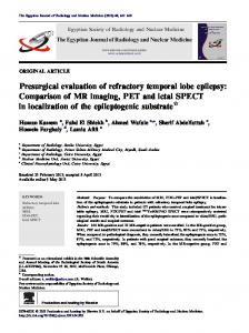
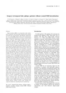
One-way ANOVA showed that SICI was increased significantly in the experimental group compared to the sham group (F = 10.3, p = 0.005) (Figure 2). The individual SICI level before and after c-tDCS can be seen in Figure 3.
This article is protected by copyright. All rights reserved.
Discussion
Accepted Article
This pilot study showed that c-tDCS using the 9-20-9 protocol could not only increase SICI, but also decrease seizure rates in patients with drug resistant TLE. The results of this pilot study cannot be compared to previous studies since no SICI data are available post c-tDCS and this new protocol (9-20-9) has not been applied previously to patients with drug resistant epilepsy.
Regardless of the cause of epilepsy, it has been argued that seizure networks are highly excitable as a result of unbalanced neuronal excitatory/inhibitory networks within the affected region 20. Therefore, the main aim in seizure control is manipulating these networks
in such a way that the seizure networks remain in a “sub-threshold” condition and not triggered. The excitatory/inhibitory networks can be manipulated by decreasing the excitability of the excitatory networks or increasing the function of inhibitory networks, or both at the same time 21. Drugs that block voltage-dependent ion-channels or excitatory Nmethyl-D-aspartate (NMDA)-receptors, or enhance GABA-ergic activity have been shown to have a profound ability to suppress the development of seizures 21. Anti-epileptic effects of ctDCS can be expected due to the fact that c-tDCS decreases cortical excitability by hyperpolarizing the membrane potentials and subsequently altering synaptic efficacy 22, 23. Lang et al. (2011) recorded corticospinal volleys evoked by single-pulse TMS of M1 before and after a 5-min period of c-tDCS in eight conscious patients who had electrodes implanted in the cervical epidural space 24. They showed that c-tDCS suppressed the excitability of cortical circuits generating later “indirect waves” (I waves) in the corticospinal system. They suggested that c-tDCS facilitates inhibitory connections or it produces dis-facilitation (e.g., hyperpolarization) of excitatory connections leading to a selective suppression of later I waves 24. In a recent study Dhamne et al. (2015) reported that 1 mA c-tDCS for 20 minutes
This article is protected by copyright. All rights reserved.
could reduce seizures, augment lorazepam efficacy and enhance GABAergic cortical
Accepted Article
inhibition in kindled rats 25.
The effects of tDCS are strongly dependent on electrode montage and parameters of stimulation. It has been shown that the induced excitability changes and the length of the lasting effect depend on two parameters of direct currents: intensity and duration of application 26, 27. The effects of modifications of these parameters have been tested in a few clinical applications 28-30.
Fregni et al. 31 studied the effects of c-tDCS (one session, 20 minutes, 1mA) in patients with drug resistant epilepsy and malformations of cortical development as indicated by seizure frequency and epileptiform EEG discharges. They applied the c-tDCS over the epileptogenic focus, and showed that c-tDCS could decrease cortical excitability in the epileptogenic focus of these patients. They also reported a trend (p = 0.06) for decrease in seizure frequency after active c-tDCS compared with sham treatment (mean seizure frequency decrease of -44.0% for the active treatment group and -11.1% for the sham treatment group). The seizure reduction rate was similar to that of the present study (-42.14% in experimental group and 16.98% in sham group).
Auvichayapat et al. 32 assessed the antiepileptic efficacy of c-tDCS in 36 children (6-15 years old) with drug resistant epilepsy. Twenty-seven children in the active group received a single session c-tDCS (1mA) for 20 min, while the remainder were in a sham group. In the active group, c-tDCS suppressed epileptiform discharges for 48 h, with a small (clinically negligible but statistically significant) decrease in seizure frequency. Faria et al. 33 applied c-tDCS for
30 min with the same intensity in 2 children with drug-resistant continuous spike-wave
This article is protected by copyright. All rights reserved.
discharges during slow sleep. They reported similar results in terms of safety and the efficacy
Accepted Article
of c-tDCS in reduction of the inter-ictal epileptiform EEG discharges. Varga et al. 34 however, applied c-tDCS before sleep on 5 patients with focal, drug-resistant continuous spikes and waves during slow sleep with no effects on the spikes. The c-tDCS parameters used were similar to those used in the Faria et al. study except that the duration of treatment was 10 minutes shorter 34.
Yook et al. 35 reported the results of c-tDCS application in an 11-year-old female with focal cortical dysplasia. This patient received c-tDCS over the site where the abnormal wave was observed (through EEG recordings) for 20 minutes with 2mA, five days per week for two weeks. During the two month monitoring period after this treatment they reported that not only was the duration of the seizures decreased but also the frequency of her seizures reduced from 8 seizures per month to 3 seizures per month. This protocol was reapplied for another two weeks with only one seizure reported in the following two months.
Although all the tDCS protocols that have been used in the above-mentioned clinical studies are well within safety limits 36, it is a general goal to keep current exposure as low as
possible. High-intensity stimulation can not only be painful 37, it also can affect different neuronal populations compared with low intensity stimulation. By increasing the intensity, the current may reach deeper sites that might not be the intended target.
One way to prolong the after-effects of tDCS might be the repetition of tDCS sessions. Monte-Silva et al. (2010) showed that application of c-tDCS for 18 minutes with a 20 minute break after the first 9 minutes will increase the inhibitory effects of this technique for up to 2 hours (9-20-9 protocol) 38.
This article is protected by copyright. All rights reserved.
Accepted Article
This protocol was used in this pilot study in one session on patients with drug resistance TLE.
We had an opportunity to apply c-tDCS with (9-20-9) protocol on two consecutive days in a
48 year old female patient with drug-resistant TLE while she was admitted to VEM unit at RMH 39. She had a right fronto-temporal pleomorphic astrocytoma for 10 years with 5-10 seizures per day. After receiving the c-tDCS on the last two days of her admission, she reported a seizure reduction of 0-3 seizures per day over a 4 month period. These observations suggest that c-tDCS might have accumulative inhibitory effects on the abnormal epileptic networks.
A recent review of the effect of c-tDCS on epilepsy assessed 9 original studies (3 animal, 6 human) up to 2014 40. In these studies, 109 animals and 65 humans received c-tDCS with
different parameters (including electrode montage, size of the electrodes, duration and intensity of the applied c-tDCS). Because of the different methods of c-tDCS application, and the lack of long-term follow-up, only preliminary evidence of the safety and efficacy of this technique in controlling seizures in animals and patients with epilepsy could be concluded 40.
Limitations We acknowledge the limitations of this pilot study (open label, unequal groups). However the results are encouraging. The results of this pilot study should be interpreted with caution. Even though all participants were diagnosed with drug-resistant TLE, they were not homogenous in regards to the etiology, onset of their epilepsy or the seizure rates. The seizure rate changes were measured based on baseline seizure rates that were reported by patients. TMS assessment could not be done on all participants due to unforseen reasons (e.g.
This article is protected by copyright. All rights reserved.
time constraint due to being scheduled for other assessments before they were discharged,
Accepted Article
unavailability the device etc).
Future directions This novel technique (tDCS) has, to date, shown no or minimal side effects and it can be applied by an inexpensive battery-operated device. This new technique should be assessed in a large randomised double blind controlled trial. If successful, it has the potential to be readily translated into clinical practice as a safe and well-tolerated non-medical treatment option for epilepsy management. In this way, patients may be able to control their seizures without suffering from the side effects of taking additional AEDs.
Disclosure of Conflicts of Interest None of the authors has any conflict of interest to disclose. Key Point Box •
Within session repeated (9-20-9 protocol) c-tDCS has shown no or minimal side effects in patients with drug-resistant temporal lobe epilepsy.
•
Cortical excitability was reduced, as measured by SICI, after one application of ctDCS using the 9-20-9 protocol in patients with drug-resistant temporal lobe epilepsy.
•
Seizure rates reduced by 42% after one application of c-tDCS using the 9-20-9 protocol in patients with drug-resistant temporal lobe epilepsy.
We confirm that we have read the Journal’s position on issues involved in ethical publication and affirm that this report is consistent with those guidelines.
This article is protected by copyright. All rights reserved.
References
Accepted Article
1. 2.
3. 4.
5.
6.
7.
8. 9.
10. 11.
12. 13. 14. 15. 16. 17.
18.
19.
20. 21.
Thurman DJ, Beghi E, Begley CE, et al. Standards for epidemiologic studies and surveillance of epilepsy, Epilepsia 2011;52 Suppl 7:2-26. Tellez-Zenteno JF and Hernandez-Ronquillo L. A review of the epidemiology of temporal lobe epilepsy, Epilepsy Res Treat 2012;2012:5 pages. Spencer S and Huh L. Outcomes of epilepsy surgery in adults and children, Lancet Neurol 2008;7:525-37. Kujirai T, Kurokawa K, Kujirai K, et al. Cortico-cortical inhibition in focal motor cortical lesion, Electroencephalography and Clinical Neurophysiology/Electromyography and Motor Control 1995;97:S109. Ziemann U, Reis J, Schwenkreis P, et al. TMS and drugs revisited 2014, Clin Neurophysiol 2015;126:1847-68. Macdonell RAL and Badawy RAB. Transcranial Magnetic Stimulation in Epilepsy, Atlas of Epilepsies 2010;1-3:823-828. Schrader LM, Stern JM, Koski L, et al. Seizure incidence during single- and pairedpulse transcranial magnetic stimulation (TMS) in individuals with epilepsy, Clin Neurophysiol 2004;115:2728-37. Hamer HM, Reis J, Mueller HH, et al. Motor cortex excitability in focal epilepsies not including the primary motor area: a TMS study, Brain 2005;128:811-8. Badawy RA and Jackson GD. Cortical excitability in migraine and epilepsy: a common feature?, J Clin Neurophysiol 2012;29:244-9. Badawy RA, Jackson GD, Berkovic SF, et al. Inter-session repeatability of cortical excitability measurements in patients with epilepsy, Epilepsy Res 2012;98:182-6. Badawy RA, Vogrin SJ, Lai A, et al. Cortical excitability changes correlate with fluctuations in glucose levels in patients with epilepsy, Epilepsy Behav 2013;27:45560. Badawy RA, Macdonell RA, Berkovic SF, et al. Predicting seizure control: cortical excitability and antiepileptic medication, Ann Neurol 2010;67:64-73. Badawy RA, Curatolo JM, Newton M, et al. Changes in cortical excitability differentiate generalized and focal epilepsy, Ann Neurol 2007;61:324-31. Badawy R, Macdonell R, Jackson G, et al. The peri-ictal state: cortical excitability changes within 24 h of a seizure, Brain 2009;132:1013-21. Varrasi C, Civardi C, Boccagni C, et al. Cortical excitability in drug-naive patients with partial epilepsy: a cross-sectional study, Neurology 2004;63:2051-5. Nitsche MA, Cohen LG, Wassermann EM, et al. Transcranial direct current stimulation: State of the art 2008, Brain Stimul 2008;1:206-23. Bikson M, Datta A, Rahman A, et al. Electrode montages for tDCS and weak transcranial electrical stimulation: role of "return" electrode's position and size, Clin. Neurophysiol. 2010;121:1976-8. Gil-Nagel A, Zaccara G, Baldinetti F, et al. Add-on treatment with pregabalin for partial seizures with or without generalisation: pooled data analysis of four randomised placebo-controlled trials, Seizure 2009;18:184-92. Brunoni AR, Amadera J, Berbel B, et al. A systematic review on reporting and assessment of adverse effects associated with transcranial direct current stimulation, Int J Neuropsychopharmacol 2011;14:1133-45. McCormick DA and Contreras D. On the cellular and network bases of epileptic seizures, Annu Rev Physiol 2001;63:815-46. Levy RH, Mattson RH, Meldrum BS, et al., Antiepileptic Drugs. 5th ed ed. 2002, Philadelphia, PA: Lippincott Williams & Wilkins.
This article is protected by copyright. All rights reserved.
22.
Accepted Article
23.
24. 25. 26.
27. 28. 29.
30. 31. 32. 33.
34.
35.
36. 37.
38.
39.
40.
Liebetanz D, Nitsche MA, Tergau F, et al. Pharmacological approach to the mechanisms of transcranial DC-stimulation-induced aftereffects of human motor cortex excitability, Brain 2002;125:2238 -2247. Nitsche MA, Fricke K, Henschke U, et al. Pharmacological modulation of cortical excitability shifts induced by transcranial direct current stimulation in humans, J Physiol 2003;553:293-301. Lang N, Nitsche MA, Dileone M, et al. Transcranial direct current stimulation effects on I-wave activity in humans, J Neurophysiol 2011;105:2802-10. Dhamne SC, Ekstein D, Zhuo Z, et al. Acute seizure suppression by transcranial direct current stimulation in rats, Ann Clin Transl Neurol 2015;2:843-56. George MS and Aston-Jones G. Noninvasive techniques for probing neurocircuitry and treating illness: vagus nerve stimulation (VNS), transcranial magnetic stimulation (TMS) and transcranial direct current stimulation (tDCS), Neuropsychopharmacology 2010;35:301-16. Nitsche MA and Paulus W. Excitability changes induced in the human motor cortex by weak transcranial direct current stimulation, J Physiol 2000;527 Pt 3:633-9. Ferrucci R, Bortolomasi M, Vergari M, et al. Transcranial direct current stimulation in severe, drug-resistant major depression, J Affect Disord 2009;118:215-9. Fregni F, Gimenes R, Valle AC, et al. A randomized, sham-controlled, proof of principle study of transcranial direct current stimulation for the treatment of pain in fibromyalgia, Arthritis Rheum 2006;54:3988-98. Ohn SH, Park CI, Yoo WK, et al. Time-dependent effect of transcranial direct current stimulation on the enhancement of working memory, Neuroreport 2008;19:43-7. Fregni F, Thome-Souza S, Nitsche MA, et al. A controlled clinical trial of cathodal DC polarization in patients with refractory epilepsy, Epilepsia 2006;47:335-42. Auvichayapat N, Rotenberg A, Gersner R, et al. Transcranial direct current stimulation for treatment of refractory childhood focal epilepsy, Brain Stimul 2013; Faria P, Fregni F, Sebastiao F, et al. Feasibility of focal transcranial DC polarization with simultaneous EEG recording: preliminary assessment in healthy subjects and human epilepsy, Epilepsy Behav 2012;25:417-25. Varga ET, Terney D, Atkins MD, et al. Transcranial direct current stimulation in refractory continuous spikes and waves during slow sleep: a controlled study, Epilepsy Res 2011;97:142-5. Yook SW, Park SH, Seo JH, et al. Suppression of seizure by cathodal transcranial direct current stimulation in an epileptic patient - a case report, Ann Rehabil Med 2011;35:579-82. Liebetanz D, Koch R, Mayenfels S, et al. Safety limits of cathodal transcranial direct current stimulation in rats, Clin Neurophysiol 2009;120:1161-7. Furubayashi T, Terao Y, Arai N, et al. Short and long duration transcranial direct current stimulation (tDCS) over the human hand motor area, Exp Brain Res 2008;185:279-86. Monte-Silva K, Kuo MF, Liebetanz D, et al. Shaping the optimal repetition interval for cathodal transcranial direct current stimulation (tDCS), J Neurophysiol 2010;103:1735-40. Zoghi M, O'Brien TJ, Kwan P, et al. The Effects of Cathodal Transcranial Direct Current Stimulation in a Patient with Drug-Resistant Temporal Lobe Epilepsy (Case Study), Brain Stimul 2016; San-Juan D, Morales-Quezada L, Orozco Garduno AJ, et al. Transcranial Direct Current Stimulation in Epilepsy, Brain Stimul 2015;8:455-64.
This article is protected by copyright. All rights reserved.
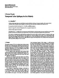
Figure 1: Experimental set-up
Accepted Article
All participants received one session of c-tDCS or sham tDCS paradigm (9-20-9 protocol). The active surface electrode (cathode) was placed over the temporal lobe in the affected hemisphere. The return (anode) electrode was placed over the supraorbital area contralateral to the stimulated hemisphere. SICI was assessed before and after tDCS intervention. Seizure rates were recorded for 4 weeks after tDCS intervention.
Figure 2: Mean SICI changes post c-tDCS vs sham tDCS The MEPs were recorded from the first dorsal interosseous (FDI) muscle. The area of the conditioned and unconditioned MEPs were measured from the averaged rectified MEPs obtained in each trial. The size of the conditioned MEPs was expressed as a percentage of the unconditioned test MEPs in order to assess the effectiveness of SICI. SICI was increased significantly in the experimental group compared to the sham group (F = 10.3, p = 0.005).
Figure 3: Individual SICI changes post c-tDCS or sham tDCS SICI changes post c-tDCS (panel A) or sham tDCS (panel B) for each participant. White circles show SICI level at baseline. Full triangles show SICI level post tDCS. In panel A, most participants show a trend of increased SICI post c-tDCS. In panel B, no trend of increased SICI is seen in any of participant.
This article is protected by copyright. All rights reserved.
Table 1: Distribution of collected data in participants
Accepted Article
Group
Number of participants
TMD data collected
Seizure diary returned
No TMS data collected/no seizure diary returned
Experimental
20
12
16
3
Sham
9
5
7
2
Total
29
17
23
5
Table 2: Seizure rates in experimental group vs. sham group before and after tDCS intervention and mean response ratio in both groups. Group
Seizure diary returned
Seizure rate before tDCS ± SD
Seizure rate after tDCS ± SD
Experimental
16
53 ± 78.95
17.18 ± 26.03
-42.14 ± 35.93
Sham
7
20.28 ± 33.69
13 ± 16.2
-16.98 ± 52.41
This article is protected by copyright. All rights reserved.
Mean response ratio [(T - B)/(T + B)] X 100
Accepted Article This article is protected by copyrigght. All rights reserved.