J. Appl. Glycosci., 60, 29 36 (2013) doi:10.5458/jag.jag.JAG-2012_013 ©2013 The Japanese Society of Applied Glycoscience
Special Issue: Starch Metabolism, Structure and Properties Review
The Characteristic Polyhedral, Sharp-edged Shape of Compound-type Starch Granules in Rice Endosperm is Achieved via the Septum-like Structure of the Amyloplast (Received October 19, 2012; Accepted November 5, 2012) (J-STAGE Advance Published Date: January 25, 2013)
Yasushi Kawagoe1,* 1
Division of Plant Sciences, National Institute of Agrobiological Sciences (2-1-2 Kannondai, Tsukuba 305-8602, Japan)
Abstract: The starch granules in rice endosperm are of the compound-type, which comprises up to several dozen granules per amyloplast. In contrast, the granules in the endosperm of maize and wheat are of the simple-type, which comprises only a single granule. The molecular mechanisms that control granuletype have long been an open question. Laser scanning confocal microscopy (LSCM) is advantageous over conventional microscopy when combined with the expression of fluorescent proteins (FPs) in the tissue under investigation. We have analyzed numerous transgenic rice expressing FPs fused to amyloplasttargeted proteins in endosperm. The fluorescent signals of these fusion proteins provided high-quality images of the internal structures of amyloplasts. Interestingly, changing expression levels of plastiddivision proteins or isoamylase3 alter both amyloplast division and granule synthesis. On the basis of these results, a model for the synthesis of compound-type granules in rice has been proposed. In this model, the septum-like structure (SLS) is synthesized beneath the outer limit envelope prior to de novo granule synthesis, where it functions like a partition-wall in the stroma, casting the growing granules into their characteristic polyhedral, sharp-edged shape. LSCM analyses using FPs fused to starch synthase I (SSIGFP) and the transit peptide of granule-bound starch synthase I (tpGBSSICherry) revealed that SSI-GFP and tpGBSSI Cherry are respectively targeted to the granule surface in the stroma and the SLS sandwiched between the inner envelope membranes. This review examines the technical issues of LSCM and transgenic analysis, summarizes models for compound-type granule synthesis, and presents future questions. Key words: rice starch granule, amyloplast division, laser scanning confocal microscopy, compound-type granule, fluorescent protein, septum-like structure Transient and storage starch are synthesized in the chloroplast and amyloplast, respectively. Transient starch is synthesized during the day and degraded the following night in the chloroplast of source tissues, such as leaf and stem.1,2) Granule numbers per chloroplast in Arabidopsis leaves are correlated with chloroplast volume.3) Storage starch is synthesized and stored in the amyloplast of sink tissues, such as the endosperm and root. It is chemically stable, which is apparent by the fact that storage starch in the seed can be metabolically inert for decades under appropriate conditions, yet can be efficiently degraded to provide carbon and energy for seedling establishment.4) The shapes of storage granules vary depending on the tissue and species from which they are derived.5) Unlike the water-soluble glycogen of bacteria and animals, starch storage granules are insoluble in water.6,7) The granules are *
Corresponding author (Tel. +81-29-838-8444, Fax. +81-29-8387417, E-mail:
[email protected]). Abbreviations: LSCM, laser scanning confocal microscopy; FP, fluorescent protein; SLS, septum-like structure; GBSSI, granule bound starch synthase I; SSI, starch synthase I; OEM, outer envelope membrane; IEM, inner envelope membrane; IMS, intermembrane space.
easily visualized with conventional light microscopy by staining thin sections of storage tissues with iodine solution.8) Scanning electron microscopy (SEM) has also been used to analyze purified granules and those exposed on the surface of cross-sections of storage tissues.9,10) These conventional microscopic analyses have revealed that storage granules can be divided into two types: the simple-type and the compound-type. A typical simple-type granule is round, oval, or lenticular, and includes those of the potato tuber and the endosperm of maize, wheat and barley.5) In contrast, the compound-type granule consists of multiple granules, ranging from two to several dozen per amyloplast, and, interestingly, often creates a round or oval shape when seen as a unit. As such, each granule in the compound-type granules is polyhedral and sharp-edged. Rice and oat are examples of species that synthesize compound-type granules. The two types of granule can also be described from the standpoint of the amyloplast; that is, an amyloplast contains either a single granule (the simple-type) or multiple granules (the compound-type). Given that cereals including maize, wheat and barley, as well as potato, make simple-type granules in their storage
30
J. Appl. Glycosci., Vol. 60, No. 1 (2013)
tissues, it is surprising that the compound-type granule turns out to be the more common granule-type in the Poaceae family.11,12) Although phylogenetic relationships in the Poaceae family are not accurate predictors of the type of granule synthesized in the endosperm, it appears that the granule-type is primarily determined by genetic factors, and not by environmental conditions. Yet, the molecular mechanisms that control granule-shape remain an open question. Laser scanning confocal microscopy (LSCM) offers many advantages over conventional light microscopy and SEM. LSCM is particularly advantageous when it is combined with the use of fluorescent proteins (FPs) in the tissue under investigation. The ability to obtain serial optical sections from thick, living tissues has generated valuable information about the dynamic development of various organelles, including the amyloplast, and has provided new insights into the functions of amyloplast-targeted proteins. Although LSCM is user-friendly and can generate hundreds of extremely high-quality images in a day, we have learned from experience that the use of appropriate marker proteins and control cells, such as those of mutants and transformants, are critical to reduce, if not eliminate, misinterpretation of confocal data. The availability of genetic resources, including collections of full-length cDNAs and mutant lines, and the development of an increasing number of FPs have provided unprecedented opportunities to study the molecular mechanisms involved in granule synthesis in the rice endosperm. In this short review, I provide an overview of LSCM and the use of FPs for amyloplast studies and present models for the synthesis of compound-type granules and the internal structure of the amyloplast in rice. Application of LSCM to amyloplast studies. FPs such as cyan (CFP), green (GFP), yellow (YFP) and red (RFP) contain specific fluorophores within the protein that can be used as a tag to study the localization of FP-fused proteins in the cell.13) These FPs, which are about 27 kDa, may be too bulky to serve as tags for some proteins, although this problem may be alleviated by tagging at the opposite end of the protein or truncating the protein to be tagged. Nevertheless, it is important to keep in mind that the fusion protein may be mislocalized in the cell for a variety of reasons. When in doubt, protein localization may be analyzed by other conventional methods including immunocytochemistry or protein fractionation to determine whether the untagged and fusion proteins are in the same fraction. The choice of promoter to drive the expression of the transgene encoding the FP requires care. First, the promoter determines where and when the gene encoding the FP is expressed in the transformant. Second, the expression level is strongly influenced by the promoter; it is common to find at least a 10-fold difference in expression levels between transformants containing the same transgene at different loci in the genome. The expression level is important because if it is too low, the fluorescent signals of the FP will be below the detection limit of LSCM. Although several settings of LSCM can be adjusted to detect weak signals, the resolution of the confocal image is inevitably compromised. On the other hand, high expression increases the likelihood of the FP-fused proteins being mislocalized to organelles to which
the endogenous proteins would not normally be targeted. Given these limitations, when selecting a protein to fuse to an FP, it is desirable to choose one that is expressed at a high level in the tissue to be analyzed. Rice transformation can be conducted by the method of Agrobacterium-mediated transformation of rice callus.14) There are large differences in the frequency of regeneration from callus derived from different cultivars. For cultivars for which it has been difficult to obtain transformants, a recently developed method with considerably higher frequencies of regeneration may reduce the labor and cost.15) We routinely use Yukihikari, a cultivar grown in the northern part of Japan, for several reasons: (1) it is relatively easy to regenerate from callus; (2) flowering does not require a short-day period and (3) the growth period till flowering is much shorter than that of other cultivars such as Nipponbare and Koshihikari. The binary Ti vector used in the Agrobacterium-mediated transformation method refers to the plasmid that contains, minimally, the origins of replication of E. coli and Agrobacterium tumefaciens, a selection marker gene, and T-DNA containing the left and right border sequences necessary for DNA transfer into the plant genome.16) Construction of a series of binary Ti vectors for rice transformation may require lengthy and careful procedures including initial designing and later, laborious cloning, which could hamper the rate of progress. To alleviate these problems, we modified a binary Ti vector with the Gateway system (Invitrogen Corporation, Carlsbad, USA), which does not use the conventional cut-and-paste technique. In addition to its speed and ease of use, the Gateway system has made it easy to make binary Ti vectors that contain up to four transcription units, each of which consists of a promoter, an ORF or inverted fragments for the induction of RNAi on a target gene, and a terminator. For example, rice plants transformed with binary Ti vectors containing four independent transcription units encoding (1) a GFP fusion protein, (2) an RFP fusion protein, (3) inverted fragments inducing RNA interference (RNAi) of a target gene and (4) the hpt gene (hygromycin selection marker gene) have been regenerated and analyzed.17) Rice endosperm cells do not contain chlorophylls or other compounds that autofluoresce to a level easily detectable by LSCM. Therefore, it is easy to differentiate even weak fluorescent signals of FPs from the background of endosperm cells. We usually stain cross-sections of developing seed with rhodamine B (Sigma R6626, Sigma-Aldrich Co., St. Louis, USA) to see protein bodies. Rhodamine B is a red fluorescent dye that binds strongly to the PB-I type of protein body, which contains prolamins, and weakly to the PB-II type of protein body, which contains glutelins and α-globulin.18) Rhodamine B also binds to the envelope of the amyloplast, but this binding is limited to the early stages of development. The excitation and emission spectra of GFP do not appreciably overlap with those of rhodamine B. Visualization of cellular structures such as protein bodies with rhodamine B helps to identify the organelle to which the fusion protein is targeted. In addition, how PB-I and PB-II are distributed in the cells greatly helps in determining the type and developmental stage of the cells being looked at under the microscope. When seed is cross-sectioned at the middle portion, the
Kawagoe: Application of LSCM to Amyloplast Studies
31
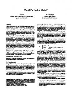
Fig. 1. Amyloplasts and PB-I protein bodies in rice endosperm. (A) Rice granule-bound starch synthase I (GBSSI, Os06g0133000, AK070431) contains a transit peptide (tp, amino acids M1V76) at the N-terminus. The N-terminal sequence (M1-T79) of GBSSI (tpGBSSI) was placed at the N-terminus of GFP to generate tpGBSSIGFP. (B) LSCM analysis of developing endosperm of rice (Nipponbare). A whole seed at 7 DAF was embedded in 5% agarose and cross-sectioned with a microslicer in 200-µm thick sections; sections were stained with 1 µM rhodamine B in PBS. The lateral side of the outer endosperm is shown. Rhodamine B and GFP were sequentially illuminated with laser beams at 543 and 488 nm, respectively. Rhodamine B binds protein bodies (PB-I) containing prolamins. Developing amyloplasts were visualized with fluorescent signals of tpGBSSIGFP. AL, aleurone cells; SE, starchy endosperm (outer endosperm/subaleurone). Bar = 10 µm.
ventral side refers to the side adjacent to the principal vascular bundle (ovular vascular trace) and the dorsal side is the opposite side of the long axis in the cross-section.19) The lateral side refers to both sides of the short axis in the crosssection. The cereal endosperm consists of several types of cell, including the aleurone, transfer cells, starchy endosperm and embryo-surrounding cells.20) In rice, the aleurone in the lateral and dorsal sides is usually a single layer of cells with thick cell walls. Aleurone cells contain many pleomorphic plastids, but they do not develop into chloroplasts containing chlorophylls or amyloplasts containing compound-type granules. The starchy endosperm of rice is further divided into the inner and outer endosperm. The outer endosperm, which is also called the subaleurone, consists of several layers of cells next to the aleurone layer in the lateral and dorsal sides. These cells are rich in both PB-I and PB-II, as well as amyloplasts. In contrast, the inner endosperm is occupied almost entirely by amyloplasts; it contains a few PB-I between the amyloplasts, but does not contain PB-II. Rice granule-bound starch synthase I (GBSSI, waxy protein) is an enzyme that synthesizes amylose in the amyloplast of endosperm. The catalytic domain of rice GBSSI has been analyzed by X-ray crystallography.21) The N-terminal amino acid residue of the rice GBSSI purified from mature seed is A78,22) indicating that the N-terminal sequence from M1 to Y77 functions as a transit peptide (tp), although the ChloroP program predicts that the N-terminal sequence from amino acids M1 to V76 constitutes a tp. When the N-terminal 79 amino acid residues (M1-T79) are translationally fused to the N-terminus of GFP (Fig. 1 (A)),
the fusion protein (tpGBSSIGFP) is targeted to the amyloplasts in the endosperm. The efficiency of amyloplast-targeting is so high that only processed GFP molecules are detectable by immunoblot analysis using proteins extracted from seeds.23) When tpGBSSIGFP is expressed under the control of a maize ubiquitin (Ubi) promoter,24) the endosperm may be analyzed by LSCM from 6 or 7 days after flowering (DAF) until about 20 DAF depending on the speed of maturation, which is influenced by environmental conditions. Before 6 DAF, the endosperm is still in the milky stage and is not firm enough to preserve its original shape when a whole seed embedded in 5% agarose is sectioned with a knife. The intensity of the fluorescent signal of tpGBSSIGFP declines to a low level after 20 DAF. Figure 1 (B) shows confocal images of the lateral side of an endosperm expressing tpGBSSIGFP under the control of the maize Ubi promoter at 7 DAF. At this developmental stage, some of the subaleurone cells next to the aleurone layer are still dividing, but the inner endosperm has already stopped division. The fluorescent signal of the tpGBSSIGFP targeted to the amyloplasts in the outer endosperm is usually bright enough to generate a high-resolution confocal image. However, the tpGBSSIGFP signal in the inner endosperm is so weak by 7 DAF that it is difficult to visualize the amyloplasts at high resolution. Why the signal in the inner endosperm is weak is not clear, but it may be that there is little protein synthesis occurring in the inner endosperm at this stage. Alternatively, most of the amyloplasts in the inner endosperm may no longer be sufficiently intact to retain the imported GFP within the outer limit envelope. Proteins that leak from the amyloplasts may be degraded in the cytosol to
32
J. Appl. Glycosci., Vol. 60, No. 1 (2013)
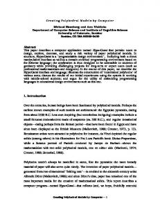
Fig. 2. The compound-type granule of rice. (A) SEM analysis of a cracked seed of Yukihikari rice (left panel) and purified granules (right panel). The samples were directly viewed without surface coating at 0.7 kV (cracked seed) and 1 kV (purified granules) using VE-8800 (Keyence). Bars = 5 µm. (B) Three possible routes of granule synthesis. The small amyloplast on the left contains granules numbered from 1 to 7. Each granule grows as the amyloplast develops. In (a), granules grow and become characteristic polyhedral, sharp-edged granules of similar size. In (b), granules grow at different rates, for example, granule no. 5 is much larger than granule no. 3 and the shape of granule no. 4 is oval. In (c), the granules fuse during the course of synthesis.
recycle the amino acids for the synthesis of storage proteins and other enzymes in the outer endosperm. What is the compound-type granule? SEM images of a section of mature seed and purified granules provide complementary sets of data, which together suggest that multiple granules are packed in such a way that they form round or oval units, and that each unit represents an amyloplast (Figs. 2 (A) and (B)). The crosssection shown in Fig. 2 (A) was prepared by simply cracking a mature seed in half with a knife. Importantly, granules are often separated from their neighbors in the same amyloplast when a seed is cracked. Granules are also separable when seed powder is homogenized in water. These findings indicate that granules located side by side in the same amyloplast are not directly connected by a large number of α-glucan polymers. Conventional microscopic analyses of starch granules suggest that the synthesis of the compound-type granule is a highly organized process. In other words, it is unlikely that compound-type granules are generated as a net result of reactions catalyzed by starch biosynthesizing enzymes that occur independently at numerous sites in an amyloplast. To visualize organized and random processes of compoundtype granule synthesis, three possible routes of granule synthesis are presented schematically in Fig. 2 (B). The first scheme (a) shows that small granules, numbered from 1 to 7, grow to form polyhedral granules, which are similar in size. In contrast, scheme (b) exhibits a situation where small granules grow at different rates, and become heterogeneous in shape. The third scheme (c) shows neighboring granules
Fig. 3. LSCM analysis of the amyloplast. (A) Schematic view of a z-series analysis. A tissue (sphere) is analyzed by optical sectioning along the z-axis from the top to the bottom. (B) The seed of Nipponbare expressing tpGBSSIGFP at 7 DAF. The seed section was stained with rhodamine B. Amyloplasts and PB-I were sequentially viewed by using z-series analysis, from which 3D images were reconstructed. The two panels are 3D images seen from different angles. Bars = 5 µm. (C) Three possible paths of amyloplast development. Imagine an experiment in which the small amyloplast on the left, visualized with tpGBSSIGFP, develops into larger ones under the microscope. In (a), the fluorescent image of tpGBSSIGFP targeted to the large amyloplast is almost equal to that of an enlarged image of the small amyloplast. In (b), the fluorescent image of the large amyloplast is dissimilar to the enlarged image of the small one, indirectly indicating that granule shapes are variable. In (c), the fluorescent signal of tpGBSSIGFP in the large amyloplast can be described as discontinuous short lines. Such fluorescent images would be anticipated if the growing granules fuse during the course of development.
fuse in the course of granule synthesis. Whatever the mechanism for the organized process is, it restricts the latter two schemes, thereby allowing scheme (a) to dominate. 3D image of the amyloplast. The major advantage of LSCM is the ability to collect serial optical sections. Such image acquisition is possible because out-of-focus light in specimens is eliminated by the use of spatial filtering. A z-series is a sequence of optical sections collected along the z-axis from the top to the bottom of the specimen (Fig. 3 (A)). The sequential images of a z-series can be processed to reconstruct three-dimensional (3D) structures. Figure 3 (B) shows 3D images of amyloplasts, which were reconstructed from a z-series visualized with tpGBSSIGFP. The generated 3D image can be rotated and seen from different angles. The two panels of Fig. 3 (B) show the 3D images of amyloplasts and PBs seen from different angles. Although the confocal images using tpGBSSIGFP do not directly reveal the shape of the granules, the granule shape can be indirectly inferred because the imported GFP is excluded from the granules, which are semicrystalline. In brief, the shape of the granule is revealed by the lack of light in the amyloplast. However, this is an assumption that may be only partially correct, as will be discussed again later. LSCM analyses using tpGBSSIGFP have revealed that small amyloplasts already contain compound-type granules, although the granules are very small. Now, let us imagine that such a small amyloplast visualized with tpGBSSIGFP can be monitored under the microscope for several days until it fully grows. If this experiment were possible, three possible outcomes may be anticipated, as schematically shown in Fig. 3 (C). Scheme (a) illustrates a situation in which the fluorescent signal of tpGBSSIGFP detected from the developed amyloplast resembles an enlarged image of the small
Kawagoe: Application of LSCM to Amyloplast Studies
33
Fig. 4. Visualization of the outer envelope membrane (OEM) of the amyloplast. (A) A full-length cDNA (AK105326) of rice OEP7 (Os02g0715400) was obtained from NIAS. The ORF was amplified by PCR with primers 5 -ATAGGTACCTCCCATCGCAATCCAATC-3 and 5 -TATCTCGAGTTCTCCTCGTCGG-3 , using the cDNA as the template. The PCR fragment was inserted at the N-terminus of GFP, generating the OEM-targeted fluorescent protein OEP7GFP. tpGBSSICherry was constructed by replacing the GFP of tpGBSSIGFP with mCherry (Clontech), a bright monomeric RFP.28) (B) Localization of two fusion proteins in the developing endosperm at 7 DAF as analyzed by use of LSCM. OEP7-GFP was targeted to the OEM of the outer limit envelope of the amyloplast containing compound-type granules. The fluorescent signal of OEP7-GFP was also detected at unidentified organelles, which did not show detectable levels of the tpGBSSICherry signal (arrows). Bars = 5 µm
amyloplast. In contrast, scheme (b) represents a situation in which the developed amyloplast is not a uniform enlargement of the small one. Such a result may be anticipated if the rates of granule synthesis are different between the granules in the amyloplast. Scheme (c) shows an amyloplast in which the fluorescent signal of tpGBSSIGFP is seen as discontinuous short lines. This type of image may be observed if neighboring granules fuse in the course of granule synthesis. Although monitoring amyloplast development under the microscope has not yet been successful, based on the numerous fluorescent images of large amyloplasts that have been obtained to date, it is plausible that scheme (a) dominates in vivo. Here again, these data suggest that the synthesis of compoundtype granules does not occur by chance. Visualization of the OEM of the amyloplast. The chloroplast is enclosed by double envelopes consisting of an outer envelope membrane (OEM), an inner envelope membrane (IEM) and the intermembrane space (IMS) between the two. A number of nuclear-encoded proteins are imported to the stroma by passing through protein complexes called Toc in the OEM and Tic in the IEM.25) In addition to the Toc/Tic importing machinery, vesicle transport of secretory proteins, including α-amylases, from the endoplasmic reticulum (ER) through the Golgi to the chloroplast has been demonstrated.26) Because the amyloplast contains heavy granules, it is difficult to purify intact amyloplasts from rice endosperm for protein import assays in vitro. Although not proven by in vitro assays, amyloplasts should also have the Toc/Tic complexes for importing nuclear-encoded proteins into the stroma. GFP fused with a transporter called Brittle1 (BT1), which imports ADP-glucose from the cytosol in the endosperm cells,27) localizes in the IEM of amyloplasts. BT1-GFP
signals are easily detected in the outer limit envelope,18,28) which encloses all of the granules that together constitute a unit of compound-type granule. In addition, simultaneous observations of BT1-GFP and tpGBSSI fused with Cherry, a bright RFP, have revealed that the amyloplast contains an internal structure between the granules, which we call the septum-like structure (SLS). Whether the SLS contains the OEM is not yet known. GFP fused to the membrane protein OEP7 has been used to visualize the OEM of chloroplasts in the Arabidopsis leaf.29) A rice orthologue (Os02g0715400) of Arabidopsis OEP7 was similarly used as a marker protein for the OEM of the amyloplast (Fig. 4 (A)). Simultaneous observations of OEP7-GFP and tpGBSSICherry revealed that OEP7-GFP was targeted to the outer limit envelope of amyloplasts (Fig. 4 (B)). The OEP7-GFP signal was also detected in a number of cellular structures that did not contain the tpGBSSICherry signal (Fig. 4 (B), arrows). These results suggest that tpGBSSICherry may be targeted to a subgroup of amyloplasts in the rice endosperm. Alternatively, amyloplast-targeted tpGBSSICherry may be retained in the amyloplast to different degrees depending on the developmental stage or physiological status of the amyloplast. The two large amyloplasts shown in Fig. 4 (B) did not show a detectable level of the OEP7-GFP signal at the SLS between the granules, although this signal was easily detected at the outer limit envelope. These results suggest the OEM containing OEP7-GFP was not associated with the SLS. However, we also obtained evidence that may suggest otherwise. Figure 5 shows a z-series analysis of developing amyloplasts in the subaleurone cells at 7 DAF. Judging from their shape and size, the organelles indicated by arrowheads and asterisks are apparently amyloplasts, although they did not contain a detectable tpGBSSICherry signal. These putative
34
J. Appl. Glycosci., Vol. 60, No. 1 (2013)
Fig. 5. z-series analysis of amyloplasts visualized with OEP7-GFP and tpGBSSICherry. Asterisks show putative amyloplasts, which had a detectable signal from OEP7-GFP but not from tpGBSSICherry. Similarly, putative amyloplasts indicated by arrowheads were not visualized with tpGBSSICherry, but these putative amyloplasts contained structures that looked like disks or partition-walls within the outer limit envelope. Bars = 5 µm.
amyloplasts indicated by arrowheads contained a strong OEP7-GFP signal at structures that look like disks or partition-walls inside the amyloplasts. It remains to be seen whether these internal structures represent the SLS being synthesized within the amyloplast. Amyloplast division and compound-type granule synthesis. The chloroplast differentiates from the proplastid and increases in number by dividing.30) The chloroplast division apparatus evolved primarily from the cell division apparatus of a cyanobacterial progenitor.31) It is therefore not surprising that the chloroplast divides by binary fission, as do cyanobacteria. However, to date, evidence indicating that large granules in the amyloplast are degraded at the middle of the granule before or during amyloplast division has not been obtained. Instead, the amyloplast in the rice endosperm divides simultaneously at multiple sites.32) These results clearly indicate that the division mechanisms of the chloroplast and amyloplast differ. Interestingly, amyloplast division and granule morphology can be modulated by changing the expression levels of plastid-division proteins such as ARC5, FtsZ1, FtsZ2-1, MinD, MinE and PDV2.28,32) In addition to these typical components of the division machinery, the debranching enzyme isoamylase3 (ISA3) plays an important role in both amyloplast division and the synthesis of compound-type granules.33) These results have suggested that amyloplast division and compound-type granule synthesis are related processes. Although conventional microscopic studies have revealed the wide diversity in shape of starch granules, the mechanism for the synthesis of compound-type granules remains an open question. On the basis of the results of LSCM analyses using numerous transgenic rice expressing FPs fused with amyloplast-targeted proteins, we have proposed a model for the synthesis of compound-type granules in rice.28) The most essential part of this model is that SLS synthesis occurs
beneath the outer limit envelope. As a result, a stromal section is created between the new SLS and the outer limit envelope, where granule synthesis initiates de novo and the granules grow outward. If these processes are repeated at multiple sites on the surface of the growing amyloplast, we believe that a compound-type granule consisting of up to several dozen granules can be produced in an amyloplast.28) Because SLS synthesis precedes granule synthesis, each granule is surrounded by either SLSs on all sides or SLS(s) and the outer limit envelope. Importantly, small amyloplasts are known to already contain compound-type granules, which suggest that the SLS would have to enlarge to accommodate the growing granules. The proposed model may present the basic principles of the mechanism for compound-type granule synthesis, since it is compatible with the synthesis of characteristic polyhedral, sharp-edged granules. The SLS physically divides the stroma into sections and thus prevents the fusion of granules located side by side in the same amyloplast. Because each granule is surrounded by either SLSs on all sides or SLS(s) and the outer limit envelope, the SLS functions like a mold that casts the growing granule into the characteristic polyhedral, sharp-edged shape. In other words, granules are synthesized in the partitioned stroma until the stromal sections are filled with growing granules. In short, it is the shape of the partitioned stroma that determines the shape of the granule being synthesized. The proposed model contrasts with another school of thought that all of the granules that constitute a compound-type granule are synthesized in a single stromal space. However, LSCM analyses have not provided evidence that exclusively supports the latter model. Future questions. Little is known about the structure and components of the SLS between granules. Because some SLSs contain IEM-targeted BT1-GFP,28) it is possible that the SLS and
35
Kawagoe: Application of LSCM to Amyloplast Studies
IMS are structurally related, at least in origin. Although GFP fused to OEP7, an OEM protein, is detectable at structures that look like disks or partition-walls inside some amyloplasts (Fig. 5), the lack of signal at the SLS in developed amyloplasts suggests that the OEM may not be preserved at the SLS once it is synthesized. Although the resolution of confocal images may not be sufficiently high, the SLS appears to be directly connected to the IMS of the outer limit envelope. One current structural model of the SLS is that it is like a partition-wall sandwiched between two IEMs that are continuous from the IEM of the outer limit envelope, as schematically shown in Fig. 6 (A). It is important to note that IEM containing BT1-GFP is prone to detach from the outer limit envelope when the expression of FtsZ2-1 or PDV2 is repressed by RNAi.28) These results suggest that the IEM is tightly associated with some components in the IMS and SLS, and that this association is stabilized by protein interactions involving FtsZ 2-1, PDV2 and an as yet unidentified IEM protein (compare Figs. 6 (A) and (B)). The 3D images of amyloplasts shown in Fig. 3 (B) indicate that tpGBSSIGFP is mostly present between granules. ISA3-GFP is also mostly present between granules.33) We have assumed that these FPs are imported to the stroma, and that they are excluded from the growing granules in the stroma. However, we recently obtained evidence that suggests this assumption may be only partially correct. Preliminary results of studies using GFP fused to starch synthase I (SSI-GFP) indicate that SSI-GFP mostly localized to the surface of granules rather than being colocalized with tpGBSSICherry (unpublished data). If SSI-GFP and tpGBSSI-
Cherry were both targeted to the stroma with similar efficiency, the two FP signals should have colocalized in the amyloplast. The fact that SSI-GFP and tpGBSSICherry showed distinct localization patterns may therefore indicate that tpGBSSIFPs are present primarily in the IMS of the outer limit envelope and the SLS sandwiched between two IEMs, but not in the stroma (Fig. 6 (A)). Protein fractionation studies using the developing endosperm of maize have revealed that SSI is highly enriched in the amyloplast stroma, whereas GBSSI is fully insoluble.34) The reason for the insolubility of GBSSI is not clear, but this lack of solubility has made it difficult to use GBSSI in protein fractionation analysis to pinpoint where in the amyloplast it functions. The finding of distinct localization patterns for SSI-GFP and tpGBSSICherry in the rice amyloplast has raised a number of questions. First, it is not clear whether the transit peptide or the rest of the protein or both determine the final destination of nuclear-encoded starch synthases in the amyloplast. An obvious question is whether the transit peptide of SSI is necessary and sufficient for targeting FPs to the stroma. Another related question is whether FPs fused with the full-length GBSSI are localized to the IMS and SLS or to the stroma. Second, it remains unknown whether the IMS and SLS persist intact throughout the period of granule growth. If not, the localization, hence the site of action of starch synthases including GBSSI may alter within the amyloplast according to the developmental stage of amyloplast. Studies on chain-length distributions of rice starch have indicated that GBSSI is responsible for the biosynthesis of not only amylose but also extra-long unit chains of amylopectin.35) Although these biochemical studies may suggest that GBSSI functions in the stroma, it should be kept in mind that where amylose molecules are distributed in granules is still a major question in the field of starch synthesis. Structural studies on the amyloplasts of waxy mutants using LSCM and GBSSI fused to FPs may shed further light on the function of GBSSI and the distribution of amylose molecules in compound-type granules. ACKNOWLEDGMENTS
Fig. 6. A model for the internal structure of a rice amyloplast containing compound-type granules. (A) In the wild-type amyloplast, the outer limit envelope consists of the outer envelope membrane (OEM), inner envelope membrane (IEM), and intermembrane space (IMS). The septum-like structure (SLS) functions as partition-walls in the stroma and is directly connected to the IMS. Each wall of the SLS is sandwiched between two IEMs. The OEM may be present in the SLS, as indicted by the broken lines, but the OEM in the SLS may not remain intact in the developed amyloplast. Starch granules are synthesized in the stroma. The growing granule is enclosed by the IEM on all sides and grows until the stromal space is completely filled, enabling the synthesis of a characteristic polyhedral, sharp-edged granule. The IEM is physically attached to the IMS and SLS. This IEM attachment is stabilized by a protein complex involving FtsZ2-1 in the stroma, PDV2 in the OEM, and as yet unidentified IEM proteins, which may include ARC6. When tpGBSSIGFP is expressed in the rice endosperm, it is efficiently targeted to the IMS and SLS, but not to the stroma. In contrast, when SSI-GFP is similarly expressed, the fusion protein localizes mostly to the granule surface, suggesting that the fusion protein is imported to the stroma through Toc/Tic complexes. (B) Amyloplasts in transformants in which the expression of FtsZ2-1 or PDV2 is repressed by RNAi. The IEM tends to detach from the IMS and SLS due to the absence of the proteins that are necessary to make the protein complex across the OEM and IEM.
I thank Mutsumi Sakurai for SEM analysis. This work was supported by the NIAS Strategic Research Fund and by the Ministry of Agriculture, Forestry and Fisheries of Japan (Genomics for Agricultural Innovation, IPG-0023).
REFERENCES ) 1 A.M. Smith: Starch in the Arabidopsis plant. Starch/Stärke, 64, 421-434 (2012). ) 2 M. Stitt and S.C. Zeeman: Starch turnover: pathways, regulation and role in growth. Curr. Opin. Plant Biol., 15, 282-292 (2012). ) 3 M. Crumpton-Taylor, S. Grandison, K.M.Y. Png, A.J. Bushby and A.M. Smith: Control of starch granule numbers in Arabidopsis chloroplasts. Plant Physiol., 158, 905-916 (2012). ) 4 I.J. Tetlow: Starch biosynthesis in developing seeds. Seed Sci. Res., 21, 5-32 (2011). ) 5 J.L. Jane, T. Kasemsuwan, S. Leas, H. Zobel and J.F. Robyt: Anthology of starch granule morphology by scanning electronmicroscopy. Starch/Stärke, 46, 121-129 (1994). ) 6 S.C. Zeeman, J. Kossmann and A.M. Smith: Starch: its metabolism, evolution, and biotechnological modification in plants. Annu. Rev. Plant Biol., 61, 209-234 (2010). ) 7 Y. Nakamura: Towards a better understanding of the metabolic
36
) 8
) 9 ) 10 ) 11 ) 12
) 13 ) 14 ) 15 ) 16
) 17
) 18
) 19 ) 20 ) 21
) 22
J. Appl. Glycosci., Vol. 60, No. 1 (2013) system for amylopectin biosynthesis in plants: Rice endosperm as a model tissue. Plant Cell Physiol., 43, 718-725 (2002). R. Matsushima, M. Maekawa, N. Fujita and W. Sakamoto: A rapid, direct observation method to isolate mutants with defects in starch grain morphology in rice. Plant Cell Physiol., 51, 728-741(2010). A.D. Evers: Scanning electron microscopy of wheat starch. 3. Granule development in endosperm. Starch/Stärke, 23, 157-192 (1971). H.J. Kang, I.K. Hwang, K.S. Kim and H.C. Choi: Comparison of the physicochemical properties and ultrastructure of Japonica and Indica rice grains. J. Agric. Food Chem., 54, 4833-4838 (2006). T. Tateoka: Starch grains of endosperm in grass systematics. Bot. Mag. Tokyo, 75, 377-383 (1962). R. Matsushima, J. Yamashita, S. Kariyama, T. Enomoto and W. Sakamoto: A phylogenetic re-evaluation of morphological variations of starch grains among Poaceae species. J. Appl. Glycosci., 60, 37 44 (2013). N.C. Shaner, P.A. Steinbach and R.Y. Tsien: A guide to choosing fluorescent proteins. Nature Methods, 2, 905-909 (2005). S. Toki: Rapid and efficient Agrobacterium-mediated transformation in rice. Plant Mol. Biol. Rep., 15, 16-21 (1997). K. Ozawa and F. Takaiwa: Highly efficient Agrobacteriummediated transformation of suspension-cultured cell clusters of rice (Oryza sativa L.). Plant Sci., 179, 333-337 (2010). S. Lee, G.Y. Su, E. Lasserre, M.A. Aghazadeh and N.Murai: Small high-yielding binary Ti vectors pLSU with co-directional replicons for Agrobacterium tumefaciens-mediated transformation of higher plants. Plant Sci., 187, 49-58 (2012). Y. Onda, A. Nagamine, M. Sakurai, T. Kumamaru, M. Ogawa and Y. Kawagoe: Distinct roles of protein disulfide isomerase and P5 sulfhydryl oxidoreductases in multiple pathways for oxidation of structurally diverse storage proteins in rice. Plant Cell, 23, 210-223 (2011). Y. Onda, T. Kumamaru and Y. Kawagoe: ER membrane-localized oxidoreductase Ero1 is required for disulfide bond formation in the rice endosperm. Proc. Natl. Acad. Sci. USA, 106, 1415614161 (2009). S. Krishnan and P. Dayanandan: Structural and histochemical studies on grain-filling in the caryopsis of rice (Oryza sativa L.). J. Biosci., 28, 455-469 (2003). P.A. Sabelli and B.A. Larkins: The development of endosperm in grasses. Plant Physiol., 149, 14-26 (2009). M. Momma and Z. Fujimoto: Interdomain disulfide bridge in the rice granule bound starch synthase I catalytic domain as elucidated by X-ray structure analysis. Biosci. Biotechnol. Biochem., 76, 1591-1595 (2012). T. Taira, N. Fujita, K. Takaoka, M. Uematsu, A. Wadano, S. Kozaki and S. Okabe: Variation in the primary structure of waxy proteins (granule-bound starch synthase) in diploid cereals. Biochem. Genet. 33, 269-281 (1995).
) Y. Kawagoe, A. Kubo, H. Satoh, F. Takaiwa and Y. Nakamura: 23 Roles of isoamylase and ADP-glucose pyrophosphorylase in starch granule synthesis in rice endosperm. Plant J., 42, 164-174 (2005). ) H. Nakamura, M. Hakata, K. Amano, A. Miyao, N. Toki, M. 24 Kajikawa, J. Pang, N. Higashi, S. Ando, S. Toki, M. Fujita, A. Enju, M. Seki, M. Nakazawa, T. Ichikawa, K. Shinozaki, M. Matsui, Y. Nagamura, H. Hirochika and H. Ichikawa: A genomewide gain-of-function analysis of rice genes using the FOX-hunting system. Plant Mol. Biol., 65, 357-371 (2007). ) F. Kessler and D. Schnell: Chloroplast biogenesis: Diversity and 25 regulation of the protein import apparatus. Curr. Opin. Cell Biol., 21, 494-500 (2009) ) A. Kitajima, S. Asatsuma, H. Okada, Y. Hamada, K. Kaneko, Y. 26 Nanjo, Y. Kawagoe, K. Toyooka, K. Matsuoka, M. Takeuchi, A. Nakano and T. Mitsui: The rice alpha-amylase glycoprotein is targeted from the Golgi apparatus through the secretory pathway to the plastids. Plant Cell, 21, 2844-2858 (2009). ) N.J. Patron, B. Greber, B.E. Fahy, D.A. Laurie, M.L. Parker and 27 K. Denyer: The lys5 mutations of barley reveal the nature and importance of plastidial ADP-Glc transporters for starch synthesis in cereal endosperm. Plant Physiol., 135, 2088-2097 (2004). ) M.S. Yun and Y. Kawagoe: Septum formation in amyloplasts 28 produces compound granules in the rice endosperm and is regulated by plastid division proteins. Plant Cell Physiol., 51, 1469-1479 (2010). ) W. Bae, Y.J. Lee, D.H. Kim, J. Lee, S. Kim, E.J. Sohn and I. 29 Hwang: AKR2A-mediated import of chloroplast outer membrane proteins is essential for chloroplast biogenesis. Nature Cell Biol., 10, 220-227 (2008). ) 30 S. Miyagishima: Mechanism of plastid division: from a bacterium to an organelle. Plant Physiol., 155, 1533-1544 (2011). ) 31 C.X. Chan, J. Gross, H.S. Yoon and D. Bhattacharya: Plastid origin and evolution: New models provide insights into old problems. Plant Physiol., 155, 1552-1560 (2011). ) M.S. Yun and Y. Kawagoe: Amyloplast division progresses 32 simultaneously at multiple sites in the endosperm of rice. Plant Cell Physiol., 50, 1617-1626 (2009). ) M.S. Yun, T. Umemoto and Y. Kawagoe: Rice debranching 33 enzyme isoamylase3 facilitates starch metabolism and affects plastid morphogenesis. Plant Cell Physiol., 52, 1068-1082 (2011). ) Y. Yu, H.H. Mu, C. Mu-Forster and B.P. Wasserman: Polypep34 tides of the maize amyloplast stroma: Stromal localization of starch-biosynthetic enzymes and identification of an 81-kilodalton amyloplast stromal heat-shock cognate. Plant Physiol., 116, 1451-1460 (1998). ) I. Hanashiro, K. Itoh, Y. Kuratomi, M. Yamazaki, T. Igarashi, J.I. 35 Matsugasako and Y. Takeda: Granule-bound starch synthase I is responsible for biosynthesis of extra-long unit chains of amylopectin in rice. Plant Cell Physiol., 49, 925-933 (2008).