Appl Microbiol Biotechnol (2007) 75:599–607 DOI 10.1007/s00253-007-0869-0
APPLIED GENETICS AND MOLECULAR BIOTECHNOLOGY
The development of a flagellin surface display expression system in a moderate thermophile, Bacillus halodurans Alk36 Michael Crampton & Eldie Berger & Sharon Reid & Maureen Louw
Received: 3 October 2006 / Revised: 29 January 2007 / Accepted: 29 January 2007 / Published online: 21 February 2007 # Springer-Verlag 2007
Abstract This study relates to the development of an alkaliphilic, thermotolerant, Gram-positive isolate, Bacillus halodurans Alk36, for the over-production and surface display of chimeric gene products. This bacterium continuously over-produces flagellin. To harness this ability, key genetic tools, such as gene targeted inactivation, were developed for this strain. The hag gene, which codes for flagellin, was inactivated on the chromosome giving rise to the B. halodurans BhFC01 mutant. Polylinkers were inserted as in-frame, chimeric, flagellin sandwich fusions to identify the permissive insertion sites corresponding to the variable regions of the flagellin protein. Flagellin expression and motility were evaluated for these constructs. Two sites were identified for possible peptide insertion in the flagellin gene, one of which produced functional flagella and was able to restore the motility phenotype to a non-motile mutant. Peptides encoding a poly-histidine peptide and the HIV-1 subtype C gp120 epitope were, respectively, incorporated into this site as in-frame fusions. The peptides were found to be successfully displayed on the cell surface and functional through metal binding and immunological studies, respectively. Keywords Flagellin . Cell surface display . Bacillus halodurans . Fusion protein . Integration M. Crampton and E. Berger contributed equally to this manuscript. M. Crampton : E. Berger : M. Louw (*) CSIR Biosciences, P.O. Box 395, Pretoria 0001, South Africa e-mail:
[email protected] S. Reid Department of Molecular and Cell Biology, University of Cape Town, Cape Town, South Africa
Introduction Microbial cell surface display can be defined as the presentation of a heterologous protein or peptide of interest (passenger protein) by attaching it to the bacterial cell wall through an anchoring motif (carrier) so that it is exposed to the outside of the bacterial cell. This area of research has gained enormous interest over the past 20 years with potential applications in a number of research fields, including immunology, applied microbiology and biotechnology. Various display systems have been extensively reviewed for both Gram-positive and Gram-negative bacteria (Lee et al. 2003; Li 2000; Samuelson et al. 2002; Wernérus et al. 2002; Westerlund-Wikström 2000). The protein or peptide of interest can be fused to the carrier protein at either the C-terminal or N-terminal domains, or may be presented as a tri-hybrid in-frame or “sandwich” fusion. Sandwich fusions have mostly been associated with the display of heterologous proteins or peptides in Gramnegative bacteria using two major classes of carrier proteins, the outer membrane proteins or protein monomers of extracellular appendages such as flagella and pili (Lee et al. 2003). Flagella are predominantly composed of a single protein, flagellin, product of the hag gene. Flagellin display systems have only been successfully reported in Gramnegative bacteria, such as Escherichia coli or Salmonella, and involved the use of the flagellin (FliC) protein as a carrier protein, which is able to display the passenger protein as a genetic fusion in the surface-exposed central variable region (Cattozzo et al. 1997; Westerlund–Wikström et al. 1997). The first heterologous peptide to be displayed in this manner was an 11 amino acid epitope from eggwhite lysozyme (Kuwajima et al. 1988). Other examples utilising this system in Gram-negative bacteria include the display of peptide libraries (Lu et al. 1995), the develop-
600
ment of vaccine delivery systems (Stocker and Newton 1994; Levi and Arnon 1996; Cattozzo et al. 1997; Westerlund-Wikström 2000) and determining the structure and function of adhesive epitopes (Hynonen et al. 2002; Majander et al. 2005). Comparison of the FliC proteins from different Gram-positive and Gram-negative bacteria showed high sequence similarity at the N and C termini, whereas the central domains were highly variable in size and sequence (Beatson et al. 2006). It is this region which is therefore targeted as the site for insertion of foreign peptides in both E. coli and Salmonella. Bacillus halodurans Alk36 is an alkaliphilic, thermotolerant Gram-positive bacterial isolate that requires a pH of greater than 7.5 and sodium ions for efficient growth. This isolate was also found to have the ability to grow over a wide pH range (7.5–10) and temperature (30–55°C). This bacterium continuously expresses a ∼34 kDa cell surface protein at a high level, subsequently identified as FliC. This unique feature was utilised for the development of a surface display system giving rise to a high concentration of heterologous peptides on the cell surface. Gram-positive bacteria are more robust than their Gramnegative counterparts and the flagella as well as the chimeric peptides can be easily isolated from the cell surface. In addition, this system is extremely versatile as peptides or proteins can be displayed over a range of temperatures or pH depending on the requirements of the applications. This report focuses on the identification of the optimal display site within the FliC protein and the successful expression and display of a metal-binding polypeptide and a HIV antigenic peptide as chimeric flagellin fusions. This is the first report of flagellin surface display in a Gram-positive organism, which has the added advantage of thermotolerance.
Appl Microbiol Biotechnol (2007) 75:599–607
Transformants were selected using 100 μg/ml ampicillin or 10 μg/ml chloramphenicol, respectively. DNA techniques Plasmid DNA was isolated using a Plasmid Midi Kit (Qiagen). Restriction enzymes were used as specified by the manufacturer (Fermentas and Roche Diagnostics). All mini-preps were done using Perfectprep Plasmid Mini Kit (Eppendorf). All DNA manipulations were done in E. coli, which was transformed using electroporation (Dower et al. 1988). Transformation into B. halodurans Alk36 was carried out according to Kudo et al. (1990) with modifications, using polyethylene glycol (PEG) 4000 (final concentration, 22.5%; w/v). For the DM3 protoplast regeneration medium (pH 7.8) PEG (4000) was added at 1% (w/v) final concentration and chloramphenicol (5 μg/ml). Thermus aquaticus DNA polymerase was used for polymerase chain reaction (PCR) as recommended by the supplier (BIOLINE). Construction of temperature sensitive shuttle vector pSEC194 Plasmid pE194 was digested with TaqI. The 1.35 kb fragment containing the temperature sensitive ori was ligated to pSK (ClaI) and transformed into E. coli DH10B to create pSE194 (4.313 kb). Plasmid pJM103 (Perego 1993) was digested with BglII/PvuII to obtain the chloramphenicol resistance gene (1.2 kb). This fragment was ligated to pSE194 (BamHI/SmaI) and transformed into E. coli DH10B to create pSEC194 (Fig. 1).
Materials and methods
Construction of pSECFliC and pSECFlg− Plasmid pSECFliC carrying the complete hag gene (accession number D10063, Sakamoto et al. 1992) and the σD promoter was constructed with primers SigDKpn and DownRev (Table 1) using B. halodurans Alk36 genomic DNA as the template (Fig. 2). Construction of pSECFlg− (defective hag gene, Fig. 2) involved PCR primers UpFor, UPRev, and DownFor, DownRev (Table 1).
Bacterial strains, plasmids and growth conditions A Bacillus species isolated from a soil sample in South Africa (Louw et al. 1993) was identified as B. halodurans Alk36 (NCIMB 41348) using 16S rDNA sequence analysis (Weisburg et al. 1991). B. subtilis 1A46 was obtained from BGSC (Bacillus Genetics Stock Centre, Ohio, USA). Plasmid pE194 was obtained from the Deutsche Sammlung von Mikroorganismen und Zellkulturen. pSKBluescript (pSK) was obtained from Stratagene. E. coli DH10B (F− mcrA Δ(mrr–hsdRMS–mcrBC) (φ80dlacZΔM15) ΔlacX74 endA1 recA1deoR Δ(ara–leu)7697 araD139 galU galK nupG rpsL λ−) was obtained from Invitrogen. E. coli and B. subtilis cultures were grown at 37°C in LuriaBertani (LB) medium pH 7. B. halodurans Alk36 was grown in LB medium pH 8.5 at 30, 42 or 52°C as specified.
Fig. 1 Plasmid map of pSEC194 (5.496 kb). Temperature sensitive ori (pE194) and ColE1 ori used for replication in Bacillus and E. coli, respectively. Restriction enzyme sites (bold ) useful for cloning. Plasmid map created with DNAMAN, Version 4.1
Appl Microbiol Biotechnol (2007) 75:599–607
601
Table 1 List of primers and their corresponding nucleotide sequences Primer name
Nucleotide sequence
Restriction enzyme sites
DownRev SigDKpn UpFor UpRev DownFor FliN–terRev FliDNR2 Up–Forward CterF2 VNR2 VCF NC5F NC5RX VCF6 VNR6 FliCR
5′CGCTGCAGAAGAGGAACGTAAACG3′ 5′CTCGGTACCCTCGCGTTACGCTCTTTCTGT3′ 5′GCGGATCCGTGTGGTGACATTTGAC3′ 5′GCTCTAGACGATGCGCATTCATTGCTGG3′ 5′GCTCTAGAGAGTCTCGTATCCGTG3′ 5′CTCCTCGAGCGACCTTCTGAAACAGC3′ 5′CGAGGATCCAAGACCGGCAGAGTTAATGTC3′ 5′GTGTGGTGACATTTGAC3′ 5′CACGAATTCTCGAGCCCGGGATCCTCTTACCTAGGAGCTATGCAAAAC3′ 5′CGGCAGCTGTTCACCAGAATTAGCACCAAC3′ 5′CACGTCGACTCGAGCCCGGGATCCTTAATTGAACTTGATTTAACAAAAG3′ 5′CACGTCGACTCGAGCCCGGGATCCTTTAATACGCAAAAATTACTC3′ 5′CACCTCGAGTGAGTTGTATCTTTGATTC3′ 5′CACGTCGACTCGAGCCCGGGATGGATCCAGAATGCACAATCAGCTATTGAC3′ 5′GACGTCGACAGTGTGGTCAGTAATATCCTC3′ 5′CAACAAAGTAACGGTTGAGCG3′
PstI KpnI BamHI XbaI XbaI XhoI BamHI – XhoI – SalI XhoI XhoI SalI SalI –
Restriction enzyme sites used are underlined.
Integration and inactivation of the hag gene on the chromosome of B. halodurans Alk36 All pSEC194 constructs were stably maintained in B. halodurans Alk36 at 30°C with antibiotic selection. Integration was a combination of methods by Biswas et al. (1993) and Poncet et al. (1997). To screen for a double cross-over (dco) event colonies were transferred to motility plates (LB pH 8.5, 0.4% (w/v) agar, 0.8% (w/v) gelatin). A non-motile phenotype would indicate a dco event resulting in a flagellin minus (Δhag) integration mutant (BhFC01). The dco event was confirmed with PCR using two chromosomal primers Up–Forward and FliDNR2 (Table 1). Identification of a display site within the FliC protein of B. halodurans Alk36 To identify the optimal site for the insertion of heterologous peptides and proteins, polylinkers (Table 2) were inserted as in-frame fusions in the hag gene
Fig. 2 Schematic presentation of the different gene constructs in plasmid pSEC194. Lines represent untranslated DNA regions, filled bars ( grey), the coding regions of the hag gene, the dotted region the putative variable region as defined from this study and white bars, inserted linkers. The symbol Δ denotes deletions and the numbers inside the blocks denote the size in nucleotides of the hag gene fragments. σD represents the σD-dependent promoter of the hag gene
on pSECFliC in four different sites within the putative variable region based on sequence alignments between the Salmonella, E. coli, B. subtilis and B. halodurans hag genes (LaVallie and Stahl 1989; Sakamoto et al. 1992; Fig. 2). Primers used for the construction of pSECNC2 were SigDKpn/FliN-terRev and CterF2/DownRev, for pSECNC3 SigDKpn/VNR2 and VCF/ DownRev, for pSECNC5 SigDKpn/NC5RX and DownRev/NC5F and for pSECNC6 SigDKpn/VNR6 and FliCR/VCF6 (Table 1). All constructs were verified with sequencing. Construction of hybrid flagella displaying the poly-His petide and HIV-1 subtype C gp120 V3 loop peptides The synthetic oligonucleotides were derived from the HIV-1 subtype C gp120 V3 loop (Hewer and Meyer 2003; Table 3). Oligonucleotides were annealed according to the method described by Integrated DNA Technologies (http:// www.idtdna.com), digested with the appropriate restriction enzymes and ligated into pSECNC6. This gave rise to pSECNHivC6. Complementary oligonucleotides were designed for the insertion of a poly-His peptide into pSECNC6 to give pSECNHisC6. All constructs were confirmed by PCR and sequencing analysis. Integration of the poly-His peptide fusion construct in the chromosome of B. halodurans BhFC01 Integration of pSECHisC6 into the B. halodurans chromosome was carried out as for the hag gene, resulting in strain BhFHC02 (hag::his). After the dco event, colonies unable to grow on chloramphenicol plates were confirmed to contain the integrated poly-His peptide fusion by PCR analysis using two chromosomal primers UpFor and FliDNR2 (Table 2).
602
Appl Microbiol Biotechnol (2007) 75:599–607
Table 2 Nucleotide and amino acid sequences of inserted polylinkers Construct name
Polylinker nucleotide sequence
Amino acid sequence
pSECNC2 pSECNC3 pSECNC5 pSECNC6
5′ 5′ 5′ 5′
Ser–Ser–Pro–Gly–Ser Gln–Leu–Pro–Val–Ser–Ser–Pro–Gly–Ser Val–Asp–Ser–Ser–Pro–Gly–Ser Val–Asp–Ser–Ser–Pro–Gly–Trp–Ile–Gln
TCG AGC CAG CTG GTC GAC GTC GAC
CCG GGA TCC 3′ CCG GAC TCG AGC CCG GGA TCC 3′ TCG AGC CCG GGA TCC 3′ TCG AGC CCG GGA TGG ATC CAG 3′
Protein extractions and analysis Cell surface proteins were isolated as follows: cultures were grown at 30°C in 30 ml LB (pH 8.5) to stationary phase (16–24 h). The cultures were centrifuged at 7,000×g for 10 min, the cell pellet was resuspended in 2.5 ml sterile water and an equal volume of 0.2 M NaOH was added and stirred vigorously for 30 min on ice. The cell suspension was centrifuged at 8,000×g for 10 min to remove cell debris. Proteins were precipitated with an equal volume of 5% trichloroacetic acid with 30 min shaking at room temperature. The precipitate was pelleted at 15,000×g for 30 min, air dried and resuspended in 400 μl phosphate buffer (pH 7.5). For whole flagella preparations, bacterial cells were grown in 50 ml LB pH 8.5 to late log phase (OD540 ∼1.2), harvested and resuspended in 10 ml phosphate-buffered saline (PBS; pH 7.5). The cell suspension was subjected to three times 5-s pulses using an Ultra-Turrax (Janke & Kunkel, IKA-WERK) homogeniser and centrifuged at 8,000×g for 10 min to remove cell debris. The supernatant was centrifuged at 40,000×g for 1 h to pellet the flagella and was resuspended in 50 μl PBS buffer. Protein concentrations were determined according to the Bradford method (Bradford 1976). All proteins were separated on sodium dodecyl sulfate polyacrylamide gel electrophoresis (SDS-PAGE) gels as described by Laemmli (1970). Western blot analysis Western blot analysis was carried out according to Gallagher et al. (1997). Polyclonal rabbit antiflagellin antibodies were generated using B. halodurans Alk36 flagellin protein according to the method of Cooper and Paterson (2000). Anti-6His antibodies were obtained
from AEC Amersham and MEIV3b 4 antibodies from D. Meyer (Hewer and Meyer 2003). Metal binding analysis of chimeric flagellin proteins Metal binding ability of the poly-His peptide displayed on whole and denatured chimeric flagella, (cell surface protein fraction), was assayed directly using Ni-NTA alkaline phosphatase (AP) conjugate according to the manufacturer (QIAGEN). Cell surface proteins (2 μg) were immobilised in each well of a Maxisorp Nunc-Immuno plate. After incubation of the Ni-NTA AP conjugate (QIAGEN), 200 μl of substrate solution [50 μl p-nitrophenyl phosphate sodium salt (250 mg/ml), 3 ml diethanolamine (1 M) and 15 μl MgCl2 (0.005 mM), pH 9.8] was added. The reaction was incubated at 37°C for 1 h and stopped with 50 μl NaOH (3 M). Colour development was read at OD405.
Results The highly expressed FliC protein produced by B. halodurans Alk36 Cell surface protein fractions of both B. halodurans Alk36 and B. subtilis 1A46 grown in LB were compared on a SDS-PAGE gel at different time intervals (Fig. 3). It was clear from these profiles that B. halodurans Alk36 overproduced flagellin protein in comparison to B. subtilis 1A46. An important observation was the significant amount of protein present on the cell surface of B. halodurans Alk36 48 h after inoculation which remained evident in samples up to 144 h (results not shown).
Table 3 HIV-1 subtype C gp120 V3 loop and His-peptide oligonucleotides used for annealing and subsequent insertion into pSECNC6 Oligo name
Oligo sequence
Peptide sequence
Restriction enzyme sites
HIVCF
5′-GGG CGT CGA CAC GCG TCC AAA TAA TAA TAC GCG TAA ATC AAT TCG TAT TGG ACC AGG ACA AAC GTT TTA TGC AAC GGG AGA TTG GAT CCG CGG-3′ 5′-CCG CGG ATC CAA TCT CCC GTT GCA TAA AAC GTT TGT CCT GGT CCA ATA CGA ATT GAT TTA CGC GTA TTA TTA TTT GGA CGC GTG TCG ACG CCC-3′ 5′-TCG AGA CAT CAT CAT CAT CAT CAC A-3′ 5′-GAT CCT GTG ATG ATG ATG ATG ATG T-3′
Val–Asp–Thr–Arg–Pro–Asn– Asn–Asn–Thr–Arg–Lys–Ser– Ile–Arg–Ile–Gly–Pro–Gly–Gln– Thr–Phe–Tyr–Ala–Thr–Gly– Asp–Trp–Ile–Gln
SalI/BamHI
Val–Asp–Ser–Arg–His–His–His– His–His–His–Arg–Ile–Gln
XhoI/BamHI
HIVCR
HisF2 HisR2
Amino acid sequence: NC6 linker. Sequence of the inserted heterologous peptide in bold. Restriction digest sites are underlined.
Appl Microbiol Biotechnol (2007) 75:599–607
603
Characterisation of an integration site for peptide display
Fig. 3 SDS-PAGE gel (10%) comparing cell surface protein profiles of B. halodurans Alk36 (pH 8.5, 42°C) and B. subtilis 1A46 (pH 7.0, 37°C) grown in LB broth at different times. Lanes 2–4, B. halodurans Alk36 and lanes 5–7, B. subtilis 1A46. Lane 1, molecular mass marker. The arrows point to the FliC protein
Construction of a B. halodurans Alk36 Δhag mutant (BhFC01) A defective hag gene was constructed with 641 bp of the internal region deleted and was cloned into the thermosensitive vector (pSEC194) to create pSECFlg− (Fig. 2). The dco event to generate the deleted hag gene was confirmed by PCR analysis, and the absence of a motility halo on motility plates confirmed that this isolate was a Δhag mutant (strain BhFC01). Complementation of the Δhag non-motile phenotype was obtained after transformation of strain BhFC01 with plasmid pSECFliC (Fig. 2; results not shown).
Fig. 4 Motility plate showing the presence or absence of motility haloes of the various constructs. Colony 1, B. halodurans Alk36; 2, BhFC01; 3, BhFC01(pSECNC2); 4, BhFC01(pSECNC3); 5, BhFC01 (pSECNC5); 6, BhFC01(pSECNC6) and 7, BhFC01(pSEC194)
The B. halodurans Alk36 flagellin is significantly smaller than that of E. coli (30 kDa vs ∼52 kDa) with a correspondingly small variable region. It was therefore important to determine whether it was possible to disrupt the variable region by insertion of small peptides and still obtain functional flagella. In an attempt to identify the variable domain, polylinkers (Table 2) were inserted at four different sites in plasmids pSECNC2, pSECNC3, pSECNC5 and pSECNC6 (Fig. 2). All the constructs were shown to be stably maintained through mini-prep analysis. No motility haloes were observed for transformants containing plasmids pSECNC2, pSECNC3 or pSECNC5 (Fig. 4). However, transformants with pSECNC6 were shown to be motile, indicating that in this strain, the individual chimeric flagellin monomers were polymerised to form functional flagella. Cell surface protein fractions were isolated and analysed on a SDS-PAGE gel. There were no detectable flagellin protein bands of the correct size for constructs pSECNC2, or pSECNC5 protein fractions (Fig. 5a). However, there were bands of the correct size present in the cell surface
Fig. 5 a SDS-PAGE gel (12%) of cell surface protein fractions of BhFC01 containing different plasmid constructs. Lane 1, pSECNC2; lane 2, pSECNC3; lane 3, pSECNC5; lane 4, pSECNC6; lane 5, BhFC01; lane 6, B. halodurans Alk36 and lane 7, low molecular mass ladder. b Western blot of a
604
Appl Microbiol Biotechnol (2007) 75:599–607
Surface display as a vaccine delivery system
Fig. 6 SDS-PAGE gel (10%) of cell surface proteins demonstrating expression of the poly-His peptide in the different constructs. Lane 1, B. halodurans Alk36; lane 2, BhFC01; lane 3, BhFC01 (pSECNC6); lane 4, BhFC01 (pSECNHisC6); lane 5, BhFHC02 and lane 6, Fermentas low molecular mass marker
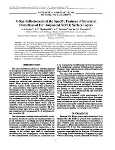
protein fractions of pSECNC3 and pSECNC6 (Fig. 5a) indicating that the chimeric peptide flagellin fusions are over-produced on the cell surface of these strains. Western blot analysis using polyclonal rabbit anti-FliC antibodies (Fig. 5b) confirmed that pSECNC2 was not expressed even at low levels (Fig. 5b, lane 2). The band observed for pSECNC5 ran higher than predicted and was only expressed at low levels on the cell surface of BhFC01 (Fig. 5b, lane 3). BhFC01 carrying pSECNC3 and pSECNC6 (Fig. 5b, lane 2 and 4) and the B. halodurans Alk36 positive control (Fig. 5b, lane 6) were all positive for FliC production. Expression and functionality of the poly-His peptide Expression of the 13 amino acid poly-His peptide as a polyHis fusion peptide on the cell surface of B. halodurans strain BhFCO1 was clearly evident (Fig. 6, lane 4). The integration of the DNA fragment encoding the poly-His fusion peptide into the chromosome of BhFC01 also resulted in the successful expression of the poly-His fusion on the cell surface (Fig. 6, lane 5). Both whole flagella and denatured flagellin subunits carrying the poly-His peptide were able to bind a significant amount of Ni2+ as compared to the wild type B. halodurans Alk36 flagella (Fig. 7).
Fig. 7 Histogram representation of results from Ni2+-binding assay. Results demonstrate binding of a poly-His::flagellin monomers and b whole flagella, to Ni–NTA conjugate. Bar 1, B halodurans Alk36; bar 2, BhFC01(pSECNC6); bar 3, BhFC01(pSECNHisC6) and bar 4, BhFHC02
Expression of the 29 amino acid antigenic peptide from HIV-1 subtype C was successful, as can be seen from the cell surface fraction of B. halodurans BhFC01 carrying pSECNHivC6 (Fig. 8a). Western blot analysis showed that MEIV3b 4 antibodies bound successfully to the exposed HIV-1 C gp120 epitope (Fig. 8b).
Discussion In this paper, we report on the development of a flagellin surface display system for a moderately thermophilic Bacillus isolate. This system is based on the continuous over-production of flagellin, product of the hag gene, by B. halodurans Alk36. As each bacterial flagellum may be made up of approximately 20,000 flagellin monomers, this system would enable the highly efficient immobilisation of a significant number of heterologous peptides on the bacterial cell surface. In addition, as flagellin production is continuous throughout the growth cycle of the bacterium, an inducible promoter system is not required for overproduction of the chimeric flagellin, which is the case in most surface display systems. To exploit the over-production of flagellin by B. halodurans Alk36, genetic techniques were developed for this strain. They included an integration system enabling gene-targeted inactivation based on a thermo-sensitive ori of replication located on plasmid pE194 derived from S. aureus (Gryczan et al. 1982). This was used to successfully inactivate the hag gene of B. halodurans Alk36, resulting in a non-motile strain. The modified hag gene encoding the poly-His fusion peptide was also integrated into the chromosome through a double crossover event using this methodology. At the nucleotide level, the B. halodurans Alk36 hag gene and the up- and down-stream regions were found to show 100% identity with the corresponding regions from B. halodurans C-125 (Sakamoto et al. 1992). The genome of B. halodurans C-125 has been sequenced and is available
Appl Microbiol Biotechnol (2007) 75:599–607
605
Fig. 8 a SDS-PAGE gel (12%) showing expression and display of the HIV-1 subtype C gp120 epitope in cell surface protein fractions of strain BhFC01. Lane 1, BhFC01 (pSECNC6); lane 2, strain BhFC01; lane 3, BhFC01 (pSECNHivC6) and lane 4 Bio-Rad low molecular mass marker. b Western blot analysis of cell surface protein fractions using the MEIV3b 4 antibodies. Lane 1, BhFC01 (pSECNC6); lane 2, BhFC01 (pSECNHivC6) and lane 3, Low molecular mass marker
in the DNA Data Bank of Japan (http://www.gib.genes.nig. ac.jp). The hag gene from B. subtilis is transcribed by the σD form of RNA polymerase (LaVallie and Stahl 1989; Mirel and Chamberlin 1989), and we have identified a similar σD promoter sequence upstream of the alkaliphilic B. halodurans Alk36 hag gene. Transcription of the hag gene in wild-type B. subtilis cells demonstrated a peak of expression as the cells entered stationary phase and then decreased to zero within 4 h after the onset of sporulation (Mirel and Chamberlin 1989). This is consistent with our findings for B. subtilis 1A46 flagellin production, but is significantly different from our results obtained for flagellin production by B. halodurans Alk36, where high levels of FliC protein can be detected up to 144 h. Thus, despite the presence of the σD promoter sequence in B. halodurans Alk36, the mechanism controlling flagellin expression appears to differ from that of B. subtilis, leading to the over-production of this protein. It was also found in B. subtilis that expression of the hag gene on a multicopy vector from the σD promoter, resulted in the formation of long filamentous cells and the accumulation of flagellin intracellularly (LaVallie and Stahl 1989). However, complementation studies of the B. halodurans Alk36 BhFC01 strain with the plasmid pSECFliC (carrying the complete hag gene and σD promoter from B. halodurans Alk36) did not result in filamentous cells or intracellular flagellin accumulation indicating a possible difference in mechanisms of flagellin expression. Sequence alignments indicated that the variable domain occurred between amino acids 127–200 of the B. halodurans Alk36 flagellin. This region of 73 amino acids is much smaller than that postulated for E. coli (187 amino acids) and S. typhimurium flagellin (241 amino acids). This is not surprising considering that the total size of the flagellin monomers from E. coli (497 amino acids) and S. typhimurium (494 amino acids) are much larger than that from B. halodurans (272 amino acids; Kuwajima et al. 1986; Yonekura et al. 2003). To further define the variable
region, polylinkers were inserted at four different sites revealing two permissive insertion sites. Only in one instance did the insertion of a polylinker (construct pSECNC6) restore the motile phenotype to BhFC01. In all other instances, either no or negligible chimeric flagellin production was observed, indicating the importance of inserting peptides optimally within the variable domain. A recent study by Beatson et al. (2006) aligning 202 flagellin sequences narrowed down the variable region to 24 amino acids between amino acids 159– 185, thereby confirming our results. To demonstrate the applicability of this system, we evaluated the expression of polypeptides ranging in size from 13 to 29 amino acids as displayed chimeric flagellin fusions. We also evaluated the biological functionality of these polypeptide fusions. Flagella are potent immunogens, and numerous reports show that immunisation with purified hybrid flagella or whole Salmonella cells carrying hybrid flagella induces cellular and humoral immune responses in laboratory animals against the chimeric flagellin epitope (Cattozzo et al. 1997; Westerlund-Wikström 2000). We have successfully demonstrated the potential of this application by inserting a HIV-1 subtype C gp120 synthetic epitope of 29 amino acids in the variable region of the B. halodurans Alk36 flagellin and shown production of large amounts of the flagellin::peptide fusions on the bacterial cell surface. The combination of flagella to act as an effective adjuvant (McSorley et al. 2002) and the potential for largescale chimeric flagellin over-production make this a potentially attractive candidate as a vaccine delivery system. Histidine-rich metal-binding peptides have been previously shown to function as effective bio-adsorbants. Both E. coli and Staphyloccus have been shown to harbour increased Niand Cd-binding capacity when these poly-His peptides were surface displayed (Samuelson et al. 2002; Xu and Lee 1999). A poly-His peptide fusion was successfully displayed in B. halodurans, and could be expressed either from a plasmid or from a gene integrated into the chromosome. The ability to integrate heterologous constructs is a great advantage of this
606
system, as the construct can then be stably maintained without antibiotic pressure and can be continuously expressed. The amount of chimeric flagellin produced was not noticeably decreased by integration of the gene, and an increased metal binding was observed with both constructs using both whole and denatured flagella. The construction of a surface display expression system in a thermotolerant Gram-positive bacterium capable of carrying a foreign peptide provides a tool for the display of numerous peptides and proteins, which may have value in the field of bioremediation, biomining, biotransformation, vaccine development and biosensors. Taken together, the robustness of Gram-positive bacteria, the ability of this strain to grow at high temperatures and pH, and its ability to continuously over-produce flagellin displaying heterologous peptides without impairing cell growth, make this strain a promising candidate for application in a number of biotechnological applications. Acknowledgment We would like to thank Dr Gerhard Pietersen (Plant Protection Institute, ARC, Pretoria, South Africa) for preparation of the anti-flagellin antibodies. We would also like to thank Dr D. Meyer for kindly supplying the MEIV3b 4 antibodies. This research was supported through funding from a CSIR Thematic grant PPTH/2004/002.
References Beatson SA, Minamino T, Pallen MJ (2006) Variation in bacterial flagellins: from sequence to structure. Trends Microbiol 14:151– 155 Biswas I, Gruss A, Ehrlich SD, Maguin E (1993) High-efficiency gene inactivation and replacement system for Gram-positive bacteria. J Bacteriol 175:3628–3635 Bradford MM (1976) A rapid and sensitive method for the quantitation of microgram quantities of protein utilizing the principle of protein dye binding. Anal Biochem 72:248–254 Cattozzo EM, Stocker BAD, Radaelli A, De Giuli Morghen C, Tognon M (1997) Expression and immunogenicity of V3 loop epitopes of HIV-1 isolates SC and WMJ2, inserted in Salmonella flagellin. J Biotechnol 56:191–203 Cooper HM, Paterson Y (2000) Preparation of polyclonal antisera. In: Ausubel FM, Brent R, Kingston RE, Moore DD, Seidman JG, Smith JA, Struhl K (eds) Current protocols in molecular biology, Wiley, New York, pp 11.12.1–11.12.7 Dower WJ, Miller JF, Ragsdale CW (1988) High efficiency transformation of E. coli by high voltage electroporation. Nucleic Acids Res 16:6127 Gallagher S, Winston SE, Fuller SA, Hurrell JGR (1997) Immunoblotting and immunodetection. In: Ausubel FM, Brent R, Kingston RE, Moore DD, Seidman JG, Smith JA, Struhl K (eds) Current protocols in molecular biology. Wiley, New York pp 10.8.1–10.8.21 Gryczan TJ, Hahn J, Contente S, Dubnau D (1982) Replication and incompatibility properties of plasmid pE194 in Bacillus subtilis. J Bacteriol 152:722–735 Hewer R, Meyer D (2003) Peptide immunogens based on the envelope region of HIV-1 are recognized by HIV/AIDS patient polyclonal antibodies and induce strong humoral immune responses in mice and rabbits. Mol Immunol 40:327–335
Appl Microbiol Biotechnol (2007) 75:599–607 Hynonen U, Westerlund-Wikström B, Palva A, Korhonen TK (2002) Identification by flagellum display of an epithelial cell- and fibronectin-binding function in the SlpA surface protein of Lactobacillus brevis. J Bacteriol 184:3360–3367 Kudo T, Hino M, Kitada M, Horikoshi K (1990) DNA sequences required for the alkalophily of Bacillus sp. strain C-125 are located close together on its chromosomal DNA. J Bacteriol 172:7282–7283 Kuwajima G, Asaka JI, Fujiwara T, Fujiwara T, Node K, Kondoh E (1986) Nucleotide sequence of the hag gene encoding flagellin of Escherichia coli. J Bacteriol 168:479–1483 Kuwajima G, Asaka JI, Fujiwara T, Fujiwara T, Nakano K, Kondoh E (1988) Presentation of an antigenic determinant from hen eggwhite lysozyme on the flagellar filament of Escherichia coli. Biotechnol 6:1080–1083 Laemmli UK (1970) Cleavage of structural proteins during the assembly of the head of bacteriophaege T4. Nature 33:267–272 LaVallie ER, Stahl ML (1989) Cloning of the flagellin gene from Bacillus subtilis and complementation studies of an in vitroderived deletion mutation. J Bacteriol 171:3085–3094 Lee SY, Choi JH, Xu Z (2003) Microbial cell-surface display. Trends Biotech 21:45–52 Levi A, Arnon R (1996) Synthetic recombinant influenza vaccine induces efficient long-term immunity and cross-strain protection. Vaccine 14:85–92 Li M (2000) Applications of display technology in protein analysis. Nat Biotechnol 18:1251–1256 Louw ME, Reid SJ, Watson TG (1993) Characterization, cloning and sequencing of a thermostable endo-(1,3-1,4) β-glucanase-encoding gene from an alkaliphilic Bacillus brevis. Appl Microbiol Biotechnol 38:507–513 Lu Z, Murray KS, Van Cleave V, LaVallie ER, Stahl ML, McCoy JM (1995) Expression of thioredoxin random peptide libraries on the Escherichia coli cell surface as functional fusions to flagellin: a system designed for exploring protein–protein interactions. Biotechnol 13:366–372 Majander K, Korhonen TK, Westerlund-Wikström B (2005) Simultaneous display of multiple foreign peptides in the FliD capping and FliC filament proteins of the Escherichia coli flagellum. Appl Environ Microbiol 71:4263–4268 McSorley SJ, Ehst BD, Yu Y, Gewirtz AT (2002) Bacterial flagellin is an effective adjuvant for CD4+ T cells in vivo. J Immunol 169:3914–3919 Mirel DB, Chamberlin MJ (1989) The Bacillus subtilis flagellin gene (hag) is transcribed by the sigma 28 form of RNA polymerase. J Bacteriol 171:3095–3101 Perego M (1993) Integrational vectors for genetic manipulation in Bacillus subtilis. In: Sonenshein AL, Hoch JA, Losick R (eds) Bacillus subtilis and other gram-positive bacteria, biochemistry, physiology, and molecular genetics. American Society for Microbiology, Washington DC, pp 615–624 Poncet S, Bernard C, Dervyn E, Cayley J, Klier A, Rapoport G (1997) Improvement of Bacillus sphaericus against dipteran larvae by integration, via homologous recombination, of the Cry11A toxin gene from Bacillus thuringiensis subsp israelensis. Appl Environ Microbiol 63:4413–4420 Sakamoto Y, Sutherland KJ, Tamaoka J, Kobayashi T, Kudoh T, Horikoshi K (1992) Analysis of the flagellin (hag) gene of alkaliphilic Bacillus sp C-125. J Gen Microbiol 138:2159–2166 Samuelson P, Gunneriusson E, Nygren P, Stahl S (2002) Display of proteins on bacteria. J Biotechnol 96:129–154 Stocker BAD, Newton SMC (1994) Immune response to epitopes inserted in Salmonella flagellin. Int Rev Immunol 11:167–178 Weisburg WG, Barns SM, Pelletier DA, Lane DJ (1991) 16S Ribosomal DNA amplification for phylogenetic study. J Bacteriol 173:697–703
Appl Microbiol Biotechnol (2007) 75:599–607 Wernérus H, Lehtio J, Samuelson P, Stahl S (2002) Engineering of staphylococcal surfaces for biotechnological applications. J Biotechnol 96:67–78 Westerlund-Wikström B (2000) Peptide display on bacterial flagella: principles and applications. Int J Med Microbiol 290:223–230 Westerlund-Wikström B, Tanskanen J, Virkola R, Hacker J, Lindberg M, Skurnik M, Korhonen TK (1997) Functional expression of
607 adhesive peptides as fusions to Eschericia coli flagellin. Protein Eng 10:1319–1326 Xu Z, Lee SY (1999) Display of polyhistidine peptides on the Escherichia coli cell surface by using outer membrane protein C as an anchoring motif. Appl Environ Microbiol 65:5142–5147 Yonekura K, Yonekura SM, Namba K (2003) Complete atomic model of the bacterial flagellar filament by electron cryomicroscopy. Nature 424:643–650