Damage to the axons of adult sensory neurons results in massively increased expression of the protooncogene c-jun both in neurons and in the associated.
The Journal
The Differential Control of c-Jun Expression Neurons and Their Associated Glial Cells Carmen
De Felipe
MRC Laboratory
and Stephen
of Neuroscience,
May
in Regenerating
1994,
74(5):
291 l-2923
Sensory
P. Hunt
of Molecular Biology, Division of Neurobiology,
Damage to the axons of adult sensory neurons results in massively increased expression of the protooncogene c-jun both in neurons and in the associated Schwann cells. The role of growth factors and axon contact in mediating this expression was investigated in dissociated cultures of adult sensory neurons and glial cells that expressed c-jun within 24 hr of plating. Trk, trkB, and trkC growth factor receptor genes were expressed in discrete subpopulations of sensory neurons but addition of NGF or brain-derived neurotrophic factor (BDNF) did not inhibit the expression of c-jun. Similarly, axon contact was not sufficient to decrease c-jun expression in glial cells. However, c-jun expression could be downregulated in glial cells, but not neurons, by treatment with CAMP or forskolin and increased by raising intracellular calcium levels. The results suggest that c-jun levels are differentially regulated in neurons and glial cells but that NGF or BDNF do not regulate c-jun expression in damaged neurons. [Key words: c-jut?, regeneration, sensory neurons, glial cells, neurotrophins, second messengers]
A number of recent studieshave implicated the c-jun protooncogenein the control of growth and differentiation in a variety of cell types including neurons and glial cells (Bos et al., 1990; de Groot et al., 1990; Yamaguchi-Iwai et al., 1990; Castellazzi et al., 1991; Jenkins and Hunt, 1991; Leah et al., 1991; De Felipe et al., 1992; Wong et al., 1992; Jenkins et al., 1993a-c). Following axon damageor block of axonal transport in peripheral sensory or motor neurons in the rat, there is a delayed expressionof c-jun protein and mRNA that is maintained until the nerve has fully regenerated(Jenkins and Hunt, 1991; Leah et al., 1991; Herdegen et al., 1993). Similar modifications in c-jun expressionoccur following axotomy of rubrospinal (Jenkins et al., 1993a)or nigrostriatal (Jenkinset al., 1993b)neurons but thesechangeslargely reversed within 14 d in parallel with the failure of these central neurons to regenerate.These observations suggestedthat the expressionof c-jun was in someway important, if not essential,for the regenerationof adult neurons and implied that the mechanismsby which c-jun expression could be regulatedin neuronsand glial cellscould be crucial for
Received Aua.- 2. 1993: revised Oct. 14, 1993; accepted Oct. 26, 1993. We thank Stuart Ingham for excellent photographic assistance. This work was supported by Nato and Wellcome Trust fellowships to C. De F. Correspondence should be addressed to Dr. S. P. Hunt, MRC Laboratory of Molecular Biology, Division of Neurobiology, Hills Road, Cambridge CB2 2QH, UK. Copyright 0 1994 Society for Neuroscience 0270-6474/94/14291 l-13$05.00/0
Cambridge
CB2 2QH, United Kingdom
our understanding of the regenerative processin adult mammals. c-jun is the cellular homolog of v-jun the transforming oncogeneof the avian sarcomavirus 17 (Maki et al., 1987; Sakai et al., 1989) and encodesfor a 39 kDa nuclear phosphoprotein that is the major component of the AP- 1 complex of transcriptional regulators(Bohmann et al., 1987; Halzonetis et al., 1988; Abate et al., 1990; Kerppola and Curren, 1991). c-jun is one of a family of closely related proteins (including Jun B and Jun D) that possess a leucine zipper though which they form homo- or heterodimersthat will bind to DNA (Chiu et al., 1988)and act as transcriptional regulators (seeLamb and McKnight, 1991). However, the control of c-jun geneexpressionand protein activity is complex and may involve transcriptional, combinatorial, temporal, and posttranslational mechanismsthat vary between different cell types (for reviews, seeKarin and Smeal, 1992; Jackson, 1992). c-jun hasbeenshownto heterodimerize with c-fos, other fosrelated proteins, and members of the CREB/ATF (CAMP responseelement-binding protein/activating transcription factor) family of binding proteins or to form Jun-Jun homodimers (Karin and Smeal, 1992). These different combinations of proteins can possess different DNA binding specificities, affinities, and transcriptional activities. Jun-Fos heterodimerspossess both an increasedDNA binding activity and trans-activating activity when comparedwith Jun-Jun homodimersalthough c-jun itself retains the highesttransactivating potential @mealet al., 1989, 1991; Angel and Karin, 1991). Following stimulation of neurons, neuronal cell lines, or glial cellswith phorbol esters,polypeptide hormones,growth factors, cytokines or neurotransmitters, the expressionof c-jun mRNA may follow “immediate early” genekinetics (Bartel et al., 1989; Shen and Greenberg, 1990; Sheng et al., 1990; Wisden et al., 1990; McNaughton and Hunt, 1992). This is characterized by a rapid but transient expressionof c-jun mRNA following stimulation and is not dependentupon new protein synthesis.However, in certain situations the induction of c-jun, but not c-fos, expressioncan be both delayed and prolonged (Brenner et al., 1989) for example, during the differentiation of F9 carcinoma cells with retinoic acid or in the neuronal responseto axonal damage(Yang-Yen et al., 1990b;Jenkins and Hunt, 1991; Leah et al., 1991; Sleigh, 1992). The observation that in peripheral nerve, axoplasmic block was as effective as axon section or ligation in causing an increasedc-jun expressionin neurons, led us to suggestthat the c-jun responsemay have resulted in part from deprivation of axonally transported target factors (Jenkinset al., 1993a-c).The most obvious candidatesare the neurotrophins, NGF and brain-
2912
De Felipe
and Hunt
l
Control
of c-Jun
Expression
during
Regeneration
derived neurotrophic factor (BDNF), which are known to be retrogradely transported in sensory neurons (Hendry et al., 1974; DiStefano et al., 1992). Indeed, exogenously applied NGF has been shown to reverse the changes in peptide and growth factor receptor expression seen following peripheral nerve section in rat (Fitzgerald et al., 1985; Verge et al., 1989a,b, 1992). To look at these possibilities more closely we have established primary cultures of adult dorsal root ganglion (DRG) cells and their supporting satellite and Schwann cells and investigated the signals and intracellular events that lead to and modify c-jun expression. Our results demonstrate that c-jun expression in neurons and glial cells is differentially regulated and not obviously related to neurotrophin starvation. Some of these results have been briefly reported elsewhere (De Felipe et al., 1993). Materials
and Methods
Cell cultures. Cultures of adult dorsal root ganglion cells (DRGs) were prepared as described previously (Lindsay, 1988) with minor modifications. Briefly, cleaned ganglia from adult Sprague-Dawley rats were collected in Dulbecco’s minimal Eagle’s medium (DMEM) plus 10% horse serum and treated twice for 1.5 hr with 0.25% collagenase. A single-cell suspension was then obtained by trituration (1 O- 12 passages) though a fire-polished Pasteur pipette. Cells were collected, rinsed, and plated (2000 cells/ml) on to polyomithine (100 mg/ml) and laminin (5 mg/ml) coated coverslips. After 15-20 hr cells cultures were maintained in the same serum-containing medium or more usually transferred to DMEM defined medium (Bottenstein and Sato. 1979). Cells were maintained in culture at 37°C; 7.5% CO, for up to’30 d. ‘Experiments were routinely done on 7-lo-d-old cultures. Treatments were always carried out without replacing the culture medium to avoid the possibility that changing the medium may induce changes in protooncogene expression. Aliquots of 100 times concentrated stock substances were added directly to the culture medium (1 ml). Peripheral nerve lesions. Adult male Sprague-Dawley rats (200-300 gm) were deeply anesthetized by intraperitoneal injection of 4 ml/kg Equithesin. Sciatic nerve ligation and section was performed through a 1 cm incision over the sciatic notch. In sham controls the nerve was exposed but not damaged. The animals survived for 1 d (n = 3), 4 d (n = 2), and 7 d (n = 2). Animals were deeply anesthetized with chloral hydrate and perfused transcardially with 4% paraformaldehyde in sodium phosphate buffer (PB: 0.1 M, pH 7.4); nerves and ganglia were removed, postfixed for 2 hr, and cryoprotected overnight in PB plus 30% sucrose. Nerves were sectioned at 40 Nrn and incubated in antibodies, free floating, for 1848 hr. Zmmunohistochemistry. Cells were fixed in methanol (-20°C) for 5 min and freshly depolymerized paraformaldehyde (4% in 0.05% sodium phosphate buffer, pH 7.4, for 5 min) and incubated overnight in antibodies. Antiserum to c-jun was generated by immunizing rabbits with synthetic peptide conjugated to the carriers keyhole limpet hemoglobin or thyroglobin. Subsequent characterization of this serum was by enzyme-linked immunoabsorbance assay (ELISA), Western blotting, immunoprecipitation, and immunocytochemistry. This indicated reactivity with a protein of the predicted molecular weight, and subcellular localization in response to various stimuli. A DRG cell culture protein extract analyzed by Western blotting revealed two bands in the region of 40-44 kDa that reacted with the c-jun antibody as predicted from previous studies (data not shown). The cljun antiserum was raised against the sequence COLMLTOOLOTF from the C-terminus of the c-iun peptide, which did not re&&ze jun-B or jun-D (H. Hurst, and 6. I. Evan, unpublished observations) (diluted 1:2000). Sections were then processed with either an avidin-biotin complex horseradish peroxidase kit (Williams et al., 1990; Wisden. et al., 1990) or with an avidinfluorescein isothiocyanate (FITC) tertiary step (all from Vector). Sections were rinsed after all incubations in 0.1 M PB for 30 min and finally either reacted with 0.1% diaminobenzidine (DAB, Sigma) and 0.012% hydrogen peroxide to reveal a brown reaction product or mounted and viewed with conventional epifluorescence or with a Bio-Rad MRC-600 confocal microscope. Occasionally, a double labeling procedure was used. The first antibody, usually E-jun was used first-and binding revealed with the HRP-linked method described above. The second antibody was then applied overnight (usually 3A 10 and or RT97) and the
procedure repeated except that 5-chloro-1-naphthol, which gives a blue reaction product, was used as the second chromogen. Alternatively, a double fluorescence procedure was used, the first antibody detected with an avidin-FITC probe and the second with an avidin-Texas red probe (both from Vector). The c-jun antiserum has been used extensively in other studies (Wisden et al., 1990; Jenkins and Hunt, 1991; Jenkins et al., 1993a-c). Controls, which were all negative, included selective adsorption of antisera with 10 mg of native peptide and omission of the primary antibody. Because of the potential problems of cross-reactivity, immediate early gene immunoreactivity should be regarded as being-“-like-activity.” Similarly, changes in the exnression of c-iun mav reflect increased synthesis of messageor other mechanisms, s&h as increased stability of mRNA or protein. The methods used here are unable to distinguish between these possibilities. The rabbit antisera to c-jun does not recognize either iun B or jun D. Other antibodies used included sensory neuron-specific monoclonal antibody 3A 10 (diluted 1:2), which recognizes a neurofilament-related antigen (gift of J. Dodd and T. Jessell); RT97, which recognizes the 200 kDa phosphorylated neurofilament antigen (gift of B. Anderton); a mouse monoclonal antibodv to GAP43 (1:25.000: Boehrineerl: rabbit antisera to c-fos (1:3000; Hunt et al., 198;) anh NcFI-A (l::&$ Waters et al., 1990; Wisden et al., 1990); rabbit antisera to calcitonin gene-related peptide (CGRP, gift of Dr. K. Stemini; diluted 1:3000). Glial cells were identified with antibodies to glial acidic fibrillary protein (GFAP) and SlOO (Dako). In situ hybridization. For in situ hybridization, cells grown on 19 mm coverslips were fixed in 4% paraformaldehyde for 5 min, dehydrated in alcohol, and stored in 100% alcohol until used. A 60-mer oligonucleotide probe that recognized c-jun mRNA or 45-mer oligonucle 30 mm). Double labeling studies with 3AlO or RT97 revealed that many of these
large c-jun-negative
neurons had extensive arborizations
(Fig.
3B). cJun protein and mRNA were also highly expressed in pure cultures of Schwann cells derived from sciatic nerve as well as in satellite cells plated with neurons, indicating that the presence of neurons is not required for c-jun expression in peripheral glial cells (not shown). No c-jun expression was seen in fibroblasts. Glial c-jun immunoreactivity was unaffected by contact with overlying axons (Fig. 4).
Expression of growth factor receptors in DRG cultures Before monitoring the effects of growth factors on c-jun levels in our cultures, we looked for the presence of trk receptor mRNAs that mediate the effects of the neurotrophins. Growth factor receptor mRNA was differentially expressed by sensory neurons in our cultures (Fig. 5). Of 120 labeled sensory neurons, trk mRNA was present on 40%, trkB mRNA on 25%, and trkC mRNA on 30% of neurons. No attempt was made to assess whether these were expressed in the same or overlapping populations of neurons or whether different ganglia expressed different proportions of the three trk receptor mRNAs. However, larger-diameter neurons appeared to preferentially labeled by trkC (Fig. X). There was also evidence for the presence of trkB and trkC on glial cells.
Eflect of growth factors and cytokines on c-jun levels in DRG cultures Cultures maintained for 7 d in defined medium were treated for 30 min, 2 hr, or 24 hr before fixation with a number of neurotrophins and cytokines. In addition, some cultures were maintained in defined medium with NGF or BDNF or both for the whole 7 d period. c-jun mRNA levels were not changed by addition of growth factors at any time point in either neurons or Schwann cells. Similarly, the total number of c-jun-positive neurons (and neuronal survival) was unaffected by growth factor treatment and
The Journal
of Neuroscience,
May
1994,
74(5)
2915
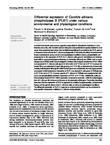
Figure 3. The majority of neuronswere c-jun immunopositivein vitro. The majority of neuronaland glial nuclei werec-jun positive (A, immunoperoxidase technique).However,a smallnumberof neuronalnucleiwerenot c-jun positive(B, solid arrow) althougha secondantibody
(3AlO) revealedthe extensivearborizationsof both c-jun-positive(A) and-negative(B) neurons.In this 7 d culture,3AlO stainingwasalsoseen to be restrictedto the perikaryalcytoplasmin a subsetof neurons(arrowheads, B). A backgroundof positiveglial cell staining(openarrows) can alsobe seen.Scalebar, 40 Frn. no changesin c-jun protein immunofluorescencelevels were found in neuronsafter treatment with any of thesecompounds. However, in Schwann cells 2 hr treatment with all the factors, except transforming growth factor-p, produced a significant increasein the c-jun protein immunofluorescencelevels (30%) (seeTable 2). This change in glial cell c-jun protein-like immunoreactivity was not maintained at the 24 hr time point except in the caseof BDNF, and this changehad disappeared by 4 d in vitro (Table 1). Finally, the addition of the tyrosine kinase inhibitor lavendustin for 24 hr at 1 HM(O’Dell et al., 1991) was found to be without effect on either c-jun mRNA or protein levels in neurons or glial cells, suggestingthat this group of receptors was not involved in the maintenance of c-jun expression.
Effects of second messenger stimulation mRNA
on c-jun protein
and
To determine the influence of secondmessengercandidateson the expressionof c-jun in DRG cells in culture, levels of c-jun mRNA and protein were studied after phorbol 12-myristate 13acetate(PMA) treatment, which stimulatesprotein kinaseC, or stimulation of protein kinase A (PKA) with forskolin, which elevates CAMP though adenylate cyclase activation, or by addition of the membrane-permeableanalog of CAMP, dibutyryl cyclic adenosinemonophosphate(dBcAMP). The effects of artificially raising or lowering internal calcium levels and of depolarization were also studied. Calcium levels and depolarization. Glial cell but not neuronal
Figure 4. Axon contactin vitro does
not downregulatec-jun expressionin glial cells.In a 7 d DRG culture, an axon labeledwith the antibody RT97 growsover stronglyc-jun-immunopositive glial cells(arrowheads) without reducinglevelsof expression of theprotooncogene. Scalebar, 40 Mm.
2918
De Felipe
and
Hunt
- Control
of oJun
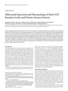
Figure 5. Trk receptor mRNAs were expressed in subpopulations of DRG neurons in vitro. Cultures (7 d in vitro) were hybridized for trk (A), trkB (B), and trkC (C) using Y3 antisense oligonucleotide probes. Subpopulations of DRGs were labeled with each probe. Arrow&&indicates glial cell. Exposure time, 8 weeks. Scale bar, 40 pm.
Expression
during
Regeneration
The Journal
Table 1. Long-term effect of neurotrophins
May
1994,
14(5)
2917
on c-jun expression in neurons
Control Total number of neurons counted % Neurons not expressing c-jun protein cJun protein levels
of Neuroscience,
BDNF
NGF
478 f 46
450 1- 40
496 + 65
19.6 t- 1.1 134.6 k 2.0
21.1 k 0.2 136.5 f 6.5
16.6 * 2.0 125.3 + 4.3
Cultures were continuously exposed to NGF or BDNF from plating to fixation (5 d) in defined medium. Neurons from three coverslips in three separate experiments were analyzed by tracking across the maximum diameter of each 19 mm coverslip and scoring each neuron and measuring c-jun immunofluorescence. The results indicate that there was no effect of growth factors on either cell survival or the percentage of c-jun-positive neurons.
levels of c-jun protein and mRNA were influenced by changing intracellular calcium levels. Intracellular levels of calcium were raised using the calcium ionophore A-23178, or lowered by buffering the medium with ethylene glycol-his@-aminoethyl ether) N, N, N’,N’-tetra-acetic acid (EGTA). After 24 hr of treatment with A-23178 (10 and 100 nM), levels of c-jun protein in glial cells, but not neurons, were significantly increased(Fig. 6) and mRNA levelsincreased twofold (Fig. 7). Higher dosesof the ionophore (1 FM) led to massive cell death in both cell populations. Treatment for 30 min or 2 hr with low-dose ionophore had no effect on c-jun protein levels. When extracellular Caz+levels were buffered to 0.8 mM (but not 1.7 or 1.3 mM) with EGTA for 24 hr, c-jun protein levels were reduced in glial cells but not in neurons(Table 3). Lower Ca*+levels led to cell death after 24 hr treatment. mRNA levels respondedin a similar fashion. High K+ concentrations(54 mM), veratridine (10 PM), or TTX (1 PM) were added to 7-d-old DRG cultures in order to ask whether depolarization or Na+ channel activity could induce changesin c-jun geneexpression.No differencesin c-jun protein or mRNA were found 24 hr after thesetreatments. PMA and CAMP. PMA treatment (200 nM) for 2 hr or 24 hr had no effect on either c-jun protein or mRNA in either of the
cell populations studied (Fig. 7, Table 4). However, forskolin (10 PM) and, to a greaterextent, dBcAMP (OS-1 .OmM) induced a time-dependant decreasein c-jun mRNA (Fig. 7, Table 4) and c-jun protein immunoreactivity in satellite and Schwann cells. This downregulation of c-jun expression was specific for glial cells, having no effect on c-jun levels in neurons. In Figure 8, B, D, and F showthe virtually complete disappearanceof c-jun protein and mRNA (85-90% decrease)after 24 hr of dBcAMP treatment in glial cells but not in neurons. Lower dosesof dBcAMP (10 PM) did not downregulate the expressionof c-jun (Table 5). We were able to antagonize partially the forskolin effect with the specific PKA blocker H8 (N-([2-methylamino] ethyl)-5-isoqinoline sulfonamidedihydrochloride; 30 PM) given 2 hr prior and during 24 hr forskolin treatment (Fig. 9). CAMP treatment did not changejun B or jun D gene expression in either of the cell types (data not shown). Mitogenic q@cts qf CAMP. Becauseof the known mitogenic effect of CAMP on Schwann cells (Wood and Bunge, 1975) we examined the relationship between the reduction of c-jun expressionand cell division in DRG cultures maintained in serumfree medium. The effects of dBcAMP on c-jun levels and cell division in Schwann cells or satellite cells were dissociable. dBcAMP had a significant mitogenic effect at concentrations of
Table 2. Effect of growth factors on c-jun protein levels in glial cells from DRG cultures Treatment
2 hr
Control TGFfl IGF I EGF PDGF NGF IFG II FGF BDNF
140.3 156.7 200.8 187.4 181.1 218.8 189.6 182.3 206.0
24 hr + k k k -t k k k +
4.6 4.9 8.0* 8.0* 9.5* 7.5* 7.2* 5.3* 7.6*
150.0 145.7 133.0 126.0 165.0 142.8 154.6 145.7 190.0
t 9.4 k 11.5 -t 10.7 f 8.1 + 10.6 +- 9.2 + 17.4 + 10.7 + 10.8*
Cultures were maintained for 7 d in defined medium and treated for 30 min (not shown), 2 hr, or 24 hr before fixation with either nerve growth factor (NGF, 50 @ml, Sigma), epidermal growth factor (EGF, Bachem, 100 @ml), insulin-like growth factor I (IGF I, Bachem, 200 t&ml), IGF II (Bachem, 200 rig/ml), plateletderived growth factor (PDGF, Calbiochem, 100 rig/ml), basic or acidic fibroblast growth factor (FGF) (Bachem, 100 rig/ml), transforming growth factor-b (TGFB) (Sigma, 2 ngml), or brain-derived neurotrophic factor (BDNF, gift of Drs. J. Wood and H. Luebbert, Sandoz). BDNV was supplied in supematant from a recombinant cell line containing a functional BDNF gene. By bioassay supematant was found to be effective at a dilution of 1:40. In this study it was used at a concentration of 1:20. The immunofluorescence from at least 50 glial cells taken from each of three separate experiments was analyzed. *p < 0.05, Dunn’s test.
Schwann Cells Neurons
IONOPHORE
A-23178
(nM)
Figure 6. c-Jun immunofluorescence is enhanced in glial cells but not neurons by raising intracellular calcium levels. Treatment of DRG cultures with A-23 178 for 24 hr caused a dose-dependent increase in c-jun immunofluorescence in satellite glial cells.
2919
De Felipe
and
Hunt
- Control
of c&n
Expression
during
Regeneration
*
-T
!
T
T
-
E E 3
5 u
extracellular calcium levels reduces the immunofluorescence in glial cells but not in
Extracellular Ca2+ (mM)
cJun immunofluorescence (relative intensity) Schwann cells Neurons
1.8 1.7 1.3 0.8
112 100 115 84
chelator
k + f k
4.2 4.9 5.3 5.3*
171 * 11 184 k 15 201 + 10 167 of: 10
EGTA
in defined medium,
different concentrations of the calcium to buffer extracellular Ca*+ levels. The con-
centrationsof EGTA were determinedwith an EQCAL program(Biosoft); 1.8rnM represents the normal concentration * p -C 0.05, Dunn’s test.
T
zE s
Buffering of c-jun
SevendayDRG cultureswere treatedwith
*
*T nil
Table 3. expression neurons
of CW+ in the medium
used.
* T
zE s: 3P
-Z Q 2ii a
Figure 7. Differential effect of second messengers on c-jun mRNA expression in glial cells. DRG cultures were treated for various times with dBcAMP (0.5 mM), forskolin (10 PM), PMA (200 nM), and the calcium ionophore A-23 178 (100 nM) and hybridized with a c-jun antisense oligonucleotide probe. The exposure time was 3 weeks. Grain counts on individual cells were performed with a 60x objective. *, p i 0.05, Dunn’s test. 10 I.LM but which had no effect on c-jun expression. dBcAMP concentrations of 0.5 mM were not mitogenic (Raff et al., 1978) but had a maximal effect in reducing c-jun levels (Table 5).
Efects of dexamethasone and retinoic acid It has been shown that in some cell systems addition of dexamethasone or retinoic acid blocks positive feedback of AP 1 onto the c-jun promoter (see Discussion). However, no effect of these compounds on c-jun protein or mRNA levels in neurons or glial cells was seen, suggesting that autoregulation of c-jun expression as a means of raising levels of expression may not be operative in this system (data not shown).
Discussion The results of these experiments are complex but demonstrate that the increased expression of c-jun by neurons and glial cells following axotomy can be reproduced and maintained in vitro and that. the levels of c-jun protein and mRNA in neurons and glial cells can be differentially regulated by intracellular second messengers. We previously suggested that the effectiveness of axoplasmic block in initiating the increased expression of c-jun in DRG cells may have been due to loss of a target-derived factor such as NGF (Jenkins and Hunt, 199 1; Leah et al., 199 1; Jenkins et al., 1993~). In culture, sensory neurons expressed the tyrosine kinase receptor mRNAs trk, trkB, and trkC, which code for
high-affinity receptors for the neurotrophins NGF, BDNF (and NT4), and NT3, respectively, and mediate their functions in vivo (see review by Chao, 1992). NGF high-affinity binding sites have been identified on approximately half of lumbar DRG cells including those containing substance P and CGRP and some of the larger-diameter neurons (Verge et al., 1989; Ernfors et al., 1990, 1993). Furthermore, the loss of trk from sensory neurons following axotomy can be partially reversed by NGF infusion (Verge et al., 1992) as can the loss of substance P immunoreactivity both in iliro and in vitro (Fitzgerald et al., 1985; Lindsay and Harmar, 1989; Lindsay et al., 1989). BDNF, but not NGF, is expressed by some DRG cells in \ivo (Ernfors and Persson, 199 1), and in cultured adult sensory neurons BDNF enhances neurite outgrowth but is not required for survival (Lindsay, 1988). It has been shown that NGF, BDNF, and NT3 display distinct patterns of retrograde axonal transport in peripheral neurons (DiStefano et al., 1992), which suggests but does not prove a functional role for these neurotrophins and their receptors. Our results also suggest that neurotrophin receptors are expressed on sensory neurons even when maintained in serum-free medium but, importantly, that the addition of NGF or BDNF to these cultures does not affect the expression of c-jun mRNA in neurons or glial cells. These results strongly suggest that the expression oft-jun is not related to growth factor starvation but to loss of unknown target-derived factors that act to repress c-jun expression in the intact nerve. This is not a particularly surprising result if it is accepted that the stimulus for regeneration is the loss of transported factors that follows axon damage. However, while NGF and probably BDNF are able to sustain and encourage cell growth and differentiation and to reverse many of the neurochemical events associated with axon damage, it seems unlikely that absence ofthese factors could account for the changes in c-jun expression seen in axotomized neurons. A change in c-jun protein but not mRNA levels was seen in glial cells 2 hr following treatment with a variety of growth factors. These changes may reflect a change in mRNA stability (Sherman et al., 1990) or be associated with an increased stability of the protein or a specific inhibition of the degradative machinery. The long-lasting effect of BDNF is of interest as there is a marked increase in the synthesis of this neurotrophin mRNA in peripheral nerve following nerve section and in isolated Schwann cells and at levels 10 times greater than those previously reported for NGF mRNA (Meyer et al., 1992). Taken together with the observation that Schwann cells and satellite cells express trkB and trkC mRNA (Carroll et al.,
The Journal
of Neuroscience,
May
1994,
14(5)
2919
Figure 8. CAMP treatment reduces levels of c-jun expression in glial cells but not neurons. At 7 d in culture c-jun immunofluorescence (A) and mRNA (C, E) were seen in both glial satellite cells (arrowheads) and neurons (arrows). Twenty-four hours following treatment with dBcAMP the levels of c-jun were substantially reduced in glial cells but not neurons. This was true for both protein (B) and mRNA (0, F’). E and Fare dark-field photomicrographs. Scale bars, 30 pm.
2920 De Felipe and Hunt
l
Control
of c-Jun Expression
during Regeneration
Table 4. Effect of second messenger activation on c-jun protein immunofluorescence levels in rat DRG cultures T
*F* -I-
Treatment
Schwanncells
Control Forskolin16hr dBcAMP 4 hr dBcAMP 8 hr dBcAMP 24 hr PMA 2 hr PMA 24 hr
107.5+ 4.0 40.8 72.3 26.9
f 3.0* + 4.2* k 3.1*
18.1Z!Z2.1* 114.0+ 9.2 112.1+ 7.0
Neurons 183.5* 11.9 190.0 170.0 185.2
k 15.0 k 15.1 k 15.8
187.0+ 18.0 162.5+ 6.1 173.0+ 8.5
Forskolin, dBcAMP, or PMA was added to cultures for various periods of time before fixation, immunofluorescence labeling for c-jun protein, and subsequent semiquantitative analysis. * p < 0.05, Dunn’s test.
brooks et al., 1983; Martini and Schachner, 1988; Martini et al., 1990).Finally, treatment of DRG culturesgrown on Schwann cell beds with forskolin or dBcAMP substantially retards the growth of dissociated sensory neurons (Palmer et al., 1993). Much of this data would fit with the suggestionthat axon-glial cell contact activates a CAMP secondmessengersystemwithin FK HI+FK H8 Control the glial cell (seeLemke, 1990)that then inhibits the expression Figure 9. The forskolin-mediated reductionof c-jun immunofluores- of numerous genessuch as c-jun and BDNF and discourages cencein &al cellscanbepartiallyblockedwith a PKA antagonist.The further axon growth. However, in our cultures, while axons were PKA antagonistH8 (30 PM) wasgiven 2 hr beforeand during24 hr mostly seenrunning over glial cells, they were without effect on forskolintreatment.The effectof forskolintreatmenton reducingc-jun levelsin glialcellswaspartiallyantagonized. *, forskolinversuscontrols, c-jun expression in these glial cells. This suggeststhat other and **, Hl+forskolin versusforskolin,p < 0.05,Dunn’stest. conditions beyond contact between axons and glial cells need to be met before downregulation of geneexpressionoccurs or that the axon must securea target before its influence on as1992),this suggests that BDNF might have an auto- or paracrine sociated glial cells is registered. However, the close correlation effect on c-jun expression in Schwann cells as well as being between the pattern of gene expression seen in denervated available to encourage the regeneration of those axotomized Schwann cells, the coordinate expression of the transcription sensory neurons expressingtrkB receptors. The rather widefactor c-jun, and the coregulation of many of these genesby spread but transient effect of other factors on glial cell c-jun CAMP suggeststhat c-jun may serve a pivotal role in the reprotein levels suggeststhat multiple pathways may converge generative process. upon the c-jun protein asa key mediator in the cellular response It was previously shown that within hours of axotomy there to denervation. is a transient expressionof c-fos in Schwann cells and a later A previous report had indicated that c-jun expressionin neoincreasein NGF expression (Hengerer et al., 1990). Further natal Schwanncellsin vitro could be downregulatedby forskolin analysis suggestedthat c-fos could mediate this increasedNGF (Monuki et al., 1989).Here we showthat the effectsof increased expression(at least in fibroblasts) via one of the intronic APl intracellular calcium levels or stimulation with secondmessen- sitespresenton the NGF gene,presumablyafter dimerizing with ger candidates such as CAMP on c-jun levels were entirely on a secondleucine zipper containing protein suchasc-jun. Howthe glial cell population even in mixed neuron-glial cultures. ever this expressionof c-fos is transient and wasnot seenunder Stimulation with PMA had no effect at the time points used. the conditions describedhere. Moreover, in vitro, the expression However, addition of stable CAMP analog or forskolin to the of NGF mRNA is upregulated by dBcAMP whereasc-jun excultures resulted in a massiveand rapid loss of c-jun mRNA pression is, like BDNF, downregulated, again suggestingthat from glial cells but with no effect on neuronal levels. These rather different mechanismsare at work resultsare similar to thosedescribedfor the regulation of BDNF The molecular mechanismsthat result in the long-term exexpression in Schwann cell cultures but the reverse of those describedfor NGF mRNA regulation (Meyer et al., 1992). Table 5. The effects of CAMP stimulation on c-jun expression and Denervation of Schwanncells following peripheral nerve seccell division in glial cells are dissociable tion hasbeenshown to result in a substantially changedpattern of geneexpressionthat is thought to encouragethe regeneration Number cJun of the damagedneuron. Thus, there is an upregulation of lowLevels of cells protein affinity NGF receptor, an increasedexpressionof BDNF and Control 17.9f 0.6 154& 7 NGF as well as c-jun described here (Taniuchi et al., 1988; dBcAMP 10PM 32.4 ? 2.7* 169 AI 6 Meyer et al., 1992)and an increasein the production of matrix dBcAMP 500PM 16.5? 1.5 50 f 3* factors (Martini and Schachner, 1988). It hasalsobeenreported that denervated nerve provides a better substratefor growth of explanted adult DRG cells than intact nerve (Bedi et al., 1991) perhaps becauseof a changed complement of adhesion factor moleculeson the surface of denervated Schwann cells (Com-
DRG cultures (24 hr after plating) were plated at the same density and stimulated with dBcAMP for 3 d. Two coverslips from each of three separate experiments were analyzed. The mean number of&al cells in four squares of 0.1 mm2/coverslip was counted and measured. *p < 0.05, Dunn’s test.
The Journal
pression of c-jun in neurons and glial cells are unknown but may well be cell type specific. Experimental investigation of the maintained expression of c-jun in non-neuronal cells has suggested that the positive autoregulation oft-jun expression through an API site on the promoter of c-jun itself may play a key role in translating a transient stimulus into a long-term response (Angel et al., 1988; Schutte et al., 1989; Yang-Yen et al., 1990b). In fibroblasts, there is evidence to suggest that this response can be inhibited by the coexpression of a second member of the Jun family, Jun B (Chiu et al., 1989; Schutte et al., 1989; Yang-Yen et al., 1990b), while data from the study of collagenase expression have demonstrated that the interaction of API with its DNA binding site can be inhibited by either dexamethasone (Jonat et al., 1990; Schule et al., 1990; Yang-Yen et al., 1990a) or retinoic acid (Yang-Yen et al., 1990b, 1991; Schule et al., 199 l), thus blocking the positive feedback onto the c-jun promoter. However, in both neurons and glial cells we failed to find either an induction of Jun B expression following stimulation with CAMP or forskolin or an effect of either retinoic acid or dexamethasone, suggesting that such mechanisms may not be operative in these cells. Dexamethasone (5 mg/kg) likewise failed to antagonize the effects of axon section on c-jun expression in viva (C. De Felipe and S. P. Hunt, unpublished observations). The mechanism by which c-jun expression is downregulated in glial cells but not neurons is also particularly obscure in that most previous reports have suggested that CAMP operates through a CREB protein acting at the CAMP response element (CRE) on the c-jun promoter causing increased gene expression (Lamph et al., 1990; de Grost and Sassoni-Corsi, 1992). PKAmediated phosphorylation of CREB activates gene transcription, while dephosphorylation inhibits c-jun expression. However, recent studies have suggested that the presence of other members of the CREB family could act as transcriptional repressors (Benbrook and Jones, 1990) and genes encoding CAMPresponsive element modulator (CREM) proteins, with antagonist properties, have been described (Foulkes et al., 199 1). The presence of IPl, a trans-activating inhibitor of API binding to DNA, is likewise inactivated by phosphorylation following stimulation with CAMP (Auwerx and Sassone-Corsi, 199 1) and unlikely to explain the present results although a novel inhibitory interaction may well be operative (Baichwal and Tjian, 1989; Baichwal et al., 1991) at least in Schwann cells. The function of c-jun in neuronal regeneration is also unclear. Not all neurons in culture were clearly labeled with the c-jun antibody although all neurons were expressing c-jun mRNA. This suggests that either there is no direct correlation between neuronal growth and differentiation and c-jun expression, at least in this subpopulation, or that in larger-diameter neurons there is a more rapid turnover of c-jun protein. In the sensory ganglion in vivo, section of the dorsalroot resultsonly in a weak and variable expressionof c-jun protein, yet the damagedaxons will regenerateto the spinal cord (Jenkins et al., 1993c). However this regeneration is greatly facilitated by a conditioning crush to the peripheral branch of the neuron, which alsoresults in a massiveexpression of c-jun protein in damagedneurons (Richardson and Issa, 1984;Jenkins et al., 1993).This suggests that c-jun expressioncan facilitate but is not an absolute requirement for growth in all nerve cells. The mechanismsby which c-jun .can facilitate growth are unknown. We recently pointed out that the suggestionof a causalrelationship between other changesin gene expression seenin sensoryneurons following axotomy and c-jun expressionmay be an oversimplifi-
of Neuroscience,
May
1994,
14(5)
2921
cation. The expressionof a number of other genesis substantially altered in DRGs following peripheral nerve section. Neurofilament and the peptides substanceP and CGRP are downregulatedwhereasincreasedlevelsof expressionof actin, tubulin, GAP43, and the neuropeptides galanin, neuropeptide Y, and vasoactive intestinal polypeptide have been reported (Barbut et al., 1981; Gibson et al., 1984; Shehab and Atkinson, 1986; Hokfelt et al., 1987; McGregor et al., 1989; Nielsch and Keen, 1989; Van der Zee et al., 1989; Villar et al., 1989; Noguchi et al., 1990; Woolf et al., 1990; Xu et al., 1990; Dumoulin et al., 1991; Wakiska et al., 1991; Wong and Oblinger, 1991). However, most of these changesin gene expressionwere restricted to subsetsof sensory neurons and occurred at different times after axotomy. For example, the upregulation of GAP43 expressionoccurs initially in small-diametersensoryneurons and involves larger-diameter DRGs only at longer survival times (Sommervaille et al., 1991). In contrast, the change in c-jun expressionwasrapid and appearedsimultaneouslyin both small and large sensoryneuronswithin 24 hr, regardlessof chemical phenotype (Jenkins et al., 1991c; Leah et al., 1991). Thus, it seemsunlikely that c-jun expressionleads directly to changes in the expressionof other downstream genessimply by direct interaction with the relevant promoter sequences. Recent studiessuggestthat genessuch asc-fos and c-jun can act as selectorsof cell responsivenessto external stimuli (Diamond et al., 1990). cJun-transformed fibroblasts, which expressup to four times their normal levels of the geneproduct, grow in medium with reducedserumcontent, suggestinga greater capacity to respond to available growth-promoting factors (Castellazzi et al., 1991, 1993). We suggestthat the activation of c-jun could prime neuronsand glial cellsto respondin a novel way to factors in their environment and so acceleraterepair.
References AbateC, Luk D, GentzR, Rauscher FJIII, CurranT (1990) Expression and purificationof the leucinezipperand DNA-bindingdomainsof Fos and Jun: both Fos and Jun contact DNA directly. Proc Nat1 Acad Sci USA 87:1032-1036. Angel P, Karin M (1991) The role of Jun, Fosandthe API complex in cell proliferation and transformation. Biochim Biophys Acta 1072: 129-157. Angel P,Hattori K, SmealT, Karin M (1988) Thejun proto-oncogene is positively autoregulated by its product, Jun/AP-1. Cell 55:875885. Atkinson ME, Shehab SAS (1986) Peripheral axotomy of the rat mandibular trigeminal nerve leads to an increase in VIP and decrease of other primary afferent neuropeptides in the spinal trigemminal nucleus. Regul Pept 16:69-82. Auwerx J, Sassone-Corsi P (1991) IP- 1:a dominantinhibitor of fos/ jun whose activity is modulated by phosphorylation. Cell 64:983993.
Auwerx J, Sassone-Corsi P (1992) AP-1 (Fos-Jun)regulationby IP1: effect of signal transduction pathways and cell growth. Oncogene 7~227 l-2280. Baichwal VR, Tjian R (1990) Control of cJun activity by interaction of a cell-specific inhibitor with regulatory domain 6: differences between v- and cJun. Cell 63:8 15-825. Baichwal VR, Park A, Tjian R (1991) v-Src and EJ Ras alleviate repression of cJun by a cell-specific inhibitor. Nature 352: 165-168. Barbut D, Polak JM, Wall PD (198 1) Substance P in spinal cord dorsal horn decreases following peripheral nerve injury. Brain Research 205: 289-298. Bartel DP, Sheng M, Lau LF, Greenberg ME (1989) Growth factors and membrane depolarization activate distinct patterns of early response genes: dissociation of fos and jun induction. Genes Dev 3:304313. Bedi KS, Winter J, Berry M, Cohen J (1991) Adult rat dorsal root ganglion neurons extend neurites on predegenerated but not on normal peripheral nerves in vitro. Eur J Neurosci 4: 193-200.
2922
De Felipe
and Hunt
l
Control
of c-Jun
Expression
during
Regeneration
Benbrook DM, Jones NC (1990) Heterodimer formation between CREB and JUN proteins. Oncogene 5295-302. Binetruy B, Smeal T, Karin M (199 1) Ha-Ras augments cJun activity and stimulates phosphotylation of its activation domain. Nature 35 1: 122-127. Bohmann D, Bos TJ, Admon A, Nashimura T, Vogt PK, Tjian R (1987) Human proto-oncogene c-jun encodes a DNA-binding protein with structural and functional urouerties of transcriotion factor AP 1. Science 238:1386-1392. - Bos TJ, Monteclaro FS, Mitsuobu F, Ball AR, Chang CHW, Nishimura T, Vogt PK (1990) Efficient transformation of chicken embryo fibroblasts by cJun requires structural modification in coding and noncoding sequences. Genes Dev 4:1677-1687. Bottenstein JE, Sato GH (1979) Growth of a rat neuroblastoma cell line in serum-free supplemented medium. Proc Nat1 Acad Sci USA 76:514-517. Brenner DA, O’Hara M, Angel P, Chojkier M, Karin M (1989) Prolonged activation of jun and collagenase genes by tumour necrosis factor-a. Nature 337:661-663. Carroll SL, Silos-Santiago I, Frese SE, Ruit KG, Milbrandt J, Snider WD (1992) Dorsal root ganglion neurons expressing trk are selectively sensitive to NGF deprivation in utero. Neuron 9:779-788. Castellazzi M, Spyrou G, La Vista N, Dangy J-P, Piu F, Yaniv M, Brun G (199 1) Overexpression of c-jun, junB, or junD. Proc Nat1 Acad Sci USA 88:8890-8894. Castellazzi M, Loiseau L, Piu F, Sergeant A (1993) Chimeric c-Jun containing an heterologous homodimerization domain transforms primary chick embryo fibroblasts. Oncogene 8: 1149-l 160. Chao MV (1992) Neurotrophin receptors: a window into neural differentiation. Neuron 9:583-593. Chiu R, Boyle WJ, Meek J, Smeal T, Hunter T, Karin M (1988) The c-fos protein interacts with c-jun/AP- 1 to stimulate the transcription of AP-1 responsive genes. Cell 54:541-552. Chiu R, Angel P, Karin M (1989) Jun-B differs in its biological properties from, and is a negative regulator of, cJun. Cell 59:979-986. Combrooks CJ, Carey DJ, Mcdonald JA, Timpl R, Bunge R (1983) In vivo and in vitro observations on laminin production by Schwann cells. Proc Nat1 Acad Sci USA 80:3850-3854. De Felipe C, Jenkins R, O’Shea R, Williams TSC, Hunt SP (1993) The role of immediate early genes in the regeneration of the central nervous system. Adv Neurol 59:263-27 1. de Groot RP, Sassone-Corsi P (1992) Activation of Jun/AP- 1 by protein kinase A. Oncosene 7~228 l-2286. de Groot RP, Kruyt FAE, van der Saag PT, Kruijer W (1990) Ectopic exuression of c-iun leads to differentiation of P19 embrvonal carcinoma cells. EMBO J 9:1831-1837. Diamond MI, Miner JN, Yoshinaga SK, Yamamoto KR (1990) Transcription factor interactions: selectors of positive or negative regulation from a single DNA element. Science 249: 1266-127 1. DiStefano PS, Friedman B, Radziejewski C, Alexander C, Boland P, Schick CM, Lindsay RM, Wiegand SJ (1992) The neurotrophins BDNF, NT-3, and NGF display distinct patterns of retrograde axonal transport in peripheral and central neurons. Neuron 8:983-993. Dumoulin FL, Raivich G, Streut WJ, Kreutzberg GW (1991) Differential regulation of calcitonin gene-related peptide (CGRP) in regenerating rat facial nucleus and dorsal root ganglion. Eur J Neurosci 3:338-342.
Emfors P, Persson H (199 1) Developmentally regulated expression of HDNF/NT-3 mRNA in rat soinal cord motoneurons and exoression of BDNF mRNA in embryonic dorsal root ganglion. Eur J Neurosci 3:953-961. Emfors P, Wetmore C, Olson L, Persson H (1990) Identification of cells in rat brain and peripheral tissues expressing mRNA for members of the nerve growth factor family. Neuron 5:5 1 l-526. Emfors P, Rosario CM, Merlio J-P, Grant G, Aldskogius H, Persson H (1993) Expression of mRNAs for neurotrophin receptors in the dorsal root ganglion and spinal cord during development and following peripheral or central axotomy. Mol Brain Res 17:2 17-226. Fitzgerald M, Wall PD, Goedert M, Emson PC (1985) Nerve growth factor counteracts the neurophysiological and neurochemical effects of chronic sciatic nerve section. Brain Res 332: 13 l-l 4 1. Foulkes NS, Laoide BM, Schlotte F, Sassone-Corsi P (1991) Transcriptional antagonist CAMP-responsive element modulator (CREM) down-regulates c-fos CAMP-induced expression. Proc Nat1 Acad Sci USA 88:5448-5452.
Gibson SJ, Polak JM, Bloom SR, Sabate IM, Mulderry PM, Ghatei MA, Mcgregor GP, Morrison JFB, Kelly JS, Evans RM, Rosenfeld MG (1984) Calcitonin gene-related peptide immunoreactivity in the spinal cord of man and of eight other species. J Neurosci 4:31013111. Halzonetis TD, Georgeopoulos K, Greenberg ME, Leder P (1988) c-jun dimerizes with itself and with c-fos forming complexes of dif: ferent DNA binding abilities. Cell 55:917-924. Hendry IA, Stockwel< K, Thoenen H, Iversen LL (1974) The retrograde axonal transport of nerve growth factor. Brain Res 68: 103-l 2 1. Hengerer B, Lindholm D, Heumann R, Ruther U, Wagner EF, Thoenen H ( 1990) Lesion-induced increase in nerve growth factor mRNA is mediated by c-fos. Proc Nat1 Acad Sci USA 87:3899-3903. Herdenen T. Fiallos-Estrada CE. Schmid W. Bravo R. Zimmermann. M (1993) The transcriptionfactors c-J& Jun D, and CREB, but not Fos and Krox 24, are differentially regulated in axotomized neurons following transection of the rat sciatic nerve. Mol Brain Res 14: 155-163. Hijkfelt T, Wiesenfeld-Hallin Z, Villar M, Molander T (1987) Increase of galanin-like immunoreactivity in rat dorsal ganglion .cells after peripheral axotomy. Neurosci Lett 83:2 17-220. Hunt SP, Pini A, Evan G (1987) Induction of c-fos like protein in spinal cord neurons following sensory stimulation. Nature 328:632634.
Jackson SP (1992) Regulating transcription factor activity by phosphorylation. Trends Cell Biol 2: 104-108. Jenkins R, Hunt SP (199 1) Long term increases in the levels of c-jun mRNA and Jun protein like immunoreactivity in motor and sensory neurons following axon damage. Neurosci Lett 129: 107-l 11. Jenkins R, Tetzlaff W, Hunt SP (1993a) Differential expression of immediate early genes in rubrospinal neurons following axotomy in the rat. Eur J Neurosci 5:203-209. Jenkins R, O’Shea R, Thomas KL, Hunt SP (1993b) cJun expression ofimmediate early genes in substantia nigra neurons following striatal 6-OHDA lesions in the rat. Neuroscience 53:447457. Jenkins R, McMahon SB, Bond AB, Hunt SP (1993~) Expression of c-Jun as a response to dorsal root and peripheral nerve section in damaged and adjacent intact primary sensory neurons in the rat. Eur J Neurosci 5:75 l-759. Jonat C, Rahmsdorff HJ, Park K-K, Cato ACB, Gebel S, Ponta H, Herrlich P (1990) Antitumor promotion and antiinflammation: down regulation of AP- 1 (Fos/Jun) activity by glucocorticoid hormone. Cell 62:1189-1204. Karin M, Smeal T (1992) Control of transcription factors by signal transduction pathways: the beginning of the end. Trends Biochem Sci 17:418-422. Kerppola TK, Curran T (199 1) Transcription factor interactions: basi& on zippers. Curr Opin Struct Biol l-7 l-79. Lamb P, M&night SL (199 1) Diversitv and suecificitv in transcriotional.regulation: the advantages of heterotypid dimerization. Trends Biochem Sci 16:4 17422. Lamph WW, Dwarki VJ, Ofir R, Montminy M, Verma IM (1990) Negative and positive regulation by transcription factor CAMP response element-binding protein is modulated by phosphorylation. Proc Nat1 Acad Sci USA 87:4320-4324. Leah JD, Herdegen T, Bravo R (1991) Selective expression of Jun proteins following axotomy and axonal transport block in peripheral nerves in the rat: evidence for a role in the regeneration orocess. Brain Res 566: 198-207. Lemke G (1990) Glial growth factors. Semin Neurosci 2:437-443. Lindsav RM (1988) Nerve growth factors (NGF. BDNF) enhance axonal regeneration but are not required for survival of adult sensory neurons. J Neurosci 8:2394-2405. Lindsay RM, Harmar AJ (1989) Nerve growth factor regulates expression of neuropeptide genes in adult sensory neurons. Nature 337: 362-364.
Lindsay RM, Lockett C, Stemberg J, Winter J (1989) Neuropeptide expression in cultures of adult sensory neurons: modulation of substance P and calcitonin gene-related peptide levels by nerve growth factor. Neuroscience 33:53-65. Maki Y, Bos TJ, Davies C, Starbuck M, Vogt PK (1987) Avian sarcoma virus 17 carries the jun oncogene. Proc Nat1 Acad Sci USA 84: 2848-2852.
Marchioni MA, Goodearl ADJ, Chen MS et al. (1993) Glial growth factors are alternatively spliced erbB2 ligands expressed in the nervous system. Nature 362:312-318.
The Journal
McGregor GP, Gibson SJ, Sabate IM, Blank MA, Christofedes ND, Wall PD, Polak JM, Bloom SR (1989) Effect of peripheral nerve section and nerve crush on spinal cord neuropeptides in the rat: increased VIP and PHI in the dorsal horn. Neuroscience 13:207-216. McNaughton LA, Hunt SP (1992) Regulation of gene expression in astrocytes by excitatory amino acids. Mol Brain Res 16:261-266. Martini R, Schachner M (1988) Immunoelectron microscopic localization of neural cell adhesion molecules (Ll, N-CAM and myelin associated glycoprotein) in regenerating adult mouse sciatic nerve. J Cell Biol 106:1735-1746. Martini R, Schachner M, Faissner A (1990) Enhanced expression of the extracellular matrix molecule J 1/tenascin in the regenerating adult mouse sciatic nerve. J Neurobiol 19:60 l-6 16. Merlio J-P, Emfors P, Jaber M, Persson H (1992) Molecular cloning of rat trkC and distribution of cells expressing messenger RNAs for members of the trk family in the rat central nervous system. Neuroscience 5 1:513-532. Meyer M, Matsuoka I, Wetmore C, Olson L, Thoenen H (1992) Enhanced synthesis of brain-derived neurotrophic factor in the lesioned peripheral nerve: different mechanisms are responsible for the regulation of BDNF and NGF mRNA. J Cell Biol 119:45-54. Monuki ES, Weinmaster G, Kuhn R, Lemke G (1989) SCIP: a glial POU domain gene regulated by cyclic AMP. Neuron 3:783-793. Nielsch U, Keen P (1989) Reciprocal regulation of tachykinin and vasoactive intestinal polypeptide gene expression in rat sensory neurons following cut or crush injury. Brain Res 481:25-30. Noguchi K, Senba E, Morita Y, Sato M, Tohyama M (1990) aCGRP and bCGRP mRNAs are differentially regulated in the rat spinal cord and dorsal horn. Mol Brain Res 7:299-304. O’Dell TJ, Kandel ER, Grant SGN (1991) Long-term potentiation in the hippocampus is blocked by tyrosine kinase inhibitors. Nature 353: 558-560. Palmer JA, De Felipe C, Hunt SP (1993) The expression of c-jun is correlated with the facilitation of neurite outgrowth. Brain Res Assoc Abstr 10:8. Raff MC. Homby-Smith A, Brockes JP (1978) Cyclic AMP as a mitogenid signal for cultured rat Schwann cells. Nature 273:672-673. Richardson. P. Issa VMK (1984) Peripheral injury enhances central regeneration’ of sensory neurons. Nature 309179 l-793. Sakai M, Okuda A, Hatayama I, Sato K, Nishi S, Muramatsu M (1989) Structure and expression of the rat c-jun messenger RNA: tissue distribution and increase during chemical hepatocarcinogenesis. Cancer Res 49:563-567. Schule R, Rangarajan P, Kliewer S, Ransone LJ, Bolado J, Yang N, Verma IM, Evans RM (1990) Functional antagonism between oncoprotein c-jun and the glucocorticoid receptor. Cell 62: 12 17-l 226. Schule R, Rangarajan P, Yang N, Kliewer S, Ransone LJ, Bolado J, Verma IM, Evans RM (199 1) Retinoic acid is a negative regulator of AP-l-responsive genes. Proc Nat1 Acad Sci USA 88:6092-6096. Schutte J, Viallet J, Nau M, Segal S, Fedorko J, Minna J (1989) jun-B inhibits and c-fos stimulates the transforming and trans-activating activities of c-jun. Cell 59:987-997. Shehab SAS, Atkinson ME (1986) Vasoactive intestinal polypeptide increases in areas of the dorsal horn of the spinal cord from which other neuropeptides are depleted following peripheral axotomy. Exp Brain Res 62:422-430. Sheng M, Greenberg ME (1990) The regulation and function of c-fos and other immediate early_-genes in the nervous system. Neuron 4:477485. Sheng M, McFadden G, Greenberg ME (1990) Membrane depolarization and calcium induce c-fos transcription via phosphorylation of transcription factor CREB. Neuron 4:57 l-582. Sherman ML, Stone RM, Datta R, Bernstein SH, Kufe DW (1990) Transcriptional and post-transcriptional regulation of c-jun expression during monocytic differentiation of human myeloid leukemic cells. J Biol Chem 265:3320-3323. Sleigh MJ (1992) Differentiation and proliferation in mouse embryonal carcinoma cells. Bioessays 14:769-775. Smeal T, Angel P, Meek J, Karin M (1989) Different requirements for formation of Jun:Jun and Jun:Fos complexes. Genes Dev 3:209 l2100. Smeal T, Binetruy B, Mercola DA, Birree M, Karin M (1991) Oncogenic and transcriptional cooperation with Ha-Ras requires phosphorylation. Nature 354:494-496. Sommervaille T, Reynolds ML, Woolf CJ (199 1) Time-dependant
of Neuroscience,
May
1994,
74(5)
2923
differences in the increase in GAP-43 expression in dorsal root ganglion cells after peripheral axotomy. Neuroscience 45:2 13-230. Taniuchi M. Clark HB. Schweitzer JB. Johnson EM (1988) Exnression of nerve growth factor receptors by Schwann cells of axoiomized peripheral nerves: ultrastructural location, suppression by axonal contact, and binding properties. J Neurosci 8:664-68 1. Van der Zee CEEM, Nielander HB, Vos JP, Lopes da Silva S, Verhaagen J, Oestreicher B, Schrama LH, Schotman P, Gispen WH (1989) Expression ofgrowth-associated protein B-50 (GAP43) in dorsal root ganglia and sciatic nerve during regenerative sprouting. J Neurosci 9:3505-3512. Verge VMK, Richardson PM, Benoit R, Ropelle RJ (1989a) Histochemical characterisation of sensory neurons with high affinity receptors for nerve growth factor. J Neurocytol 18:583-59 1. Verge VMK, Riopelle RJ, Richardson PM (1989b) Nerve growth factor receptors on normal and injured sensory neurons. J Neurosci 9:914-922. Verge VMK, Tetzlaff W, Richardson PM, Bisby MA (1990) Correlation between GAP43 and nerve growth factor receptors in rat sensory neurons. J Neurosci 10:926-934. Verge VMK, Merlio J-P, Grondin J, Emfors P, Persson H, Riopelle RJ, Hokfelt T. Richardson PM (1992) Colocalization of NGF bindine sites, trk mRNA, and low-alhnity NGF receptor mRNA in prima6 sensory neurons: responses to injury and infusion of NGF. J Neurosci 12:401 l-4022. Villar MJ, Cortes R, Theodorsson E, Wiesenfeld-Hallin Z, Schalling M, Fahrenkrug J, Emson PC, Hokfelt T (1989) Neuropeptide expression in rat dorsal root ganglion cells and spinal cord after peripheral nerve injury with special reference to galanin. Neuroscience 33:587604. Wakiska S, Kajander KC, Bennett GJ (199 1) Increased neuropeptide (NPY)-like immunoreactivity in rat sensory neurons following peripheral axotomy. Neurosci Lett 124:200-203. Waters CM, Hancock DC, Evan GI (1990) Identification and characterization of the egr-1 gene product as an inducible, short lived, nuclear phosphoprotein. Oncogene 5:669-674. Williams TSC, Evan GI, Hunt SP (1990) Changing patterns of c-fos induction in spinal neurons following thermal cutaneous stimulation in the rat. Neuroscience 36:73-g 1. Wisden W, Errington ML, Williams S, Dunnett SB, Waters C, Hitchcock D. Evan G. Bliss TV. Hunt SP (1990) Differential exoression of immediate early genes in the hippocampus and spinal cord. Neuron 4:603-6 14. Wong J, Oblinger MM (1991) NGF rescues substance P expression but not neurofilament or tubulin gene expression in axotomized sensory neurons. J Neurosci 11:543-552. Wona W-Y. Havarstein L. Moraan IM. Voet PK (1992) c-Jun causes focus formation and anchorage-independint growth in culture but is non-tumorigenic. Oncogene 7:2077-2080. Wood PM, Bunge RP (1975) Evidence that sensory axons are mitogenie for Schwann cells. Nature 256:662-664. Woolf CJ, Reynolds ML, Molander C, O’Brien C, Lindsay RM, Benowitz LI (1990) The growth-associated protein GAP-43 appears in dorsal root ganglion cells and in the dorsal horn of the rat spinal cord following peripheral nerve injury. Neuroscience 34:465-478. Xu X-J, Weisenfeld-Hallin Z, Villar MJ, Fahrenkrug J, Hiiklelt T (1990) On the role ofgalanin, substance P and other neuropeptides in primary sensory neurons of the rat: studies on spinal reflex excitability and peripheral axotomy. Neuroscience 2:733-743. Yamaguchi-Iwai Y, Satake M, Murakami Y, Sakai M, Muramatsu M, Ito Y (1990) Differentiation of F9 embryonal carcinoma cells induced by c-jun and activated c-Ha-ras oncogenes. Proc Nat1 Acad Sci USA 87:8670-8674. Yang-Yen H, Chambard J, Sun Y, Smeal T, Schmidt TJ, Drouin J, Karin M (1990a) Transcriptional interference between c-Jun and the glucocorticoid receptor: mutual inhibition of DNA binding due to direct protein-protein interaction. Cell 62: 1205-l 2 15. Yang-Yen H-F, Chiu R, Karin M (1990b) Elevation of API activity during F9 cell differentiation is due to increased c-jun transcription. New Biol 2:351-361. Yang-Yen H-F, Zhang X-K, Graupner G, Tzukerman M, Sakamoto B, Karin M, Pfahl M (199 1) Antagonism between retinoic acid receptors and AP- 1: implications for tumor promotion and inflammation. New Biol 3: 1206-l 2 19. ,
I