RESEARCH ARTICLE
The effect of Tmem135 overexpression on the mouse heart Sarah Aileen Lewis1☯, Tetsuya Takimoto1,2☯, Shima Mehrvar3, Hitoshi Higuchi1¤a, AnnaLisa Doebley1¤b, Giangela Stokes1, Nader Sheibani4,5, Sakae Ikeda1,5, Mahsa Ranji3, Akihiro Ikeda1,5*
a1111111111 a1111111111 a1111111111 a1111111111 a1111111111
1 Department of Medical Genetics, University of Wisconsin-Madison, Madison, Wisconsin, United States of America, 2 Institute for Innovation, Ajinomoto Co., Inc., Tokyo, Japan, 3 Department of Electrical Engineering, Biophotonics Laboratory, University of Wisconsin, Milwaukee, Wisconsin, United States of America, 4 Department Ophthalmology and Visual Sciences, Biomedical Engineering, and Cell and Regenerative Biology, University of Wisconsin-Madison, Madison, Wisconsin, United States of America, 5 McPherson Eye Research Institute, University of Wisconsin-Madison, Madison, Wisconsin, United States of America ☯ These authors contributed equally to this work. ¤a Current address: Department of Dental Anesthesiology, Okayama University Hospital, Okayama, Japan ¤b Current address: Molecular and Cellular Biology Program, University of Washington, Seattle, Washington, United States of America *
[email protected]
OPEN ACCESS Citation: Lewis SA, Takimoto T, Mehrvar S, Higuchi H, Doebley A-L, Stokes G, et al. (2018) The effect of Tmem135 overexpression on the mouse heart. PLoS ONE 13(8): e0201986. https://doi.org/ 10.1371/journal.pone.0201986 Editor: Guo-Chang Fan, University of Cincinnati College of Medicine, UNITED STATES Received: April 20, 2018 Accepted: July 25, 2018 Published: August 13, 2018 Copyright: © 2018 Lewis et al. This is an open access article distributed under the terms of the Creative Commons Attribution License, which permits unrestricted use, distribution, and reproduction in any medium, provided the original author and source are credited. Data Availability Statement: RNAseq data are available from the Gene Expression Omnibus (GEO) (accession number GSE99522). Funding: The research herein was funded by the National Institutes of Health (NIH) (R01 EY022086; https://www.nih.gov/) to AI, a research agreement with Ajinomoto Co., Inc. (https://www.ajinomoto. com/en/) to AI, a professorship from the Retina Research Foundation (Walter H. Helmerich Research Chair; https://retinaresearchfnd.org/) to AI, the Timothy William Trout Professorship in Eye Research to AI, the National Eye Institute Core
Abstract Tissues with high-energy demand including the heart are rich in the energy-producing organelles, mitochondria, and sensitive to mitochondrial dysfunction. While alterations in mitochondrial function are increasingly recognized in cardiovascular diseases, the molecular mechanisms through which changes in mitochondria lead to heart abnormalities have not been fully elucidated. Here, we report that transgenic mice overexpressing a novel regulator of mitochondrial dynamics, transmembrane protein 135 (Tmem135), exhibit increased fragmentation of mitochondria and disease phenotypes in the heart including collagen accumulation and hypertrophy. The gene expression analysis showed that genes associated with ER stress and unfolded protein response, and especially the pathway involving activating transcription factor 4, are upregulated in the heart of Tmem135 transgenic mice. It also showed that gene expression changes in the heart of Tmem135 transgenic mice significantly overlap with those of aged mice in addition to the similarity in cardiac phenotypes, suggesting that changes in mitochondrial dynamics may be involved in the development of heart abnormalities associated with aging. Our study revealed the pathological consequence of overexpression of Tmem135, and suggested downstream molecular changes that may underlie those disease pathologies.
Introduction Tissues with high-energy demand, such as the retina, brain, skeletal muscle, and heart, are rich in the energy-producing organelles, mitochondria, and sensitive to mitochondrial dysfunction
PLOS ONE | https://doi.org/10.1371/journal.pone.0201986 August 13, 2018
1 / 21
Heart abnormalities caused by Tmem135 overexpression
Grant for Vision Research (P30 EY016665; https:// nei.nih.gov/), a National Science Foundation Graduate Research Fellowship (grant No. 4034030513; https://www.nsfgrfp.org/) to SAL, UWM Research Growth Initiative (RGI) 101x290 to MR (http://uwm.edu/), an unrestricted award from Research to Prevent Blindness to the Department of Ophthalmology and Visual Sciences (https:// www.rpbusa.org/), NIH (EY026078; https://www. nih.gov/) to NS, Retina Research Foundation (https://retinaresearchfnd.org/) to NS, Environmental Protection Agency (EPA) (83573701; https://www.epa.gov/) to NS, and Stein RPB Innovation Award to NS (https://www.rpbusa. org/). The funders had no role in study design, data collection and analysis, decision to publish, or preparation of the manuscript. Additionally, Ajinomoto Co., Inc., provided support in the form of salary for an author [TT], but did not have any additional role in the study design, data collection and analysis, decision to publish, or preparation of the manuscript. The specific roles of authors are articulated in the ‘author contributions’ section. Competing interests: I have read the journal’s policy and the authors of this manuscript have the following competing interests. TT was employed by Ajinomoto Co., Inc., while working on this project as a Visiting Scholar at University of Wisconsin-Madison. This does not alter our adherence to PLOS ONE policies on sharing data and materials.
[1]. In the heart muscle, 35% of the volume of each cardiomyocyte is composed of mitochondria [2]. Thus, global disruptions to mitochondrial function have detrimental effects on the function of the heart and lead to a number of heart disorders including cardiomyopathies, arrhythmias and heart failure [3,4]. Decline in the mitochondrial function is also observed in the aging heart [5,6], which is thought to reduce the ability of cardiomyocytes to counter pathological stress. It has been shown that mitochondria are dynamic organelles undergoing fission and fusion (mitochondrial dynamics). Balanced mitochondrial dynamics are particularly important in terms of the quality control of this organelle, where fusion enables compensation for mitochondrial damage by joining neighboring mitochondria, and fission allows for elimination of mitochondria with unrecoverable damage through autophagy (mitophagy) [7]. Accordingly, balanced mitochondrial dynamics are essential for tissue homeostasis. Indeed, dysregulation of mitochondrial dynamics in the heart has been shown to result in abnormalities including cardiomyopathy and heart failure [8,9]. Ablation of a mitochondrial fission factor, dynamin-related protein 1 (Drp1) in the adult mouse heart lead to mitochondrial enlargement and dilated cardiomyopathy [10]. On the other hand, impairment of mitochondrial fusion through combined ablation of mitochondrial fusion factors, mitofusin (Mfn) 1 and Mfn2, in the adult mouse heart caused mitochondrial fragmentation and eccentric cardiac hypertrophy [10]. These observations indicate that proper regulation of mitochondrial dynamics is essential for heart health. However, the molecular mechanisms through which changes in mitochondrial dynamics lead to heart abnormalities have not been fully explored. Recently, we discovered a novel regulator of mitochondrial dynamics, transmembrane protein 135 (TMEM135) [11] which is highly conserved across species [12]. TMEM135 regulates mitochondrial morphology and dynamics, presumably through promoting mitochondrial fission [11]. A mouse mutation in Tmem135, a point mutation which results in an early stop codon, causes elongated mitochondria with defective respiratory functions, which leads to age-dependent retinal abnormalities with early onset and faster progression [11]. On the other hand, overexpression of Tmem135 transgene under the control of chicken beta-actin promoter (Tmem135 TG) leads to fragmented mitochondria with reduced basic oxygen consumption rate and ATP production in fibroblasts [11]. Here, we report that Tmem135 TG mice show disease phenotypes in the heart including hypertrophy and collagen accumulation. To identify key molecules and pathways affected in Tmem135 TG hearts, we examined gene expression changes in the Tmem135 TG heart combined with gene ontology analysis. Our study identifies a pathway through which abnormal mitochondrial dynamics, particularly excessive mitochondrial fission, may lead to disease phenotypes in the heart.
Materials and methods Mice All experiments were performed in accordance with the National Institute of Health Guide for the Care and Use of Laboratory Animals and were approved by the Animal Care and Use Committee at the University of Wisconsin-Madison. Tmem135 TG mice were generated at University of Wisconsin-Madison as previously described [11]. We replaced the EGFP sequence in the pCX-EGFP vector (kindly provided by Dr. Junichi Miyazaki [13]) with the full length Tmem135 cDNA and named it pCX-Tmem135. We used pCX-TMEM135 for the transgene construct after linearization with HindIII and SalI (New England Biolabs, Ipswich, MA). The construct was micro-injected into pronuclei of FVB/NJ embryos at the Transgenic Facility of the University of Wisconsin-Madison Biotechnology Center. Transgene-positive founders were crossed to C57BL/6J mice for one generation and subsequently maintained by intercrossing (FVB/NJ—C57BL/6J mixed genetic background). For the purpose of examining retinal
PLOS ONE | https://doi.org/10.1371/journal.pone.0201986 August 13, 2018
2 / 21
Heart abnormalities caused by Tmem135 overexpression
phenotypes in separate studies, we removed the Ped6brd1 mutation in the FVB/NJ background known to cause retinal degeneration during this process [11]. The Tmem135 transgene maps to chromosome 19 between SNPs 19-025120427-M and 19-049914266-M. For experiments, Tmem135 TG mice as well as age-matched, littermate non-transgenic control (wild-type [WT]) mice were used.
Heart weight/ body weight ratio Body weights were measured in grams using an Ohaus CS 200 scale (Ohaus corporation, NJ). Heart weights were measured in mg immediately following dissection using a Pinnacle Balances P-114 scale (Denver Instruments, Bohemia, NY). Ratios were calculated by dividing the heart weight by the body weight (n = 6 WT, n = 9 TG).
Masson’s Trichrome Staining Following asphyxiation by CO2 administration, hearts were immediately removed and immersion fixed in Bouin’s fixative overnight at 4˚C. Hearts were then rinsed and dehydrated, and embedded in paraffin. Paraffin blocks were sectioned 6 μm thick on an RM 2135 microtome (Leica Microsystems, Wetzlar, Germany) and mounted on glass slides. Three non-consecutive, central sections of each heart were chosen for staining. Central sections contained all 4 chambers. The slides were stained with Masson’s Trichrome Stain Kit (American MasterTech, Lodi, CA) to distinguish heart muscle cells, nuclei and collagen according to the manufacturer’s protocol. Masson’s Trichrome-stained sections were imaged on an Axio Imager 2 Microscope and camera (Carl Zeiss MicroImaging, Thornwood, NY). Collagen positive areas were quantified using the threshold function of ImageJ 1.46r [14], and represented as % blue area of total area measured, which were averaged for each genotype (n = 4 WT, n = 4 TG).
Immunohistochemistry For immunohistochemistry on cryostat sections, 6-μm-thick sections were blocked in PBS with 0.5% Triton X-100 and 2% normal donkey serum for 20 min at room temperature. Next, sections were incubated at 4˚C overnight in primary antibody solution. Primary antibodies against Collagen III (Abcam, Cambridge, MA) and myosin (Sigma, St. Louis, MO) were used. Sections were rinsed in PBS and incubated with a secondary antibody in block solution (1:200 dilution) for 2 hours at room temperature. Following PBS rinse, sections were stained with 4’,6-diamidino-2-phenylindole (DAPI). All sections were imaged on the Nikon A1R+ confocal microscope (Nikon, Tokyo, Japan) equipped with high sensitivity GaAsP detectors; highspeed resonant scanner; six lasers at 405, 440, 488, 514, 561, and 640 nm. NIS-Elements AR software (Nikon) was used for image acquisition and image analysis.
Quantification of cardiomyocyte size Following asphyxiation by CO2 administration, hearts were immediately dissected and rinsed with PBS. Hearts were then fixed in 4% paraformaldehyde by cannulation and immersion for two hours and dehydrated before embedding in paraffin. Paraffin blocks were sectioned 6 μm thick on an RM 2135 microtome (Leica Microsystems, Wetzlar, Germany) and mounted on glass slides. Three nonconsecutive, central sections of each heart were chosen for staining. Sections were rehydrated to PBS and heated in citric acid buffer for antigen retrieval. Sections were then stained with FITC-labeled wheat germ agglutinin (WGA) (Sigma, L4895) in PBS (1:200 dilution) to distinguish the cell membrane of cardiomyocytes. The size of cardiomyocytes was measured as areas outlined by WGA staining. The surface area of at least 250
PLOS ONE | https://doi.org/10.1371/journal.pone.0201986 August 13, 2018
3 / 21
Heart abnormalities caused by Tmem135 overexpression
transverse sections of cardiomyocytes were measured per heart section and averaged for each animal (n = 5 WT, n = 6 TG).
Quantification of mitochondrial size Following asphyxiation by CO2 administration, hearts were immediately dissected into fixative (2% glutaraldehyde, 4% paraformaldehyde, 1% osmium tetroxide). The left ventricular tissue was cut into sections no larger than 1mm thick and fixed overnight at 4 degrees. The tissue was post-fixed in 1% osmium tetroxide and dehydrated to 100% ethanol. The tissue was then washed in propylene oxide before being embedded in 100% propylene oxide and baked to cure resin. Sections were then cut to 0.1 μm and stained with uranyl acetate. Sections were imaged using Philips CM120 STEM. Cardiomyocytes containing correct orientation of z lines were imaged for mitochondria. Surface area of mitochondria were measured using ImageJ software (n = 4 WT, n = 4 TG; 8655 WT mitochondria and 8950 TG mitochondria examined).
Gene expression analysis We conducted RNA sequencing analysis to compare gene expression profiles between 6-month-old Tmem135 TG and WT mouse hearts (n = 3 WT, n = 3 TG). The left ventricular tissue was dissected and snap frozen in liquid nitrogen. The heart tissue was first homogenized using the Qiagen Tissuelyser II system with stainless steel beads (Qiagen USA, Germantown, MD). RNA was extracted in TRIzol (Thermo Fisher Scientific, Waltham, MA) and chloroform (Thermo Fisher Scientific) was used to separate the aqueous layer. Samples were purified using the Qiagen RNeasy Mini Kit protocol according to manufacturer’s instructions (Qiagen). Stranded mRNA was sequenced using the sequencing platform HiSeq2000 (Illumina, San Diego, CA) at University of Wisconsin-Madison Biotechnology Center. Raw, 100 bp reads were examined for quality using FastQC v0.10.1 [15]. Reads were trimmed with fastx-toolkit v 0.0.14, aligned with tophat v2.0.9 [16,17] and converted to bam files using samtools v0.1.19 [18]. Using the Cuffdiff [17] software (v2.1.1), we detected differentially expressed (DE) genes (q-value < 0.05) in the Tmem135 TG heart compared to WT controls. Gene expression data are available at the Gene Expression Omnibus (GEO), accession number GSE99522. Using the Database for Annotation, Visualization, and Integrated Discovery Functional Annotation Tool (DAVID) for gene ontology (GO) term analysis [19,20], we categorized differentially expressed (DE) genes in the Tmem135 TG heart. We obtained gene expression data of the heart of aged (25–28 month old) C57BL/6J mice (GSE12480), and a diet-induced obese C57BL/6J mice (17 week old) (GSE47022) [21] from a public database [22]. Then, we determined DE genes in the heart of each mouse model compared with control mice (young [4–6 month old] C57BL/6J mice and non-obese age-matched C57BL/6J mice on control diet, respectively). Overlap between DE gene sets was determined using GeneOverlap simulation in R and significance was determined using fisher’s exact test.
Western blot analysis Following asphyxiation by CO2, the heart was collected and stored at -80˚C. The heart was homogenized with RIPA buffer (Thermo Fisher) containing protease/phosphatase inhibitor cocktail (Thermo Fisher) on ice. Proteins were separated by SDS-PAGE gel electrophoresis and transferred to a PVDF or PVDF-FL membrane. To detect each protein, the membrane was incubated with a primary antibody for ATF4 (Cell Signaling Technology [CST]), ATF6 (Novus Biologicals, Littleton, CO), IRE1alpha (CST), PERK (CST), Phospho-PERK (Thr981) (Elabscience Biotechnology), GRP78/Bip (CST), Pdi (CST), CHOP (CST), phospho-eIF2alpha (CST), eIF2alpha (CST), beta-actin (Hybridoma Bank, Iowa City, IA), and then with a
PLOS ONE | https://doi.org/10.1371/journal.pone.0201986 August 13, 2018
4 / 21
Heart abnormalities caused by Tmem135 overexpression
horseradish peroxidase (HRP)-conjugated secondary antibody (1:10000 dilution) or LI-COR secondary antibody (LI-COR, Lincoln, NE) for florescent detection (1:10000). LI-COR secondary antibody was detected using the LI-COR Odyssey System. HRP-conjugated secondary antibodies were detected using enhanced chemiluminescent substrates for HRP (ThermoFisher Scientific, USA), followed by exposure of the X-ray film. Films were imaged with Gel Doc XR System (Bio-Rad Laboratories Inc., Hercules, CA). The optical density of each band was measured by ImageJ.
Optical cryo-imaging Hearts were collected for the 3D cryo-imaging protocol (n = 7 WT, n = 7 TG). The snap-frozen hearts, stored at −80˚C until the day of study, were imaged in our custom-made cryo-imager at the Biophotonics Lab, University of Wisconsin-Milwaukee. We have reported extensively on the cryo-imaging system in previous studies [23–25]. In brief, a microtome blade sequentially slices the embedded tissue. For each slice, the autofluorescence images from reduced nicotinamide adenine dinucleotide (NADH) and flavin adenine dinucleotide (FAD) were captured to measure the redox state of the mitochondrial metabolism in the tissue. Major NADH and FAD signals captured originate from mitochondria with negligible contributions from cytoplasmic sources [26,27]. The 3D rendered images using z-stacks of all the image slices for both NADH and FAD signals, and the NADH to FAD redox ratio of each heart was calculated voxel by voxel as previously explained [23–25]. The intensity histogram distribution of redox ratio (NADH/FAD) for each heart was determined, and the mean value (RR) calculated as a quantitative marker for the oxidative state of the tissue.
Amino acids analysis The heart tissue (20 mg) was mixed with 150μL of 10% (w/w) trichloroacetic acid, and centrifuged immediately (4˚C, 30 min, 10000 g) to remove precipitated protein. All samples were kept on ice to minimize chemical reactions of thiol metabolites. The amino acid concentrations were measured by an automatic amino acid analyzer (L-8800; Hitachi, Tokyo, Japan). Briefly, amino acids, separated by cation exchange chromatography, were detected spectrophotometrically after post-column reaction with the ninhydrin reagent.
Statistical analysis Statistical analyses were performed using GraphPad Prism software (GraphPad Software, Inc., San Diego, CA). Significance of the difference between two groups was calculated by unpaired Student’s two-tailed t-test. Variance in the heart weight to body weight ratio was compared by F-test. Comparison of survival curves was performed by Mantel-Cox log-rank test as well as Gehan-Breslow-Wilcoxon test. The size distribution of mitochondria was compared by the Kolmogorov-Smirnov test.
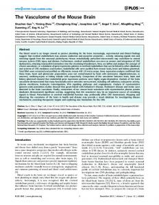
Results In order to assess the effect of Tmem135 overexpression on the heart, we compared the heart from adult transgenic mice over-expressing wild-type Tmem135 (Tmem135 TG) under the control of the ß-actin promoter [11] to that of age-matched, littermate non-transgenic control (WT) mice. Comparison of the mean heart weight to body weight ratio (n = 7 WT, n = 9 TG) at 14 months of age indicated 27% increase in the heart size relative to the body size in Tmem135 TG adult mice compared to WT mice although the difference did not reach statistical significance by unpaired t-test with Welch’s correction (p = 0.085) (Fig 1A). We observed
PLOS ONE | https://doi.org/10.1371/journal.pone.0201986 August 13, 2018
5 / 21
Heart abnormalities caused by Tmem135 overexpression
Fig 1. Tmem135 TG mice display disease phenotypes in the heart. A) Heart to body weight ratios of 14 month old mice show a trend of increase in Tmem135 TG mice indicative of mild cardiac hypertrophy (n = 7 WT, n = 9 TG; p = 0.085 by t-test). Representative Masson’s trichrome-stained hearts at 14 moths of age are shown. B) Masson’s trichrome-stained 6-month-old hearts show collagen in blue (left). The percentage of blue-stained area (% Blue) is significantly increased in Tmem135 TG hearts compared to that in WT hearts (n = 4 WT, n = 4 TG; p200 cells per sample examined, p = 0.0642 by t-test), extracellular space is significantly increased in Tmem135 TG hearts (n = 6 WT, n = 6 TG; p