Malki et al. BMC Medicine 2014, 12:73 http://www.biomedcentral.com/1741-7015/12/73
RESEARCH ARTICLE
Open Access
The endogenous and reactive depression subtypes revisited: integrative animal and human studies implicate multiple distinct molecular mechanisms underlying major depressive disorder Karim Malki1*†, Robert Keers1*†, Maria Grazia Tosto1,2, Anbarasu Lourdusamy3, Lucia Carboni4, Enrico Domenici5,6, Rudolf Uher1,7, Peter McGuffin1 and Leonard C Schalkwyk1
Abstract Background: Traditional diagnoses of major depressive disorder (MDD) suggested that the presence or absence of stress prior to onset results in either ‘reactive’ or ‘endogenous’ subtypes of the disorder, respectively. Several lines of research suggest that the biological underpinnings of ‘reactive’ or ‘endogenous’ subtypes may also differ, resulting in differential response to treatment. We investigated this hypothesis by comparing the gene-expression profiles of three animal models of ‘reactive’ and ‘endogenous’ depression. We then translated these findings to clinical samples using a human post-mortem mRNA study. Methods: Affymetrix mouse whole-genome oligonucleotide arrays were used to measure gene expression from hippocampal tissues of 144 mice from the Genome-based Therapeutic Drugs for Depression (GENDEP) project. The study used four inbred mouse strains and two depressogenic ‘stress’ protocols (maternal separation and Unpredictable Chronic Mild Stress) to model ‘reactive’ depression. Stress-related mRNA differences in mouse were compared with a parallel mRNA study using Flinders Sensitive and Resistant rat lines as a model of ‘endogenous’ depression. Convergent genes differentially expressed across the animal studies were used to inform candidate gene selection in a human mRNA post-mortem case control study from the Stanley Brain Consortium. Results: In the mouse ‘reactive’ model, the expression of 350 genes changed in response to early stresses and 370 in response to late stresses. A minimal genetic overlap (less than 8.8%) was detected in response to both stress protocols, but 30% of these genes (21) were also differentially regulated in the ‘endogenous’ rat study. This overlap is significantly greater than expected by chance. The VAMP-2 gene, differentially expressed across the rodent studies, was also significantly altered in the human study after correcting for multiple testing. (Continued on next page)
* Correspondence:
[email protected];
[email protected] † Equal contributors 1 King’s College London, MRC Social, Genetic and Developmental Psychiatry Centre, at Institute of Psychiatry, SGDP Research Centre (PO80), De Crespigny Park, Denmark Hill, London SE5 8AF, UK Full list of author information is available at the end of the article © 2014 Malki et al.; licensee BioMed Central Ltd. This is an Open Access article distributed under the terms of the Creative Commons Attribution License (http://creativecommons.org/licenses/by/4.0), which permits unrestricted use, distribution, and reproduction in any medium, provided the original work is properly cited.
Malki et al. BMC Medicine 2014, 12:73 http://www.biomedcentral.com/1741-7015/12/73
Page 2 of 14
(Continued from previous page)
Conclusions: Our results suggest that ‘endogenous’ and ‘reactive’ subtypes of depression are associated with largely distinct changes in gene-expression. However, they also suggest that the molecular signature of ‘reactive’ depression caused by early stressors differs considerably from that of ‘reactive’ depression caused by late stressors. A small set of genes was consistently dysregulated across each paradigm and in post-mortem brain tissue of depressed patients suggesting a final common pathway to the disorder. These genes included the VAMP-2 gene, which has previously been associated with Axis-I disorders including MDD, bipolar depression, schizophrenia and with antidepressant treatment response. We also discuss the implications of our findings for disease classification, personalized medicine and case-control studies of MDD. Keywords: Endogenous Depression, Reactive Depression, GENDEP, VAMP-2, DSM-IV, Stanley Brain Consortium, mRNA, Stress
Background Although antidepressants remain the first line treatment for major depressive disorder (MDD), antidepressant response varies considerably between individuals: fewer than half of all patients achieve remission following their first course of treatment [1]. The absence of robust predictors of treatment response means that the most effective antidepressant for a given patient is currently identified by trial and error. This is often a long and costly process which both delays recovery and has a negative effect on long-term outcome [2]. Clinicians have long intuited that heterogeneity in treatment response is the direct result of etiological heterogeneity in MDD [3]. Indeed, traditional diagnoses of major depression proposed that the presence or absence of stress prior to the onset of MDD results in two etiologically distinct subgroups of the disorder with different treatment recommendations. Early studies, which categorized these subtypes as ‘reactive’ (occurring as the result of a stressor) or ‘endogenous’ (occurring in the absence of stress), suggested that those with ‘endogenous’ depression responded more favorably to tricyclic antidepressants (TCAs) than selective serotonin reuptake inhibitors (SSRIs) [4]. While the validity of these subtypes remains unclear, reports continue to show that both distal stress (occurring early in life [5]) and proximal stress (occurring near the onset of a depressive episode [6]) are predictive of treatment response. It remains unclear how the presence or absence of stress in the etiology of MDD affects response to treatment. However, it has been suggested that ‘endogenous’ and ‘reactive’ subtypes of depression are associated with largely distinct biological mechanisms, which respond differentially to treatment [3]. In line with this hypothesis, a recent animal study reported that the hippocampal gene-expression profile of a ‘reactive’ model of depression (induced by chronic restraint stress) differed considerably from that of an ‘endogenous’ model [7]. While this study suggests that the gene-expression profiles of ‘reactive’ depression caused by proximal stress may indeed differ from ‘endogenous’ depression, the role of distal early-life stress in this distinction remains unknown.
Several studies have highlighted the importance of the timing of adversity and show that early and late stressors may have differential tissue-specific effects on gene-expression in the hippocampus [8-12]. The pathophysiological processes underlying MDD may therefore differ not only in the presence or absence of a stressor, but also by the timing of adversity (distal vs. proximal stress). We investigated this hypothesis by exploring hippocampal gene-expression (mRNA) differences in three animal models of depression chosen to represent ‘reactive’ and ‘endogenous’ depression. In the ‘reactive’ depression model, mice were exposed to either distal stress (maternal separation) or proximal stress (unpredictable chronic mild stress). Flinders sensitive rats, which show congenital depression-like behavior, were used to model ‘endogenous’ depression. Whole genome transcription profiles from disease relevant brain tissues in animals may provide valuable support and important information on the molecular mechanisms that may be relevant in humans. Nevertheless, the specific features of psychiatric illnesses means that molecular mechanisms uncovered in animal models are only suggestive and need to be validated in human studies [13,14]. We therefore used findings from the animal models to inform probe set prioritization in a comparable human post-mortem case-control study of depression from the Stanley Brain Consortium. Specifically, we hypothesize that a set of genes that shows concordant expression differences in response to ‘reactive’ and ‘endogenous’ depression models in the rodent studies may represent a common final pathway to MDD. These same genes may therefore also be differentially regulated in the post-mortem brain tissue of humans with the disorder.
Methods Design
Genome-wide expression profiling of the hippocampus (HIP) from two studies from the rodent arm of the Genome-based Therapeutic Drugs for Depression (GENDEP) study [15] was used to inform candidate
Malki et al. BMC Medicine 2014, 12:73 http://www.biomedcentral.com/1741-7015/12/73
gene selection in a comparable human post-mortem, case-control study on MDD from the Stanley Brain Consortium. The GENDEP project is a large-scale, multicenter human pharmacogenomics study that also includes a series of large-scale studies using animal models and in vitro experiments. The GENDEP project was designed to allow for integrative analysis of the results of the transcriptomics and proteomics on the samples from the human, the rodent and the in vitro studies, in order to gain further insight into the molecular mechanisms of MDD and identify biomarkers of antidepressant drugs (AD) treatment response. The mouse study used 144 animals from four strains of well-characterized inbred mice to model individual variation in humans. The mice were subjected to one of two stress protocols and a control condition (maternal separation (MS) - ‘early stress’, unpredictable chronic mild stress (UCMS) - ‘late stress’ - or the control condition (ENV)) to model ‘reactive’ depression. Litters of each strain were randomly allocated to the MS, UCMS or control group. Findings from the mouse study were cross validated in a parallel rat study that compared HIP mRNA differences between Flinders Sensitive and Flinders Resistant rat lines as models of ‘endogenous’ depression. Finally, genes differentially expressed in response to both stress protocols in the mouse study and in the rat study were used to inform probe set selection in comparable mRNA expression study in humans.
Page 3 of 14
use of laboratory animals, which follows the European Communities Council Directive of 24 November 1986. Additional information on the rat study is available elsewhere [18]. UCMS (Unpredictable Chronic Mild Stress)
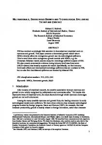
In mice, ‘reactive’ depression caused by proximal stress was modeled using an Unpredictable Chronic Mild Stress (UCMS) paradigm. A third of the 144 mice (48 male and female mice) were exposed to varying stressors on a daily basis for a period of two weeks. Exposure to UCMS commenced when the animals were 10 weeks of age. The UCSM protocols included exposure to different stressors each day in a pseudorandom order. The stressors in the UCMS regime were based on previously published protocols including two hours of home cage tilting at 45°, damp bedding for four hours, cage switching for two hours, flooded cage for 10 minutes, altered length and time of light-dark cycle and air-puff [19]. Animals were exposed to either one or two stressors each day for varying lengths of time (Figure 1). All UCMS-exposed mice were tested and maintained under standard laboratory conditions but were single-housed. Following the UCMS regimen, a set of animals was tested with a battery of behavioral tests including Porsolt as an index of UCMS-evoked depressivebehavior [19]. However, all animals used for this mRNA characterization were not behaviorally tested to control for the potential stressor effects of the tests.
Animals
A total of 144 male and female mice (72 of each sex) from four different strains ((129S1/SvImJ, C57LB/6 J, DBA/2 J and FVB/NJ) were bred in the barrier unit at the Institute of Psychiatry, London, UK. Weaning took place when the animals were 21 to 28 days old. Animals were group-housed under standard conditions with a 12:12 h light:dark cycle, 22°C ± 11°C, food and water ad libitum. A total of 144 animals were sacrificed by cervical dislocation. Animals used for the transcriptomic study were not behaviorally tested. The hippocampus, liver and spleen were dissected following previously published protocols [16,17]. All housing and experimental procedures were carried out in accordance with the UK Home Office Animals (Scientific Procedures) Act, 1986. A total of 39 animals from two cohorts of Flinders Sensitive Lines and Flinders Resistant Lines (22 FRL and 17 FSL) were bred and maintained at Karolinska Institutet (Stockholm) and housed under standard room temperature (22 ± 1°C), relative humidity (45 to 55%) and a 12 h light: dark schedule (light on at 07:00 a.m.). Food and water were available ad libitum. The study was conducted as part of a parallel GENDEP investigation. The Stockholm's Ethical Committee for Protection of Animals approved the study and all procedures were conducted in conformity with the Karolinska Institutet's guidelines for the care and
MS (Maternal Separation)
A maternal separation protocol was used to model ‘reactive’ depression caused by distal stress in a further 48 mice. A single 24-hour separation of the pup from the dam at postnatal day (PND) 9 protocol was chosen to elicit a sufficiently strong biological response. Day of birth was defined as PND 0 for that particular litter. On postnatal Day 9 the dam was removed from the litter for 24 hours. The litter was kept on a heating pad in their home cage at 33°C ± 2°C in a different room than the dam in order to avoid contact through vocalization. Separated pups did not have access to food or water during their separation period. Litters were always separated and reunited with the mother during the first half of the light phase. The first hour after reuniting the litter with the mother was videotaped. Litters were of different sizes and when possible each litter came from a different breeding pair. A more detailed description of the litters is published elsewhere [19]. ‘Endogenous’ model of depression
Flinders Sensitive Lines (FSL) and Flinders Resistant Lines (FRL) rats represent an ‘endogenous’ model of depression [20-23]. Flinders Lines are strains originally obtained by selective breeding of out-bred Sprague-Dawley rats (SD), according to their resistance or sensitivity to
Malki et al. BMC Medicine 2014, 12:73 http://www.biomedcentral.com/1741-7015/12/73
Page 4 of 14
Figure 1 This figure shows the stress administration regime for the unpredictable chronic mild stress paradigm. The duration of the stress regime was for two consecutive weeks and the order of the different stressors was randomized. This figure shows the stressors and time/ duration of administration for each of the two weeks.
anticholinesterase diisopropyl fluorophosphates (DFP) treatment [24]. FSL are congenitally more sensitive to DFP and cholinergic agonists than FRL, which is a neurobiological feature shared with depressed cases in humans [21]. They also show many behavioral similarities to human depressed patients, including decreased psychomotor activity and appetite, cholinergic hypersensitivity, immune and sleep abnormalities including delay in rapid eye movement (REM) sleep but preserved cognitive function and hedonic response [25]. Flinders rats remain a robust model of depression to date [26]. mRNA extraction and lab protocols
Mouse brains, livers and spleens were dissected from each animal and frozen on dry ice. Total RNA was extracted from frozen hippocampal tissue and 3-ug RNA was processed using the One Cycle Target Labelling kit (Affymetrix, Santa Clara, CA, USA) and hybridized to the mouse MOE430v2 Gene Expression Array (Affymetrix) following standard Affymetrix protocols. Hippocampal mRNA extraction from Flinders rats was performed by another participating group from the GENDEP project [18,22]. Briefly, cRNA probes were obtained and hybridized to Affymetrix Rat Genome 230 2.0 using Affymetrix’s One-Cycle Eukaryotic Target Labelling Assay protocol. Protocols used for the human post-mortem mRNA extraction are described in detail in the paper by Iwamoto and colleagues [27]. Briefly, total RNA was extracted from 0.1 g of frozen prefrontal cortex tissues using Trizol (Invitrogen, Groningen, The Netherlands). A total of 8 to 10 mg of mRNA was reverse transcribed and synthesized
into cDNA, hybridized onto Affymetrix HU95A oligonucleotide arrays and scanned using an HP GeneArray scanner (Hewlett-Packard, Palo Alto, CA, USA). Information on The Stanley Foundation brain collection and Neuropathology Consortium is found elsewhere [28]. Human samples
The human samples used in this study were donated to the Stanley Foundation Brain Collection at the Department of Psychiatry, University of the Health Sciences, Bethesda, MD, USA and have been made available to researchers world-wide. Human brain tissues were donated under standardized legislation according to the Uniform Anatomical Gift Act (USA). Information on Stanley Medical Research Institute (SMRI) and its research was offered to the next of kin at the time of the donation. Additional information is publically available from the Stanley Brain Consortium website [29]. The primary transcription-wide analysis was performed and described by Iwamoto and colleagues [27]. For consistency and quality assurance, the same subset has been used without additions or subtractions of cases. All data have been processed from raw files. The samples used consist of post-mortem prefrontal cortex from the Stanley Foundation Neuropathology Consortium from deceased patients affected with major depressive disorder and carefully matched controls. Exclusion criteria include poor mRNA quality and age (>65). A total of 26 samples, 11 cases and 15 controls, were used congruent with the primary data analysis (Table 1). Clinical diagnosis of MDD was made following Diagnostic and Statistical Manual of Mental Disorders – 4th Edition (DSM-IV) diagnostic
Malki et al. BMC Medicine 2014, 12:73 http://www.biomedcentral.com/1741-7015/12/73
Page 5 of 14
Table 1 Genes dysregulated by UCMS Log 2 fold change Transcript
Gene name
Pr. Rsum
c57
DBA
FVB
129
Pfp -Value
1418687_at
Arc
2,841.834
-0.242
-0.279
-0.221
-0.127
G mutation and increased expression of LARS2 gene in the brains of patients with bipolar disorder and schizophrenia. Biol Psychiatry 2005, 57:525–532. 62. Webster MJ, Elashoff M, Weickert CS: Molecular evidence that cortical synaptic growth predominates during the first decade of life in humans. Int J Dev Neurosci 2011, 29:225–236. 63. Yamada M, Takahashi K, Tsunoda M, Nishioka G, Kudo K, Ohata H, Kamijima K, Higuchi T, Momose K, Yamada M: Differential expression of VAMP2/ synaptobrevin-2 after antidepressant and electroconvulsive treatment in rat frontal cortex. Pharmacogenomics J 2002, 2:377–382. 64. Saito S, Takahashi N, Ishihara R, Ikeda M, Suzuki T, Kitajima T, Yamanouchi Y, Iwata N, Yamada M, Yoshida K, Inada T, Ozaki N: Association study between vesicle-associated membrane protein 2 gene polymorphisms and fluvoxamine response in Japanese major depressive patients. Neuropsychobiology 2006, 54:226–230. 65. Major Depressive Disorder Working Group of the Psychiatric GWAS Consortium, Ripke S, Wray NR, Lewis CM, Hamilton SP, Weissman MM, Breen G, Byrne EM, Blackwood DH, Boomsma DI, Cichon S, Heath AC, Holsboer F, Lucae S, Madden PA, Martin NG, McGuffin P, Muglia P, Noethen MM, Penninx BP, Pergadia ML, Potash JB, Rietschel M, Lin D, Müller-Myhsok B, Shi J, Steinberg S, Grabe HJ, Lichtenstein P, Magnusson P, et al: A mega-analysis of genome-wide association studies for major depressive disorder. Mol Psychiatry 2013, 18:497–511. 66. Tansey KE, Guipponi M, Perroud N, Bondolfi G, Domenici E, Evans D, Hall SK, Hauser J, Henigsberg N, Hu X, Jerman B, Maier W, Mors O, O'Donovan M, Peters TJ, Placentino A, Rietschel M, Souery D, Aitchison KJ, Craig I, Farmer A, Wendland JR, Malafosse A, Holmans P, Lewis G, Lewis CM, Stensbøl TB, Kapur S, McGuffin P, Uher R: Genetic predictors of response to serotonergic and noradrenergic antidepressants in major depressive disorder: a genome-wide analysis of individual-level data and a meta-analysis. PLoS Med 2012, 9:e1001326. 67. Keers R, Uher R, Gupta B, Rietschel M, Schulze TG, Hauser J, Skibinska M, Henigsberg N, Kalember P, Maier W, Zobel A, Mors O, Kristensen AS, Kozel D, Giovannini C, Mendlewicz J, Kumar S, McGuffin P, Farmer AE, Aitchison KJ: Stressful life events, cognitive symptoms of depression and response to antidepressants in GENDEP. J Affect Disord 2010, 127:337–342.
Page 14 of 14
68. Piva R, Belardo G, Santoro MG: NF-kappaB: a stress-regulated switch for cell survival. Antioxid Redox Signal 2006, 8:478–486. 69. Pace TW, Mletzko TC, Alagbe O, Musselman DL, Nemeroff CB, Miller AH, Heim CM: Increased stress-induced inflammatory responses in male patients with major depression and increased early life stress. Am J Psychiatry 2006, 163:1630–1633. doi:10.1186/1741-7015-12-73 Cite this article as: Malki et al.: The endogenous and reactive depression subtypes revisited: integrative animal and human studies implicate multiple distinct molecular mechanisms underlying major depressive disorder. BMC Medicine 2014 12:73.
Submit your next manuscript to BioMed Central and take full advantage of: • Convenient online submission • Thorough peer review • No space constraints or color figure charges • Immediate publication on acceptance • Inclusion in PubMed, CAS, Scopus and Google Scholar • Research which is freely available for redistribution Submit your manuscript at www.biomedcentral.com/submit