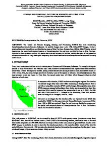
amounts of chondroitin sulphates A and C (shown by alcian blue staining) .... (A-C) Fluorescent micrographs of longitudinal sections of stage-30 MTPJ. Collagen II and ..... chondroitin lyases (chondroitinases) and endo-/S-D- galactosidase ...
Development 99, 383-391 (1987) Printed in Great Britain © The Company of Biologists Limited 1987
383
The spatial and temporal pattern of collagens I and II and keratan sulphate in the developing chick metatarsophalangeal joint
FIONA M. CRAIG, GEORGE BENTLEY and CHARLES W. ARCHER Professorial Research Unit, The Institute of Orthopaedics, Royal National Orthopaedic Hospital, Brockley Hill, Stanmore, Middlesex HA7 4LP, UK
Summary
Both intrinsic and extrinsic factors are known to be involved in the morphogenesis of diarthrodial joints. The use of specific antibodies to collagens I and II and keratan-sulphate-containing proteoglycans (KSPG) has enabled the distributions of these macromolecules to be followed during the development of the third metatarsophalangeal joint in the chicken embryo. Our study shows that cartilage differentiation occurs as a continuous rod, which is then subsequently divided into separate elements. Further development also reveals that, unlike the matrix of the cartilaginous
elements, there is a differential distribution of collagen (type II) and KSPG in the presumptive joint region. It is proposed that a decrease in KSPG in the presumptive joint region at stages 28/30 may be involved in the mechanism for the flattening of cells in formation of the interzone. Whereas, a decrease in collagen across the joint interzone region may provide an area of weakness, which might allow forces produced by the developing musculature to cause cavitation.
Introduction
intermediate interzonal layer decreases. Regional variations in collagen fibre density and arrangement have been described in joint condensations of the developing human elbow joint (Gray & Gardner, 1951). Murray (1926), Murray & Selby (1930), Fell & Canti (1934), Hamburger & Waugh (1940) & Mitrovic (1982) demonstrated with organ culture experiments that intrinsic factors are largely responsible for the determination of the general form and architecture of the joint. Environmental factors also are postulated to play an important role in the formation of joints; some workers emphasize the role of movement in both formation (Lelkes, 1958; Drachman, 1964; Drachman & Sokoloff, 1966; Persson, 1983) and maintenance of the formed joint cavity (Drachman & Coulombre, 1962; Mitrovic, 1972, & see Thorogood, 1983 for review). The relationship of intrinsic and extrinsic factors, and their influence on joint morphogenesis is unclear. The purpose of the present study was to visualize changes in the distribution of collagens I and II and keratan sulphate, (as a representative proteoglycan component) using polyclonal and monoclonal antibodies respectively, during the development of the
A characteristic feature of the cartilage skeleton of embryonic vertebrates is the formation of diarthrodial joints in a precise manner. Bernays (1878) divided joint morphogenesis into two separate phases, the first being formation of the anlage and the second being the completion of the joint. In this respect, it is of interest that studies on digit regeneration in the newt have shown that the cartilage is laid down as a solid rod and thereafter divided into separate digits (Smith, 1978). Changes in histological staining properties have been described during these two phases by a number of authors (Andersen & Bro-Rasmussen, 1961; Andersen, 19626; O'Rahilly & Gardner, 1956; Mitrovic, 1978). Prior to cavitation, Andersen (1962a), in the hip, and Andersen & Bro-Rasmussen (1961) in the joints of the hand and foot, describe large amounts of chondroitin sulphates A and C (shown by alcian blue staining) accumulated intercellularly in the three-layered interzone. When present at the site of the joint, the interzone consists of two chondrogenous layers and an intermediate layer. Just before cavitation the intercellular metachromasia of the
Key words: joint development, collagen I, collagen II, keratan sulphate, chick embryo, pattern.
384
F. M. Craig, G. Bentley and C. W. Archer
third metatarsophalangeal joint in the chick embryo in order to see if cartilage is initially a continuous rod which then subsequently segments, as in the newt. In view of their different mechanical properties, we suggest that changes in the distribution of these extracellular matrix components play a role in the initiation and completion of joint formation. Materials and methods Preparation of specimens Fertilized White Leghorn embryos were incubated, humidified at 38°C (±1°C). Stage-27 to -41 embryos (Hamburger & Hamilton, 1951) were selected and the hind limbs dissected in phosphate-buffered saline. Prior to stage 32, whole leg primordia were fixed in 95 % alcohol at 4cC