The value of specific radiological features in the ... - Semantic Scholar
Recommend Documents
portional to the tumor stage (2,5-11). Besides that, among other methods available for evaluating the patient with prostate neoplasia, we can mention prostatic ...
Objective: Evaluate the ability of serum concentration of prostate specific antigen (PSA) between 2 ... can mention prostatic acid phosphatase, alkaline phos-.
likelihood ratio test in detecting adaptive molecular evolution. Mol Biol Evol. 18:1585â1592. Baum J, Thomas AW, Conway DJ. 2003. Evidence for diversifying.
Species-Specific Features of DARC, the Primate Receptor for. Plasmodium vivax and Plasmodium knowlesi. Ann Demogines,. 1. Kimberly A. Truong,. 1.
different denominations, such as ameloblastoma of unusual type with calcification, calcifying ameloblastoma, malignant odontoma and cystic complex odontoma ...
Jan 25, 2016 - Subjects Orthopedics, Radiology and medical imaging, Surgery and ... Keywords Sacral fracture, Radiological study, Sacral alar iliac screw, ...
May 25, 2016 - Brain sar- ...... A characteristic clinical manifestation of cutaneous sar- .... Bernaciak J, Spina JC, Curros ML, Maya G, Venditti J, Chacon C.
Table 1 Contents of radionuclides in soil and plants grown in the vicinity of the wrecked Reactor. ..... neoplasm, conge
Apr 9, 2015 - of Interleukin-8 in Colorectal Cancer: A Meta- ... Union for International Cancer Control(UICC), the prognosis of advanced stage is very poor,.
Oct 8, 2016 - Dysexecutive symptoms have tre- mendous impact on ..... earlier detection of dysexecutive syndrome in AD can improve the potential benefit of ...
[2] J. W. Fisher III and T. Darrell, âSpeaker association with signal-level audiovisual fusion,â IEEE Transactions on Multimedia, vol. 6, .... McGraw-Hill, 1999, ch.
Sep 20, 2015 - terminal ileum, jejunum, and colon, enteropathy-associated. T-cell lymphoma ... enlarging radiation folds [21]; the second are usually revealed.
General Practitioner, Lecce, Italy and Member of ASSIMEFAC ... of new diagnostic instruments, e.g. computerised ... constitution and on the equipment used.
Bq/m2, respectively. Most of the ..... in terms of both activity per unit soil weight (Bq/kg) and ... 788). D3. 0-10. 7.60 ± 0.30 ( 1040) 3.75 ± 0.12 ( 513). 10-20. n.d..
HIPAA compliance. This research was presented in part at the Association of Research in Vision and Ophthalmology;. May 7th, 2012; Fort Lauderdale, FL, USA.
Jan 6, 2003 - rethorical devices in order to build an argumentative discourse. The prosodic features will be described and justified as well as the possible ...
Dec 1, 2014 - Cases with Cryptogenic Brain Abscess in Association with Patent Foramen Ovale: A Case Report and Review of the Litera- ture. International ...
mercial abattoir, employing a standard slaughter pro tocol. Oviducts were collected ..... sections of the oviducal epithelium slough off during regression. Further ...
Jan 6, 2003 - The silence is also contributing to this delay. Moreover, the Maxim of Pitch Range is also present every time a prosodic phrase starts. Figure 1: ...
primary intra-osseous carcinoma arising de novo within the jaws (PIOC) as `a squamous cell carcinoma within the jaw, having no initial connection with the.
harmful effects of radiation on the health of people" , and, therefore ... A very important aspect of the Concept is the
Apr 18, 2013 - Chesebro B, Trifilo M, Race R, Meade-White K, Teng C, et al. (2005) ... Tuzi NL, Cancellotti E, Baybutt H, Blackford L, Bradford B, et al. (2008) ...
Research, University College Dublin, Belfield, Dublin 4, Ireland, and â¡Department of Neuropathology, Beaumont Hospital, Beaumont Road, Dublin 9, Ireland.
May 28, 2015 - Citation: García-Basteiro AL, López-Varela E,. Augusto OJ, Gondo K, Muñoz J, Sacarlal J, et al. (2015) Radiological Findings in Young Children.
The value of specific radiological features in the ... - Semantic Scholar
Hospital, London,. England ... London SW17 0QT, UK. F. Dinah ..... Danny Acton for their contribution as observers, without whose time and effort this study ...
Trauma
The value of specific radiological features in the classification of acetabular fractures
V. Patel, A. Day, F. Dinah, M. Kelly, M. Bircher From St George’s Hospital, London, England
Specific radiological features identified by Brandser and Marsh were selected for the analysis of acetabular fractures according to the classification of Letournel and Judet. The method employs a binary approach that requires the observer to allocate each radiological feature to one of two groups. The inter- and intra-observer variances were assessed. The presence of articular displacement, marginal impaction, incongruity, intra-articular fragments and osteochondral injuries to the femoral head were analysed by a similar method. These factors were termed ‘modifiers’ and are generally considered when planning operative intervention and, critically, they may influence prognosis. Six observers independently assessed 30 sets of plain radiographs and CT scans on two separate occasions, 12 weeks apart. They were asked to determine the presence or absence of specific radiological features. This simple binary approach to classification yields an interκ = 0.49 to 0.88 and intra-observer agreement which ranges from moderate to near-perfect (κ and κ = 0.57 to 0.88, respectively). A similar approach to the modifiers yields only slight to κ = 0.20 to 0.34) and slight to moderate intra-observer fair inter-observer agreement (κ κ = 0 to 0.55). agreement (κ
Pelvic osteology is complex, and especially so in the peri-acetabular region. The accurate assessment of fracture lines forms the basis of a commonly-used classification proposed by Judet, Judet and Letournel1 and Letournel and Judet2 which comprises ten categories, and the process of classification requires the placement of a continuous variable into discrete categories. This process is likely to result in some disagreement.3 Previous studies have documented a variance in classification according to the Letournel and Judet classification.4-7 Predictably, both experience and training improve the ability of an observer to classify an acetabular fracture.5,6 Consequently, some authors have attempted to identify a systematic checklist of radiological features to facilitate this process for less experienced observers.6,8,9 This study analyses the inter- and intra-observer variances of one such systematic binary approach as described by Brandser and Marsh9 (Table I). It may, on occasion, be appropriate to manage an acetabular fracture non-operatively. The decision to operate is influenced by the presence of features thought to indicate a poor prognois if managed non-operatively.10,11 This study assessed the level of agreement among
observers when assessing these ‘modifiers’ (Table II).
Materials and Methods The radiographs and CT scans of 30 patients were selected from the Pelvic Reconstruction Unit database and represent a broad range of types of acetabular fracture. The plain radiographs comprise both anteroposterior (AP) and Judet views. Both radiographs and CT scans were rendered anonymous for the purpose of the study, and were catalogued according to a study reference number. Six independent observers were asked to complete a questionnaire for each set of radiographs. The observers included two consultant orthopaedic surgeons (AD, MB) with a special interest in fractures of the pelvis and acetabulum, one consultant orthopaedic surgeon (not an author) with an interest in lower limb reconstruction, and three higher surgical trainees (VP, and two who were not authors; years 6, 4, and 2, respectively). The participants were not given a copy of The Brandser and Marsh9 original paper so as to prevent inadvertent use of the summary table. The participants had varying degrees of experience, but all had worked on the Pelvic and Acetabular Reconstruction Unit, which is a tertiary THE JOURNAL OF BONE AND JOINT SURGERY
THE VALUE OF SPECIFIC RADIOLOGICAL FEATURES IN THE CLASSIFICATION OF ACETABULAR FRACTURES
73
Table I. Systematic approach to assessing acetabular fractures9 Question
Radiological feature
1 2 3 4 5 6 7
Is there a fracture of the obturator ring? Is the ilioischial line disrupted? Is the iliopectineal line disrupted? Is the iliac wing above the acetabulum fractured? Is the posterior wall fractured? Does the fracture divide the acetabulum into superior and inferior or anterior and posterior halves? Can an intact strut of bone be followed from the sacroiliac joint to the acetabular articular surface, or is a spur sign present? What is the orientation of the major fracture line on the axial CT?
8
Table II. Modifiers to assessing acetabular fractures16 Modifier
Description of radiological feature
1
Articular displacement: step > 1 mm or gap > 3 mm Marginal impaction Incongruity, including subluxation or dislocation Intra-articular fragment(s) Femoral head lesion(s)
2 3 4 5
Table III. Categorisation of κ values (after Landis and Koch15) κ value
Strength and agreement
< 0.00 0.00 to 0.20 0.21 to 0.40 0.41 to 0.60 0.61 to 0.80 0.81 to 1.00
Poor Slight Fair Moderate Substantial Near perfect
referral centre assessing around 150 cases per year. Prior to the analysis, participants received a tutorial on the use of AP and Judet views, and of axial CT reformats in the classification of fractures according to the Letournel-Judet classification. Detailed instruction was then provided on Brandser and Marsh’s systematic method, with specific attention to the definitions that underpin the questions in Table I. This analysis comprises one arm of a more extensive study12 addressing the inter- and intra-observer agreement for the classification of acetabular fractures according to both the AO13 and Letournel-Judet classifications. The same group of observers used the identical 30 sets of plain radiographs and CT scans as were used for this study. The Letournel-Judet classification produced substantial intraobserver agreement (κ = 0.61) and moderate inter-observer agreement (κ = 0.53). This is consistent with the published findings of Beaule et al.5 The intra-observer agreement on the AO type, group and subgroup were substantial (κ = 0.75), substantial (κ = 0.61) and fair (κ = 0.42), respectively. The inter-observer agreement on the AO type, group and subgroup were substantial (κ = 0.69), moderate (κ = 0.55) and poor (κ = 0.30), respectively. VOL. 89-B, No. 1, JANUARY 2007
The questionnaire requires observers to answer eight questions relating to specific radiological features9 (Table I). All observers were required in addition to determine the presence of five modifiers (Table II). The two consultant orthopaedic surgeons with a special interest in fractures of the pelvis and acetabulum were asked to select an operative approach in each case. Following completion of the firstround evaluation, the radiographs and CT scans were randomly assigned new identification numbers and were returned to the observers after an interval of 12 weeks for the second-round evaluation. Statistical analysis. Values for multiple observers were calculated using the method of Fleiss.14 The analysis was performed with Stata 5.0 (Stata Corp., College Station, Texas). The κ values were categorised according to the guidelines of Landis and Koch15 (Table III).
Results The 30 sets of radiographs and CT scans were analysed by each of the six observers on two separate occasions, as described. Nine of ten categories comprising the LetournelJudet classification were used at least once, which suggests a representative case selection. The inter- and intra-observer errors of the eight radiological features are presented in Table IV. There is moderate to near-perfect inter-observer agreement (κ = 0.49 to 0.88). The intra-observer agreement varied from moderate to near-perfect (κ = 0.57 to 0.88). The inter- and intra-observer errors for each of the five modifiers are presented in Table V. There is only slight to fair inter-observer agreement (κ = 0.20 to 0.34) and slight to moderate intra-observer agreement (κ = 0 to 0.55). There was substantial agreement in respect of the operative approach (κ = 0.64).
Discussion The classification proposed by Letournel and Judet is widely accepted by researchers and is helpful in the selection of surgical approach.16 Its application has been described elsewhere and it appears to be a largely successful classification tool.5-7 The variance observed may be a product of the classification process. Fracture configurations are generally a continuous variable, and most classification systems constrain the individual case to membership of a
74
V. PATEL, A. DAY, F. DINAH, M. KELLY, M. BIRCHER
Table IV. κ values for agreement on radiological features in systematic approach Radiological features
Inter-observer
Intraobserver
Fracture of the obturator ring Disruption of the ilioischial line Disruption of the iliopectineal line Fracture of the iliac wing above the acetabulum Fracture of the posterior wall Fracture divides the acetabulum into anterior and posterior or superior and inferior halves (assessed on axial CT) Floating acetabulum (presence of the ‘spur’ sign) Orientation of the principal fracture line on axial CT (sagittal or coronal)
0.64 0.68 0.81 0.88 0.49 0.51
0.65 0.79 0.83 0.88 0.57 0.65
0.65 0.56
0.68 0.66
Table V. κ values for agreement on modifiers Modifier
Inter-observer
Intra-observer
Articular displacement Marginal impaction Incongruity Intra-articular fragment(s) Femoral head lesion(s)
0.24 0.22 0.34 0.25 0.20
0.42 0.55 0.49 0.35 0.00
categorical dataset. This will invariably lead to some disagreement among observers in equivocal cases, and particularly so when assignment to a category requires the consideration of many factors. If the Letournel-Judet classification fracture type proposed by observers is compared with the fracture type predicted by the summary table from the collated binary data, the level of agreement is only moderate (κ = 0.44). The introduction of binary selection methods attempts, in effect, to reduce the cumulative error by clarifying the items of choice. That is, either the required feature is present or it is not. This should, in theory, smooth the transition from a continuous variable to a categorical dataset. Although the analysis of specific radiological features may be useful in assigning cases to the various categories of the Letournel-Judet classification,8,9 the consistency of this approach has not so far been determined. This study considered the systematic approach described by Brandser and Marsh,9 based on the identification of eight key radiological features (Table I). There is substantial to near-perfect agreement on five of these features (1, 2, 3, 4 and 7), but only moderate agreement on the remaining three (5, 6 and 8); this observation requires further analysis. Question 5 requires clarification. It seeks to address whether or not there is a fracture of the posterior wall. There are some fracture patterns, such as the transverse, in which there is a fracture line running through the posterior wall. This is, however, quite distinct from an isolated fracture of the posterior wall, and for that matter an associated transverse-posterior wall fracture as described according to the Letournel-Judet classification. Inexperienced observers tend not to distinguish between the two categories, and a satisfactory binary method must address this equivocation. The question may be refined by the following requirement:
the fracture line must remain within the posterior wall throughout the axial CT sequence. It should not cross the quadrilateral plate and should not extend anterior to the cotyloid fossa (Fig. 1). In the event of a transverse-posterior wall fracture, the posterior fracture is distinct from the transverse fracture and is recognised by the presence of two sagittal fracture lines, only one of which extends anterior to the cotyloid fossa. In other words, if the fragment lateral to a fracture line which crosses the posterior wall can be followed cephalad through sequential sections and into the greater sciatic buttress, there is no posterior wall fracture. Questions 6 and 8 return only moderate agreement and require the orientation of the major fracture line to be identified on axial CT. These questions aim to differentiate fractures that remain within either the anterior or the posterior column from those that traverse both. For example, a typical transverse fracture has a sagittal orientation and will effectively divide the acetabulum into superior and inferior halves. A typical anterior or posterior column fracture has coronal orientation and effectively divides the acetabulum into anterior and posterior halves. A ‘T’ fracture pattern, on the other hand, comprises fracture lines of each orientation, and complex or associated fracture patterns may contain many fracture lines of equivocal orientation. The questions may therefore be refined to address this problem. The first observation should assess whether there are one or two principal fracture lines. Where there are two, they are not mutually exclusive and should therefore be assessed independently. With reference to axial CT sequences of the acetabulum, a fracture line that crosses a coronal plane passing through the anterior extent of the cotyloid fossa and traversing both columns, is effectively transverse in orientation (Fig. 2). On the other hand, a fracture line that fails to cross a coronal plane running through the cotyloid fossa and additionally fails to exit through a column other than that in which it started, may be considered coronal in orientation (Fig. 3). The former group contains transverse and transverse-associated fracture patterns, and the latter contains isolated column and associated both-columns fractures. ‘T’ pattern fractures contain lines of each orientation. Interestingly, questions 6 and 8 seek to corroborate information that may be derived from the answers to other questions, and as such are not strictly necessary. This is THE JOURNAL OF BONE AND JOINT SURGERY
THE VALUE OF SPECIFIC RADIOLOGICAL FEATURES IN THE CLASSIFICATION OF ACETABULAR FRACTURES
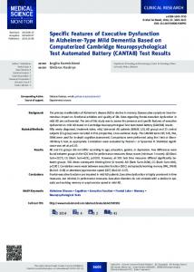
Fig. 2 Diagrammatic axial CT scans showing a transverse fracture. The fracture line crosses a coronal plane (shown by a horizontal line) passing through the anterior extent of the cotyloid fossa.
Fig. 3 Diagrammatic axial CT scans showing a posterior column fracture. The fracture line does not cross a coronal plane (shown by a horizontal line) passing through the anterior extent of the cotyloid fossa.
alluded to by Brandser and Marsh.9 They draw attention, however, to the importance of axial CT sequences in the confirmation of plain radiological observations. This technique is commonly practised by many pelvic and acetabular surgeons, and for the purpose of this study the questions have been included. If questions 6 and 8 are omitted from the analysis, the overall intra- and inter-observer agreements remain in the ‘substantial’ range, despite rising from 0.71 to 0.73 and from 0.65 to 0.69, respectively. Previous studies have shown that cases are most accurately assigned to Letournel-Judet classification criteria by those with experience or training in the use of this classification.5,6 The systematic approach examined in this study demonstrates a certain level of success when applied to those less experienced observers. When the two most VOL. 89-B, No. 1, JANUARY 2007
experienced observers were excluded from the analysis, the overall intra-observer agreement fell from 0.66 to 0.52, and the overall inter-observer agreement fell from 0.65 to 0.53. There are, however, significant flaws, some of which may be addressed by means of further clarification. The three features causing most disagreement among observers have already been addressed. The concept of a binary method is appealing, and it proves more effective than initial classification in the hands of an inexperienced observer. Subtle refinement may reduce the level of inter- and intra-observer error while retaining the elegance and simplicity of the method. Despite the usefulness of the Letournel-Judet classification, it does not address certain features of an acetabular fracture that may critically influence the prognosis.10 These features are listed in Table II and are included in the com-
76
V. PATEL, A. DAY, F. DINAH, M. KELLY, M. BIRCHER
prehensive classification proposed by the AO group.13 The ability of surgeons to identify these features consistently has not been assessed. This study analyses the level of agreement among surgeons when identifying these five modifiers.16 There is only slight to fair inter-observer agreement (κ = 0.20 to 0.34) and slight to moderate intra-observer agreement (κ = 0.00 to 0.55). The results imply that observers of variable experience are unable to agree on features that might determine prognosis and potentially influence the decision to operate. The decision should therefore be left to experienced pelvic and acetabular reconstruction surgeons. Other factors, such as age, comorbidity, associated injuries and timing of the index referral, influence the decision-making process. The two consultant orthopaedic surgeons with a special interest in fractures of the pelvis and acetabulum were asked to determine the most suitable operative approach for each fracture. It is reassuring to observe a substantial interobserver agreement (κ = 0.64). In conclusion, this study considers the use of a systematic, binary method for the classification of acetabular fractures. Five of the eight criteria appear reliable when assessed by means of inter- and intra-observer error. The remaining three require further refinement. Non-specialist observers are unable consistently to detect features that might determine prognosis and potentially influence the decision to operate. It is therefore prudent to seek the opinion of an experienced pelvic and acetabular reconstruction surgeon at the earliest possible opportunity. We would like to thank Mr Adrian Fairbank, Mr Anand Masilamani and Mr Danny Acton for their contribution as observers, without whose time and effort this study would not have been possible. We are also grateful to Professor J. M. Bland (Department of Public Health Sciences, University of York, United Kingdom) for his help with the statistical analyses. No benefits in any form have been received or will be received from a commercial party related directly or indirectly to the subect of this article.
References 1. Judet R, Judet J, Letournel E. Fractures of the acetabulum: classification and surgical approaches for open reduction. J Bone Joint Surg [Am] 1964;46-A:161546. 2. Letournel E, Judet R. Fractures of the acetabulum. Second ed. Berlin: SpringerVerlag, 1993. 3. Swiontkowski MF, Sands AK, Agel J, et al. Interobserver variation in the AO/ OTA fracture classification system for pilon fractures: is there a problem? J Orthop Trauma 1997;11:467-70. 4. Letournel E. Acetabulum fractures: classification and management. Clin Orthop 1980;151:81-106. 5. Beaule PE, Dorey FJ, Matta JM. Letournel classification for acetabular fractures: assessment of interobserver and intraobserver reliability. J Bone Joint Surg [Am] 2003;85-A:1704-9. 6. Petrisor BA, Bhandari M, Orr RD, et al. Improving reliability in the classification of fractures of the acetabulum. Arch Orthop Trauma Surg 2003;123:228-33. 7. Visutipol B, Chobtangsin P, Ketmalasari B, Pattarabanjird N, Varodompun N. Evaluation of Letournel and Judet classification of acetabular fractures with plain radiographs and three-dimensional computerized tomographic scan. J Orthop Surg (Hong Kong) 2000;8:33-7. 8. Saterbak AM, Marsh JL, Turbett T, Brandser E. Acetabular fractures classification of Letournel and Judet: a systematic approach. Iowa Orthop J 1995;15:184-96. 9. Brandser E, Marsh JL. Acetabular fractures: easier classification with a systematic approach. AJR Am J Roentgenol 1998;171:1217-28. 10. Tile M. Fractures of the pelvis and acetabulum. Second ed. Baltimore: Williams & Wilkins, 1995. 11. No authors listed. Fracture and dislocation compendium. J Orthop Trauma 1996;10(Supp 1):71-5. 12. Middleton F, Patel VR, Kelly M, et al. Classification of fractures of the acetabulum: does a systematic approach improve reliability? Poster presented at EFORT 6th Congress, Helsinki, Finland, 2003. 13. Muller ME, Nazarian S, Koch P, Schatzker J. The comprehensive classification of fractures of long bones. Berlin: Springer-Verlag, 1990. 14. Fleiss JL. Measuring nominal scale agreement among many raters. Psychological Bulletin 1971;76:378-82. 15. Landis JR, Koch GG. The measurement of observer agreement for categorical data. Biometrics 1977;33:59-74. 16. Mayo KA. Fractures of the acetabulum. Orthop Clin North Am 1987;18:43-57.