B. Jochimsen et(1997) al.: Stetteria hydrogenophila Extremophiles 1:151–156
151 © Springer-Verlag 1997
ORIGINAL PAPER Yoshihiro Hakamada · Kenzo Koike Tadashi Yoshimatsu · Hajime Mori · Tohru Kobayashi Susumu Ito
Thermostable alkaline cellulase from an alkaliphilic isolate, Bacillus sp. KSM-S237
Received: April 28, 1997 / Accepted: May 24, 1997
Key words Bacillus · Alkaliphilic · Alkaline · Thermostable · Cellulase · Purification
strains of Bacillus that produce alkaline cellulases (endo1,4-β-glucanase, EC 3.2.1.4), the properties of which fulfill the essential requirements for enzymes to be used in laundry detergents (Ito 1997). Alkaline cellulases from Bacillus strains are effective for the removal of soils from cotton fabrics without degradation of the cotton fibers (Hoshino and Ito 1997). A mutant of one of our isolates, Bacillus sp. KSM-635 (Ito et al. 1989; Yoshimatsu et al. 1990), is currently exploited for the large-scale industrial production of an alkaline cellulase (Ito et al. 1991). The alkaline cellulase based detergents are now being marketed in Japan and Asian countries. Most of the alkaline cellulases from Bacillus reported to date are not very thermostable (Horikoshi et al. 1984; Fukumori et al. 1985; Okoshi et al. 1990; Shikata et al. 1990; Yoshimatsu et al. 1990), and therefore are not economical or suitable for home laundering at high temperature. During the course of screening for detergent cellulaseproducers, we found a mesophilic, alkaliphilic strain of Bacillus that produced a thermostable, alkaline cellulase. In this report, we describe the purification and some properties of the novel enzyme from the mesophilic, alkaliphilic isolate.
Introduction
Materials and methods
Abstract Thermostable alkaline cellulase (endo-1,4-βglucanase, EC 3.2.1.4) activity was detected in the culture medium of a strictly alkaliphilic strain of Bacillus, designated KSM-S237. This novel enzyme was purified to homogeneity by a two-step column-chromatographic procedure with high yield. The N-terminal amino acid sequence of the purified enzyme was Glu-Gly-Asn-Thr-Arg-Glu-Asp-AsnPhe-Lys-His-Leu-Leu-Gly-Asn-Asp-Asn-Val-Lys-Arg. The enzyme had a molecular mass of approximately 86 kDa and an isoelectric point of pH 3.8. The enzyme had a pH optimum of 8.6–9.0 and displayed maximum activity at 45°C. The alkaline enzyme was stable up to 50°C and more than 30% of the original activity was detectable after heating at 100°C and at pH 9.0 for 10 min. The enzyme hydrolyzed carboxymethylcellulose, lichenan (β-1,3;1,4-linkage), and p-nitrophenyl derivatives of cellotriose and cellotetraose. Crystalline forms of cellulose (Avicel and filter paper), H3PO4-swollen cellulose, NaOH-swollen cellulose, curdlan (β-1,3-linkage), laminarin (β-1,3;1,6-linkage), and xylan were barely hydrolyzed at all.
The members of Bacillus often produce extracellular enzymes of industrial importance (Priest 1977; Horikoshi 1996). We have isolated many alkaliphilic and neutrophilic
Communicated by K. Horikoshi Y. Hakamada (*) · K. Koike · T. Yoshimatsu · H. Mori · T. Kobayashi · S. Ito Tochigi Research Laboratories of Kao Corporation, 2606 Akabane, Ichikai, Haga, Tochigi 321-34, Japan Tel. 181-285-68-7400; Fax 181-285-68-7403 e-mail:
[email protected]
Organism and culture conditions The organism used was Bacillus sp. KSM-S237, which was originally isolated from a soil sample collected in Okinawa, Japan. Its morphological and taxonomic characteristics were examined according to the methods of Gordon et al. (1973) and Nielsen et al. (1995). Bacillus sp. KSM-S237 was propagated at 30°C in a medium composed of (w/v): 0.1% carboxymethylcellulose (CMC; A10MC from Nihon Pulp, Tokyo, Japan), yeast extract (Difco, Detroit, MI, USA), 2.0% Polypepton S (Nihon Pharmaceutical, Tokyo, Japan), 1.0% meat extract (Wako Pure Chemical, Kyoto, Japan), 0.5% sodium glutamate,
152
0.15% K2HPO4, 0.01% CaCl2·2H2O, 0.02% MgSO4·7H2O, 0.001% FeSO4·7H2O, 0.0001% MnSO4·4H2O, and 0.5% Na2CO3. Solutions of CaCl2 and Na2CO3 were separately autoclaved. The organism was grown with shaking at 30°C for 40 h in 50-ml aliquots of medium in 500-ml flasks. Cells were harvested by centrifugation (12 000 3 g, 15 min) at 5°C. All further manipulations were also done at this temperature. The supernatant obtained was used for purification of the enzyme.
N. Matsuda et al.: EGF receptor and osteoblastic differentiation
of 0.42 ml·cm22·min21 with a 0.6-l gradient of 0–0.6 M NaCl in the equilibrating buffer. The active fractions (tubes nos. 115–118) were combined, diluted with 200 ml of 10 mM Tris-HCl buffer (pH 7.5), and then concentrated to a small volume by ultrafiltration on a PM10 membrane (Amicon, Denver, MA, USA). The resultant concentrate was used for further experiments as the final preparation of enzyme. Electrophoresis
Enzyme assays The routine assays for carboxymethylcellulase (CMCase) activity and hydrolytic activities toward cellulosic and noncellulosic polysaccharides were performed in 0.1 M glycineNaOH buffer (pH 9.0), as described previously (Ito et al. 1989). After the reaction mixture had been incubated at 40°C for an appropriate period, the reducing sugar formed was quantified as glucose by the dinitrosalicylic acid procedure (Miller 1959). Hydrolytic activities toward p-nitrophenyl (PNP) β-d-cello-oligosaccharides, including PNP β-d-glucopyranoside (PNPG1), PNP β-d-cellobioside (PNPG2), PNP β-d-cellotrioside (PNPG3), PNP β-dcellotetroside (PNPG4), and PNP β-d-cellopentaoside (PNPG5) (Seikagaku Kogyo, Tokyo, Japan), were measured at 40°C with 5 mM of each substrate in the same buffer. The p-nitrophenol liberated was determined at 400 nm, using an ε value of 15 000. One unit of enzyme activity was defined as the amount of protein that produced 1 µmol product per min under these conditions. Protein was quantified by a modification of the method of Lowry et al. (1951) using the DC protein assay kit (Bio-rad, Richmond, CA, USA), with bovine serum albumin as the standard.
Purification of the enzyme The crude enzyme was prepared by treating the supernatant of centrifuged cultures with ammonium sulfate and isolating the fraction that precipitated between 60% and 90% saturation. The precipitates were collected by centrifugation (12 000 3 g, 20 min). A small amount of 10 mM TrisHCl buffer (pH 7.5) was added to the precipitates, which were then dialyzed overnight against the same buffer. The dialyzed retentate was applied to a column (2.5 cm inner diameter (i.d.) 3 23 cm) of diethylaminoethyl (DEAE)Toyopearl 650M (Tosoh, Tokyo, Japan) that had been equilibrated with 10 mM Tris-HCl buffer (pH 7.5). After the column had been washed with 250 ml of the equilibrating buffer, proteins were eluted in 4-ml fractions at a flow rate of 0.7 ml·cm22·min21 with a 0.8-l gradient of 0– 1.0 M NaCl in the same buffer. The active fractions (tubes nos. 85–91) were pooled and diluted to 200 ml with 10 mM Tris-HCl buffer (pH 7.5). The diluted solution was put directly on a column (1.5 cm i.d. 3 20 cm) of DEAEToyopearl 650S equilibrated with 10 mM Tris-HCl buffer (pH 7.5). Elution was done in 2-ml fractions at a flow rate
Nondenaturing polyacrylamide gel electrophoresis (PAGE) was done on 7.5% polyacrylamide slab gels, according to the method of Davis (1964), with 5 mM Tris/ 38 mM glycine buffer (pH 8.5) as the running buffer. Sodium dodecyl sulfate (SDS) PAGE (SDS-PAGE) was done on 12.5% polyacrylamide slab gels with 25 mM Tris-glycine buffer (pH 8.3) plus 0.1% SDS (Laemmli 1970). Isoelectric focusing of the purified enzyme was performed on 5% polyacrylamide slab gels that contained 1.9% Biolyte (pH range 3–5; Bio-rad), essentially as described by Wrigley (1971). The standard markers with known isoelectric point (pI; Bio-rad) included amyloglucosidase (pI 3.5), methyl red (pI 3.75), phycocyanin (pI 4.65), and β-lactoglobulin (pI 5.1). Protein bands were visualized by staining with Coomassie Brilliant Blue dye G-250 (Wako). Activity staining of cellulase in the slab gel was done essentially by the Congo red-CMC procedure (Yoshimatsu et al. 1990), using CMC-agar sheets adjusted to pH 10 with 0.1 M glycine-NaOH buffer. Amino-terminal sequencing The N-terminal amino acid sequence of the purified enzyme was analyzed with an automated protein sequencer (model 477A; Applied Biosystems, Foster City, CA, USA). The sample (approximately 10 µg) was blotted onto a polyvinylidene difluoride membrane using a ProSpin cartridge (Applied Biosystems). Estimation of molecular mass Molecular mass was estimated both by gel filtration on a column of Sephacryl S-200 (1.5 cm i.d. 3 80 cm; Pharmacia, Uppsala, Sweden) and by SDS-PAGE (12.5% polyacrylamide). The Sephacryl S-200 column (1.5 cm i.d. 3 80 cm) was equilibrated with 10 mM Tris-HCl buffer (pH 7.5) plus 2 mM CaCl2 and 0.1 M NaCl, and calibrated by elution of standard protein markers which included alcohol dehydrogenase (150 kDa; Sigma, St. Louis, MO, USA), bovine serum albumin (67 kDa; Pharmacia), ovalbumin (43 kDa; Pharmacia), and carbonic anhydrase (29 kDa; Sigma). For the determination of molecular mass by SDS-PAGE, molecular mass markers (Bio-rad) were used: phosphorylase b (97.4 kDa), bovine serum albumin (66.2 kDa), ovalbumin (45 kDa), carbonic anhydrase (31 kDa), and soybean trypsin inhibitor (21.5 kDa).
B. Jochimsen et al.: Stetteria hydrogenophila
153
Results and discussion Taxonomic characterization of strain KSM-S237 The isolate, designated KSM-S237, was a facultative anaerobe, which was spore-forming (cylindrical, subterminal endospores with a swollen sporangium), Gram-positive, motile, and rod-shaped (0.6–0.8 µm 3 3.0–5.0 µm) with peritrichous flagella. It was capable of growing over the pH range of 9–12, but not at pH 7, and the range of temperature for growth was between 10°C and 40°C. The strict alkaliphile was positive for utilization of citrate (in Koser’s medium); reduction of nitrate; production of oxidase and catalase; hydrolysis of starch, casein, and esculin; and liquefaction of gelatin; and was negative for acid fastness, denitrification, formation of indole and H2S, production of urease, and growth on d-xylose. It seems likely that this isolate is a relative of Bacillus agaradhaerens (Nielsen et al. 1995).
The N-terminal amino acid sequence of the purified enzyme, determined by Edman sequencing, was Glu-GlyAsn-Thr-Arg-Glu-Asp-Asn-Phe-Lys-His-Leu-Leu-GlyAsn-Asp-Asn-Val-Lys-Arg. The sequence is identical to those of alkaline cellulases, belonging to family A5, from Bacillus sp. strain 1139 (Fukumori et al. 1986) and Bacillus sp. KSM-64 (Sumitomo et al. 1992), but is completely different from the sequence of alkaline cellulase belonging to family A2, from Bacillus sp. KSM-635 (Ozaki et al. 1990). Therefore, the Bacillus sp. KSM-S237 cellulase may belong to the cellulases of family A5.
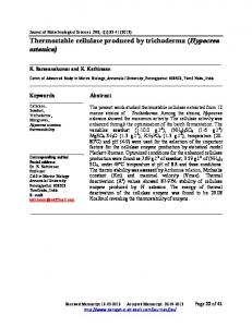
Purification of the enzyme The CMCase was purified to homogeneity from cultures of alkaliphilic Bacillus sp. KSM-S237 by precipitation with ammonium sulfate and the subsequent two-step columnchromatographic procedure. Figure 1a shows that the preparation of purified enzyme was homogeneous, as judged by nondenaturing PAGE. The band of the purified protein, detected by staining after nondenaturing PAGE, coincided fairly well with the single band seen by activity staining (Fig. 1b). The purification and total recovery of the enzyme are summarized in Table 1. Approximately 236-fold purification to a specific activity of 49.4 U/mg protein was obtained for the CMCase activity when measured at pH 9.0 and at 40°C. Molecular mass, isoelectric point, and N-terminal amino acid sequence The molecular mass of the purified enzyme was estimated to be approximately 86 kDa, as determined both by gel filtration on Sephacryl S200 and by SDS-PAGE. The pI of the enzyme was estimated to be pH 3.8 by isoelectric focusing under native conditions.
Fig. 1. Nondenaturing polyacrylamide gel electrophoresis (PAGE) (a) and activity staining (b) of the purified enzyme from Bacillus sp. KSM-S237. Approximately 5.6 µg of protein was put on each gel. Migration was from top (anode) to bottom (cathode). After electrophoresis, each gel was stained for protein and activity. After cutting the gels containing the marker dye Bromophenol blue, the gels were photographed
Table 1. Purification of carboxymethylcellulase (CMCase) activity from alkaliphilic Bacillus sp. KSM-S237 Purification step
Total protein (mg)
Total activity (units)
Crude enzyme
1892
Specific activity (units/mg)
Yield (%)
Purification (fold)
401
0.21
100
1
Ammonium sulfate (60%–90%)
64.8
270
4.1
67
19.5
DEAE-Toyopearl 650M
6.7
253
37.8
63
180
DEAE-Toyopearl 650S
3.4
168
49.4
42
236
DEAE, diethylaminoethyl.
154
N. Matsuda et al.: EGF receptor and osteoblastic differentiation
Substrate specificity As shown in Table 2, the purified enzyme hydrolyzed CMC, lichenan (β-1,3;1,4-linkage), PNPG3, and PNPG4. The enzyme rapidly decreased the viscosity of solutions of CMC. The enzyme had an apparent Km of 11.4 mg/ml for CMC with an apparent Vmax of 135 units/mg protein, when measured at 40°C and at pH 9.0 in 0.1 M glycine-NaOH buffer. Crystalline forms of cellulose (Avicel and filter paper), H3PO4-swollen cellulose, NaOH-swollen cellulose, curdlan (β-1,3-linkage), laminarin (β-1,3;1,6-linkage), xylan, PNPG1, PNPG2, and PNPG5 were barely hydrolyzed at all.
The thermal stability of the purified enzyme was assessed in 0.1 M glycine-NaOH buffer (pH 9.0) after heating for 10 min at various temperatures. In the absence of 5 mM CaCl2, the enzyme was stable up to 45°C, and above this
Effect of pH on the activity and stability of the enzyme The purified CMCase was an alkaline enzyme, having a pH optimum of 8.6–9.0 in Britton-Robinson buffer, as shown in Fig. 2a. The CMCase activity was apparent over a wide range of pH values, with more than 90% of the maximum activity detected between pH 7.5 and pH 9.5. More than 30% of the maximum activity was observed even at pH 11. To examine the stability of the alkaline CMCase at different pH values, the enzyme was preincubated at 30°C for 30 min in the various buffers (10 mM) and then residual activities were assayed at 40°C and at pH 9.0 in 0.1 M glycine-NaOH buffer (Fig. 2b). The enzyme was very stable, with more than 80% of the original activity detected throughout the wide pH range from 5 to 11. Effect of temperature on the activity and stability of the enzyme
a
b
Fig. 2. Effect of pH on the activity (a) and stability (b) of the purified enzyme. a The effect of pH on the activity (closed circles) was determined with carboxymethylcellulose (CMC) as substrate at 40°C in 40 mM Britton-Robinson buffer ranging from pH 5 to pH 11. The maximum enzyme activity, at around pH 8.6–9.0 in this buffer, was taken as 100%. b To determine the enzyme stability with changes in pH, the purified enzyme was incubated at 30°C for 30 min in various buffers (10 mM) and then the residual activities were assayed at 40°C and at pH 9.0 in 0.1 M glycine-NaOH buffer. The values are shown as percentages of the maximum activity, taken as 100%. The buffers used for preincubation were acetate (pH 4.1–6.1, open squares), phosphate (pH 6.2–8.1, closed squares), Tris-HCl (pH 7.1–9.0, open circles), glycine -NaCl (pH 8.5–11, closed circles), and phosphoric acid-NaOH (pH 11–12.3, open triangles)
The optimum temperature for the reaction with CMC as substrate was detected at around 45°C at pH 9.0 in 0.1 M glycine-NaOH buffer, as shown in Fig. 3. CaCl2 (5 mM) shifted the optimum temperature to 55°C, and the enzymatic activity was 1.15-fold greater than that at 45°C. No activity was observed at 70°C in the absence of Ca21, or at 80°C in the presence of Ca21.
Table 2. Substrate specificity of the purified enzyme Substrate (1.0%, w/v)
Relative activity (%)
Carboxymethylcellulose Avicel H3PO4-swollen cellulose NaOH-swollen cellulose Lichenan Curdlan Laminarin Xylan PNPG1a PNPG2a PNPG3a PNPG4a PNPG5a
100 0 0 0 212 0 0 0 0 1 11 4 0.5
Assays were done at 40°C and at pH 9.0 in 0.1 M glycine-NaOH buffer. a A 5-mM solution of each substrate was used for assays.
Fig. 3. Optimum temperature for the CMC-hydrolyzing activity of the purified enzyme. The purified enzyme was added to 1.0% CMC in 0.1 M glycine-NaOH buffer (pH 9.0). After a 20-min reaction at various temperatures, each activity was stopped by heating for 5 min in the presence of dinitrosalicylic acid solution. The enzymatic activities are expressed as percentages of the activity at 45°C in the absence of Ca21 ions. Closed circles, activity in the absence of CaCl2; open circles, that in the presence of 5 mM CaCl2
B. Jochimsen et al.: Stetteria hydrogenophila
155
of the enzymes conformed to the essential requirements for enzymes that can be used as effective additives in laundry detergents. In conclusion, the alkaline cellulase from Bacillus sp. KSM-S237 is unique in that it is very thermostable among the Bacillus alkaline cellulases so far reported, and may be the most suitable for use in detergents. Acknowledgments We thank Dr. K. Horikoshi of Toyo University for helpful and critical discussions about our results.
References
Fig. 4. Thermal stability of the purified enzyme. The purified enzyme was preincubated for 10 min at various temperatures in 0.1 M glycineNaOH buffer (pH 9.0). Heating was stopped by cooling in a ice bath. Aliquots suitably diluted with the same buffer were used for measurement of the residual activity at 40°C for 20 min. The enzymatic activity, after heating at 30°C for 10 min, was taken as 100%. Open circles, residual activity in the presence of 5 mM CaCl2; closed circles, that in the absence of CaCl2
temperature the residual activity decreased gradually. The response to temperature was unusual in that there was a shoulder around 65°C in the temperature stability curve and more than 30% of the original activity remained even after heating at 100°C (boiling) for 10 min, as shown in Fig. 4. CaCl2 protected the enzyme from thermal inactivation between 45°C and 65°C and consequently the enzyme was stable up to 55°C, but above 65°–70°C Ca21 had no effect on the stability of the enzyme. These results indicate that the present enzyme is apparently the most thermostable among the Bacillus cellulases reported to date (Horikoshi et al. 1984; Fukumori et al. 1985; Au and Chan 1987; Yoshimatsu et al.1990; Okoshi et al. 1990; Shikata et al. 1990). Effects of metal ions and various reagents The enzyme activity was inhibited completely by Fe21 but not by Hg21 ions (each at 1 mM), and various sulfhydryl inhibitors had either no effect or a slight inhibitory effect. It was stimulated by Co21 ions rather than Ca21 ions. The stimulation by Co21 may be a common characteristic of some Bacillus cellulases (Au and Chan 1987; Yoshimatsu et al. 1990; Okoshi et al. 1990). The enzyme was resistant to various surfactants (each added at 0.05%), such as SDS (0.01%), linear-alkylbenzene sulfonate, alkyl sulfate α-sulfonate, and polyoxyethylene alkyl ether, and to chelating agents (each at 5 mM), such as sodium citrate, zeolite (0.05%), ethyleneglycoltetraacetic acid (EGTA), and ethylenediaminetetraacetic acid (EDTA). These properties
Au K-S, Chan K-Y (1987) Purification and properties of the endo1,4-β-glucanase from Bacillus subtilis. J Gen Microbiol 133:2155– 2162 Davis BJ (1964) Disk electrophoresis II. Methods and application to human serum proteins. Ann N Y Acad Sci 121:404–427 Fukumori F, Sashihara N, Kudo T, Horikoshi K (1985) Purification and properties of a cellulase from alkalophilic Bacillus sp. no. 1139. J Gen Microbiol 131:3339–3345 Fukumori F, Kudo T, Narahashi Y, Horikoshi K (1986) Molecular cloning and nucleotide sequence of the alkaline cellulase gene from the alkalophilic Bacillus sp. strain 1139. J Gen Microbiol 132: 2329–2335 Gordon RE, Hynes WC, Pang CH (1973) The genus Bacillus (Agricultural handbook no. 427). US Department of Agriculture, Washington DC, USA Horikoshi K (1996) Alkaliphiles – from an industrial point of view. FEMS Microbiol Rev 18:259–270 Horikoshi K, Nakano M, Kurono Y, Sashihara N (1984) Cellulases of an alkalophilic Bacillus strain isolated from soil. Can J Microbiol 30:774–779 Hoshino E, Ito S (1997) Application of alkaline cellulases that contribute to soil removal in detergents. In: van Ee JH, Misset O, Baas EJ (eds) Enzymes in detergency. Marcel Dekker, New York, pp 149–174 Ito S (1997) Alkaline cellulases from alkaliphilic Bacillus: enzymatic properties, genetics, and application to detergents. Extremophiles 1:61–66 Ito S, Shikata S, Ozaki K, Kawai S, Okamoto K, Inoue S, Takei A, Ohta Y, Satoh T (1989) Alkaline cellulase for laundry detergents: production by Bacillus sp. KSM-635 and enzymatic properties. Agric Biol Chem 53:1275–1281 Ito S, Ohta Y, Shimooka M, Takaiwa M, Ozaki K, Adachi S, Okamoto K (1991) Enhanced production of extracellular enzymes by mutants of Bacillus that have acquired resistance to vancomycin and ristocetin. Agric Biol Chem 55:2387–2391 Laemmli UK (1970) Cleavage of structural proteins during the assembly of the head of bacteriophage T4. Nature 227:680–685 Lowry OH, Rosebrough NL, Farr AL, Randall RJ (1951) Protein measurement with the Folin phenol reagent. J Biol Chem 93:265– 275 Miller GL (1959) Use of dinitrosalicylic acid reagent for determination of reducing sugars. Anal Chem 31:426–428 Nielsen P, Fritze D, Priest FG (1995) Phenetic diversity of alka liphilic Bacillus strains: proposal for nine new species. Microbrol 141:1745– 1761 Okoshi H, Ozaki K, Shikata S, Oshino K, Kawai S, Ito S (1990) Purification and characterization of multiple carboxymethyl cellulases from Bacillus sp. KSM-522. Agric Biol Chem 54:83–89 Ozaki K, Shikata S, Kawai S, Ito S, Okamoto K (1990) Molecular cloning and nucleotide sequence of a gene for alkaline cellulase from Bacillus sp. KSM-635. J Gen Microbiol 136:1327–1334 Priest FG (1977) Extracellular enzyme synthesis in the genus Bacillus. Bacteriol Rev 41:711–753 Shikata S, Saeki K, Okoshi H, Yoshimatsu T, Ozaki K, Kawai S, Ito S (1990) Alkaline cellulase laundry detergents: production by alkalophilic strains of Bacillus and some properties of the crude enzymes. Agric Biol Chem 54:91–969
156 Sumitomo N, Ozaki K, Kawai S, Ito S (1992) Nucleotide sequence of the gene for an alkaline endoglucanase from an alkalophilic Bacillus and its expression in Escherichia coli and Bacillus subtilis. Biosci Biotechnol Biochem 56:872–877 Wrigley CW (1971) Gel electrofocusing. Methods Enzymol 22:559–564
N. Matsuda et al.: EGF receptor and osteoblastic differentiation Yoshimatsu T, Ozaki K, Shikata S, Ohta Y, Koike K, Kawai K, Ito S (1990) Purification and characterization of alkaline endo-1,4-βglucanases from alkalophilic Bacillus sp. KSM-635. J Gen Microbiol 136:1973–1979