BRE 22462. Time course of degeneration of the visual system induced by spontaneous glaucoma in the albino quail. (Coturnix coturnix japonica). C. weidnerl, J.
Brain Research, 419 (1987) 357-363 Elsevier BRE 22462
Time course of degeneration of the visual system induced by spontaneous glaucoma in the albino quail (Coturnix coturnix japonica) C. weidnerl, J. ~ e ~ k r a n tE. ' ~IGrpitchnikoval, ~, D. ~ i c e l iA. ~ ,~esroches'and J.P. ~ i o ~ '~aboratoireof Neurochimie-Anatornie, Institut des Neurosciences du CNRS, Universitk Paris VZ,Paris (France), 2~aboratoirede Neurornorphologie, U. 106 INSERM, H6pital de la SalpCtriBre, Paris (France) and 'Groupe de Recherche en Neuropsychologie Exptrirnentale, Unversitk de QuCbec, Trois-Rivieres (Canada) (Accepted 26 May 1987)
Key words: Glaucon~a;Sex-linked recessive gene; Buphtalmos; Albino quail
The retinal projection of an imperfect albino quail mutant with a sex-linked recessive gene was examined 2 weeks-16 months posthatch using various histological methods. During the first weeks the visual system was normal. Initial signs of buphthalmos, a form of spontaneous glaucoma, appeared between the 3th and 5th months. Its development induced a degeneration of the retinal projections according to a relatively precise sequence progressing from the mesencephalic tegmentum to the optic tectum, then from the pretectum to the thalamus. The data suggest that the degeneration of the optic axons results from their mechanical compression due to the increased intraocular pressure.
The spontaneous appearance of glaucoma some months after birth has recently been described in an albino mutant of the Japanese quail (Coturnix coturnix japonica)15. The latter is an imperfect albino mutation (gene symbol al) involving a sex-linked recessive gene8. The progressive increase of the intraocular pressure (IOP) produces a buphthalmos which in the long term, results in severe retinal lesion^'^. The aim of the present study was to precisely characterize the spatiotemporal evolution of the degeneration of the optic axons and their endings using a large sampling of a1quails aged 2 weeks-16 months. Thirty a1 albino quails were used in the present study. The birds were obtained from the controlled breeding facilities of INRA (Laboratory of Factorial Genetics). Two complementary light microscopic methods were employed to analyze the time course of optic fiber and terminal degeneration. Histophysiological techniques based on the orthograde axonal transport of tracers ([3~]prolineor RITC) after intraocular injection provided a means of detecting the retinal projections which were still intact during the
different postnatal periods. The Fink-Heimer degeneration technique carried out concomitantly in both normal and tracer-injected animals was used to examine the various stages of degeneration of the optic tract and its arborizations. The experimental procedures of these techniques have been fully described in a previous article5. In addition, the retinas of most of the specimens were removed, flatmounted and stained with Cresyl violet. In some cases, samples of retina and optic nerve were embedded in Araldite, thin-sectioned in the transverse plane and stained with Toluidine blue. The primary optic system (POS) of the 1-6-weekold albino quail showed no signs of histopathology, as revealed by the degeneration technique. In addition, histophysiological analysis of the retinal projections suggested that the pattern of organization in the mutant quail is very similar to that observed in the pigmented bird of the same age1'. The principal contralateral optic tract gave rise to two major contingents, the marginal optic tract (MOT) and the basal optic tract (BOT). The MOT successively innervated the
Correspondence: J. RepCrant, Laboratoire de Neuromorphologie, U. 106,47 Bd de l'Hdpital, 75651 Paris Cedex 13, France. OOO6-8993/87/$03.5001987 ~lsevie;Science Publishers B.V. (Biomedical Division)
hypothalamus (nucleus opticus hypothalami, nOH), the thalamus (nuclei: geniculatus lateralis pars ventralis, GLv; lateralis anterior, LA; ventrolateralis thalami, VLT; dorsolateralis anterior thalami, DLA), the pretectum (nuclei: lentiformis mesencephali, LM; geniculatus pretectalis, GP; griseus tectalis, GT; area pretectalis, AP) and the superficial layers of the optic tectum (layers 1-7 of Ramon y Cajal). The BOT projected essentially upon a nucleus of mesencephalic tegmentum, the nucleus ectomamillaris (EC). Intraocular injections of the fluorescent axonal tracer RITC revealed the presence of a weak ipsilateral visual component in all 1-6-weekold specimens which was however, less developed than that normally seen in the pigmented form17. After the seventh week this contingent in the albino appeared even more faint and by the third month after birth it was no longer visible. The earliest visible signs of buphthalmos were observed between the 4th and 5th month. Correspondingly, during this same period the first signs of degeneration of the POS were identified by using the Fink-Heimer method. By 10 months of age, the highly elevated IOP produced a sizeable distention of the ocular globe. The POS concomitantly showed clear signs of degeneration. The BOT and the MOT in their internal portions, the dorsal region of the chiasma and the external areas of the nerve were populated with degenerated fibers and cavernous spaces. The terminal degeneration of the optic axons in the various visual centers was massive, particularly in the nEC and layers 1-7 of the lateral optic tectum (see Fig. 8). Examination of the retinal flat-mounts revealed a marked decrease in the number of ganglion cells, mainly in the dorsal hemi-retina. The histophysiological examination of the POS of 10-monthold animals showed zones with a scarcity of optic terminals (pretectal optic nuclei) as well as regions to-
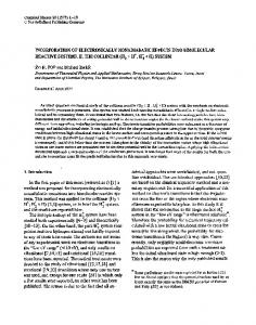
tally devoid of retinal endings (superficial layers of the lateral and ventrolateral optic tectum and the central region of the nEC. The label was localized in the lateral portion of the MOT, the ventral portion of the chiasma and the internal aspect of the optic nerve (Figs. 3,4). The degeneration of the POS was even more pronounced in 14-16-month-old animals. The optotegmentomesencephalic projection was observed to have nearly disappeared. In addition, and with the exception of a narrow band of optic terminals found within layers 1-7 of the dorsal tectum, there was an almost total absence of retinal afferents to the optic tectum (Figs. 7, 9-10). A weak projection was observed to dorsal portions of the pretectal optic nuclei (LM, GP, GT). The heaviest projections were localized within the thalamus, however, the distribution of the labeling was not homogeneous. The optic endings were essentially confined to the external portion of the nDLA pars lateralis, the dorsal region of the nGLv and the periphery of the nLA (Figs. 5,6). The regions of the MOT, chiasma and nerve that contained intact optic axons resembled those observed in the 10-month-old animals except that they appeared considerably thinner. Examination of the retinas revealed a dramatic disappearance of ganglion cells in the dorsal hemiretina (Figs. 1, 2). Those retinal cells that survived were situated mostly in the ventral hemiretina and particularly in the temporal quadrant. The cells in the latter region were large and were highly stained and exhibited a thick dendritic branching pattern (Fig. 2). Furthermore, some of the cells clearly displayed peripherally displaced nuclei, an early sign of degeneration. Takatsuji et a1.15 have previously described various histopathological charges within the peripheral optic system related to the appearance of glaucoma in the albino quail. The present study provides com-
Figs. 1 and 2. The ganglion cell layer of the whole flat-mounted retina following methylene blue staining in a 14-month-old albino quail. Fig. 1: Microphotograph showing the presence of large strongly basophilic ganglion cells in the temporal quadrant of the ventral hemiretina ( ~ 1 9 0 )Fig. . 2. Highly stained ganglion cells as viewed at higher magnification exhibiting a thick dendritic branching pattern ( ~ 7 5 0 ) . Figs. 3 and 4. Coronal sections of the optic nerve and chiasma in a 10 month-old albino quail. Fig. 3. Silver staining method. Fig. 4. Dark-field radioautographic labeling after binocular injections of [3~]proline.These two techniques revealed a heavy labeling only in the internal region of the nerve and in the ventral portion of the TOM. The arrowheads indicate the degenerative zones in the nerve and the optic chiasm ( ~ 2 3 0 ) .
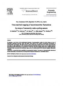
Figs. 5-10. Microphotographs of retinal projections in 10- (Fig. 8) and 16-month-old albino quails (Figs. 5-7, 9, 10). Figs. 5 and 6. Bright-field and dark-field photomicrographs of the thalamic region after intraocular injection of [3~]prolineshowing the partial la. 7. Bright-field radiobeling of the nucleus dorsolateralis (n DLA) and nucleus geniculatus lateralis pars ventralis (n GLv) ( ~ 2 3 0 )Fig. autograph of retinal projections to the dorsal part of the rostra1 tectum. This photomicrograph shows a strong labeling restricted to layers 1-7 of the dorsorostral tectum and a weak labeling of the nucleus geniculatus pretectalis pars dorsalis (n GPd) ( x 160). Fig. 8. Photomicrograph of a tectum coronal section stained by the Fink-Heimer method. Spontaneous degeneration is found in the superficial layers ( ~ 4 2 0 )Figs. . 9 and 10. Bright-field and dark-field photomicrographs of the posterior tectum showing that the optotectal projection has almost completely disappeared ( ~ 3 8 ) .
plementary information on the spatiotemporal evolution of the degeneration of the POS at the central level. The data indicate that the axonal degeneration of the retinal projections does not develop randomly but according to a relatively precise sequence progressing from the mesencephalic tegmentum to the optic tectum, then from the pretectum to the thalamus. By 16 months of age the ogtotegmental, mesencephalic and optotectal projections have almost completely disappeared. The optopretectal projection becomes scarcely visible. Although clearly altered, the optothalamic projection remains nevertheless fairly heavy. The degeneration of the POS undoubtedly continues beyond the period of our observations. Indeed, Takatsuji et a1.I' have reported the total disappearance of retinal ganglion cells in 24month-old quails. Since the end of the 19th century the study of glaucoma has been the subject of a large amount of anatomo- and physio-pathological research (refs. 1, 16 for review). Almost all of this work has been carried
out in mammals, most particularly in man. Although the etiology of glaucoma is very diverse, the disease is generally characterized by a progressive increase in the intraocular pressure followed secondarily by the degeneration of the optic neurons and consequently the loss of visual f~nction'~'~. Two major hypotheses have been proposed to explain the mechanisms which produce the degeneration of the optic neurons in mammals. One theory suggests that the augmented IOP induces a severe retinal ischemia resulting in a large reduction of blood flow, notably in the capillaries which irrigate the ganglion cell layer and the optic disc. Such a circulatory insufficiency would have the principal effect of triggering in the mid-term, the degenerating processes in the optic neuron^^,'^. The second hypothesis suggests that the elevation of the IOP brings about a mechanical compression of the optic axons and consequently the blocking of axonal flow, the factor initiating the degenerative process. The compression would act preferentially at the level of the lamina cri-
brosa, the region where the nerve fibers leave the
region might reveal a similar architectural arrangement susceptible to the effects of IOP. In mammals, the hereditary basis of certain glaucomas is well e~tablished~-~. In addition, it has been shown that this ocular neuropathy, rarely linked to albinism. results from a recessive autosomal mutation4. In the quail, the buphthalmic glaucoma appears only in the albino mutant. This recessive mutation is either sex-linked (ref. 15 and present material) or is autosomal15. In our breedings, the hereditary character of glaucoma is confirmed by its transmission with complete penetrance across generations arising from the crossing of a homozygotic male albino and a heterozygotic female of the same strain. Whether the glaucoma in the albino quail is due to the pleotropic effect of the albino gene or to a distinct gene remains unknown. A search for recombinants in the descendants of crosses between glaucomatous albino quails and normal pigmented quails should allow us to answer this question.
In birds, the avascular retina is irrigated by the vessels emanating from the pecten and choroid9. In the aged albino quail we have not observed occlusions of the blood vessels either at the level of the pecten or the choroid (Weidner et al., unpublished data). Thus it seems that the ganglion cell degeneration in these birds afflicted with glaucoma is not directly linked to the existence of circulatory perturbations at the level of the retina but rather to a blockade of axonal flow by compression of the axons at the point where they leave the ocular globe. Takatsuji et al.15have, in fact, observed the first signs of axonal degeneration in the albino quail in this area. A similar selective degeneration of the optic fibers is seen in monkeys subjected to experimentally induced The first axonal alterations appear at the level of the lamina cribrosa, particularly in the bundles of fibers situated at the periphery of the superior and inferior poles of the nerve12. The vulnerability of this group of axons to high intraocular pressure would result principally from the fact that they are less well protected by the , ~the , ~ a~1 .quail, no informaperifascicular ~ h e e t ~ In tion is available regarding the fine structure of the optic nerve at its point of emergence from the eyeball. It is thus difficult to explain why the ganglion cells of the dorsal hemiretina and the axons situated in the external portion of the optic nerve are the first to degenerate. It is possible that, as in mammals, ultrastructural examination of the same optic nerve
We thank Dr. A. Perramon and his coworkers of the Laboratoire de G6nCtique Factorielle I.N.R.A. (Centre de Recherche Zootechnique, Jouy-en-Josas) for having graciously provided the quails. We would also like to thank M. Amouzou, S. Arnold, G. Chailloux, F. Roger and G. Sanchez for their technical support and D. Le Cren for his skilful1 photographic assistance. This research was supported by the C.N.R.S., I.N.S.E.R.M., M.R.T. (Grant 85C0349) and C.R.S.N.G.
1 Caprioli, J., The pathogenesis and medical management of glaucoma, Drug Dev. Res., 6 (1985) 193-215. 2 Duggan, 5. and Minckler, D.S% Morphometry of human optic nerve head, Invest. Ophthal. Vis. Sci., Suppl. 25 (1984) 224 (ARVO Abst.). 3 Duggan, J. and Minckler, D.S., Computer reconstruction of optic nerve axonal canals, Invest. Ophthal. Vis. Sci., Suppl. 26 (1985) 41 (ARVO Abst.). 4 Frangois, J., Multifactorial or polygenic inheritance in ophthalmology, Dev. Ophthal., 10 (1985) 1-39. 5 Gelatt, K.N. and Gum, G.G., Inheritance of primary glaucoma in the beagle, Am. J. Vet. Res., 42 (1981) 1691-1693. 6 Hanna, B.L., Sawin, P.B. and Sheppard, L.B., Recessive buphthalmos in the rabbit, Genetics, 47 (1962) 519-529. 7 Harrington, P.O., The pathogenesis of the glaucoma field. Clinical evidence that circulatory insufficiency in the optic nerve is the primary cause of visual field loss in glaucoma, Am. J. Ophthalmol., 47 (1959) 177-185. 8 Lauber, J.K., Sex-linked albinism in the Japanese quail, Science, 146 (1964) 948-949.
9 Meyer, D.B., The avian eye and its adaptations. In H.J.A. Dartnall (Ed.), Handbook of Sensory Physiology, VIIl3, Springer, Berlin, 1977,pp. 575-582. 10 Minckler, D.S. and Doheny, E.S., Morphometry of optic nerve, Invest. Ophthal. Vis. Sci., Suppl. 20 (1981) 83 (ARVO Abst.). 11 Quigley, H.A. and Addicks, E.M., Chronic experimental glaucoma in primates. 11. Effect of extended intraocular pressure elevation on optic nerve head and axonal transport, Invest. Ophthalmol. Vis. Sci., 19 (1980) 137-157. 12 Quigley, H.A. and Addicks, E.M., Regional differences in the structure of the lamina cribrosa and their relation to glaucomatous optic nerve damage, Arch. Ophthal., 99 (1981) 137-143. 13 Quigley, H.A., Flower, R.W., Addicks, M. and Scott McLeod, D., The mechanism of optic nerve damage in experimental acute intraocular pressure elevation, Invest. I Ophthalmol. Vis. Sci., 19 (1980) 505-521. 14 Spaeth, G.L., The Pathogenesis of Nerve Damage in Glaucoma: Contributions of Fluorescence Angiography. Grune
and Stratton, New York, 1977. 15 Takatsuji, K., Ito, H., Watanabe, M., Ikushima, M. and Nakamura, A , , Histopathological changes of the retina and optic nerve in the albino mutant quail Coturnix coturnix japonica, J. Comp. Pathol., 94 (1984) 387-404. 16 Trevor-Roper, P.P., The Eye and its Disorders, Blackwell, Oxford, 1974. 17 Weidner, C., Reperant, J., Haby, M. and Kirpitchnikova,
E., La projection rttinienne ipsilatkrale chez la caille sauvage et le mutant albino, C. R. Acud. Sci. Paris, 300 (1985) 581-586. 18 Weidner, C . ! Reperant, J., Miceli, D., Haby, M. and Rio, J.P., An anatomical study of ipsilateral projections in the quail using radioautographic horseradish peroxidase fluorescence and degeneration techniques, Bruin Research, (1985) 99-108.