microcalcification represent in situ or invasive breast cancer to ... its noninvasive counterpart lobular carcinoma in situ (LCIS) ex- hibit the same pathophysiology.
MEDINFO 2004 M. Fieschi et al. (Eds) Amsterdam: IOS Press © 2004 IMIA. All rights reserved
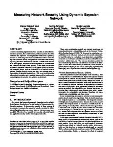
Using a Bayesian Network to Predict the Probability and Type of Breast Cancer Represented by Microcalcifications on Mammography Elizabeth S. Burnsidea, Daniel L. Rubinb, Ross D. Shachterc aDepartment
of Radiology, University of Wisconsin Medical School, USA Stanford Medical Informatics, Stanford University, CA, USA c Management Science and Engineering, Stanford University, CA, USA b
Elizabeth S. Burnside, Daniel L. Rubin, Ross D. Shachter malignant microcalcifications may also indicate the presence (or future potential development) of invasive malignancy. Invasive breast cancer has an increased risk of axillary node metastasis and depending on the size and grade of the malignancy, axillary node sampling is usually necessary. Microcalcifications can also represent several benign conditions including fibrocystic changes, a fibroadenoma and fat necrosis.
Abstract Since the widespread adoption of mammographic screening in the 1980’s there has been a significant increase in the detection and biopsy of both benign and malignant microcalcifications. Though current practice standards recommend that the positive predictive value (PPV) of breast biopsy should be in the range of 25-40%, there exists significant variability in practice. Microcalcifications, if malignant, can represent either a non-invasive or an invasive form of breast cancer. The distinction is critical because distinct surgical therapies are indicated. Unfortunately, this information is not always available at the time of surgery due to limited sampling at image-guided biopsy. For these reasons we conducted an experiment to determine whether a previously created Bayesian network for mammography could predict the significance of microcalcifications. In this experiment we aim to test whether the system is able to perform two related tasks in this domain: 1) to predict the likelihood that microcalcifications are malignant and 2) to predict the likelihood that a malignancy is invasive to help guide the choice of appropriate surgical therapy.
It has been surmised and confirmed in the literature that the mammographic appearance as described by the radiologist can predict the histology of breast cancer.[5] Unfortunately, there is significant variability in this predictive ability; subspecialist, fellowship-trained mammographers perform superiorly. We have demonstrated that our probabilistic expert system, a Bayesian network (BN), can predict the most likely diagnoses and therefore the likelihood of malignancy based on demographic factors and mammography findings as well as expert mammographers.[6] Our system uses predictive imaging features to determine the likely underlying breast disease by using the standardized lexicon established in breast imaging, the Breast Imaging Reporting and Data System (BI-RADS), which defines mammogram feature distinctions and the terminology used to describe them.[7]. BI-RADS arose in part from a study of the common terms used to describe mammography abnormalities. The descriptors most highly associated with a benign or malignant diagnosis were considered the most predictive.[8] Subsequently, these terms were incorporated in the BI-RADS lexicon.
Keywords: Expert system, Bayesian analysis, mammography
Introduction Early diagnosis of breast cancer through screening mammography is the most effective means of decreasing the death rate from this disease. [1, 2] The widespread adoption of mammography screening in the 1980’s introduced the diagnosis and management of clinically occult abnormalities that signified cancer that had never been dealt with before. A large proportion of these abnormalities were microcalcifications. Malignant microcalcifications on mammography most commonly represent ductal carcinoma in-situ (DCIS). Prior to the adoption of mammography, DCIS was a rare diagnosis. In the late 1990’s DCIS accounted for approximately 18% of breast cancer diagnosis.[3] In fact, in 1993, the total number of DCIS cases in the US was 200% higher than expected based on trends established in the previous decade; the majority of these cases attributable to mammographic screening.[4] DCIS is a non-invasive malignant condition with a very favorable prognosis. Involvement of the axillary lymph nodes is rare. Surgical therapy consists of lumpectomy without axillary node sampling. Unfortunately,
For these reasons, we believe that our expert system will be able to predict the likelihood of benign and malignant disease underlying microcalcifications on mammography. This is a more challenging task than our first experiment in which we tested the BN on cases in a teaching atlas.[6] The performance for the teaching cases was equal to that of an expert mammographer as described in the literature but the cases were not representative of true clinical practice. In this experiment, we chose to test our system on a more challenging dataset: a consecutive series of patients selected to undergo biopsy for microcalcifications. This retrospective review of clinical cases tested the hypothesis that our system would be able to accurately predict the likelihood that microcalcifications are malignant and assess whether the microcalcification represent in situ or invasive breast cancer to aid in preoperative planning.
13
ES. Burnside et al.
Materials and Methods
microcalcifications and masses. In our experiment, the characterization of microcalcifications is of interest. When microcalcifications are identified, the radiologist must describe the morphology of the microcalcifications as well as their distribution in the breast.
The Model Some of the details of the construction of our BN have been reported previously, but are repeated here in part for the convenience of the reader.[6] We subsequently refined our system by modifying our probability assessments. From the literature, we identified 26 diseases of the breast (Table 1) that represent the most likely diagnoses to be made on mammography. Twelve of these diseases are malignant and fourteen are benign. Malignant
Benign
Ductal carcinoma (DC) c Ductal carcinoma in situ
To construct our belief net and perform inference we used the GeNIe modeling environment developed by the Decision Systems Laboratory of the University of Pittsburgh (http:// www.sis.pitt.edu/~dsl). We began construction assuming that all of the BI-RADS descriptors except breast density would be children of the disease node. (Figure 1) We modeled the calcification descriptors as conditionally independent manifestations of disease. The distribution, or spatial orientation, descriptors of each type of calcifications are the mutually exclusive states of the corresponding calcification nodes when appropriate. The deterministic (double bordered) node in the belief network has four states, “Benign,” “Non-invasive,” “Invasive,” and “Mets.” The probabilities of non-invasive (analogous to in situ disease), invasive disease, and metastases comprise the total probability of malignancy.
b
Cyst Fibroadenoma Papilloma
DC/DCIS a, c
Lobular carcinoma (LC) c c Fibrocystic change Hamartoma LC/LCIS a, c Tubular carcinoma c
Lymph node
Papillary carcinoma c
Focal fibrosis
Medullary carcinoma c
Fat necrosis
Colloid carcinoma Phyllodes tumor c Metastasis
c
Figure 1 - Bayesian network structure
Mass Stability Mass Margins
Secretory disease Post-operative change
Mass Density
Skin lesion
Ca++ Round
Mass Shape
Radial scar
Ca++ Dystrophic
Mass Size
Atypical ductal hyperplasia
Breast Density
Lobular Carcinoma in situ (LCIS) a
Ca++ Lucent Milk of Centered Calcium ++ Ca Dermal
Mass P/A/O
Skin Lesion
Signify two individual diagnoses present simultaneously.
Ca++ Pleomorphic
Tubular Density
b Represents in situ disease. c Represents invasive disease.
Architectural LN Distortion
We assume that there is a single uncertain variable, “Disease,” which can take on one value corresponding to exactly one of the 26 diseases or “Normal.” We assume that it is impossible that two unrelated breast diseases occur concomitantly, but in situ and invasive breast cancers exist on a spectrum and are commonly present simultaneously. For example, the most common breast malignancy, ductal carcinoma (DC), is generally thought to develop from ductal carcinoma in situ (DCIS). Though the rate of transformation is not well known, the causal relationship between these entities is accepted. We therefore represent these two diseases in our model as three mutually exclusive states in the disease node: DCIS, DC, and DC/DCIS. The third state represents a case in which DC and DCIS are both present in the lesion seen on mammography. Similarly, lobular carcinoma and its noninvasive counterpart lobular carcinoma in situ (LCIS) exhibit the same pathophysiology.
Disease
Ca++ Popcorn Ca++ Fine/ Linear Ca++ Eggshell
Ca++ Punctate Asymmetric Density
Benign/ Ca++ Amorphous Inv./In situ/ Mets Ca++ Rod-like
Note: P/A/O = present, absent, or obscured; Ca ++ = calcifications; Inv. = invasive breast cancer, Mets = metastasis We made probability assessments from the medical literature and expert opinion. We obtained pretest probabilities, the age specific and risk factor specific distribution of diseases from census data and large randomized trials. We derived many of the joint probabilities from studies of the radiologic/pathologic correlation of individual breast diseases. Study Design Our study included 44 consecutive image-guided biopsies performed for microcalcifications detected and deemed suspicious by radiologists. The patient population consisted of women between the ages of 26 and 71 (mean=53.9; SD=10.1). Patients undergoing biopsy procedures between November 2001 and March 2002 were analyzed. 11-gauge stereotactic biopsies and needle localizations done for diagnosis were included in this project. Patients with a known cancer diagnosis undergoing ther-
The standardized lexicon for breast imaging, BI-RADS, consists of descriptors organized in a hierarchy. These terms describe the density of the breast tissue, and all possible findings on mammography. The most common findings on mammograms are
14
ES. Burnside et al.
curves. The areas under each ROC curve (AUC) were also calculated and compared.[9] The AUC can be used to measure the performance of a diagnostic tool in discriminating between patients with breast cancer from those without it for all possible cutoff values.
apeutic needle localization were excluded. Other exclusion criteria included: 1) the patient’s films not available for review, 2) calcifications not identified in the histologic specimen, and 3) mammographic follow-up of at least 12 months not available. These criteria ensured that accurate and complete evaluation of the abnormality of interest occurred and the chance of sampling error of the abnormality and possible progression were minimized
We also created a calibration curve for the radiologist and the Bayes net. This type of graphical representation has been proposed to measure the calibration or reliability of a system in demonstrating the relationship between observed and predicted outcome events. While a calibration curve does not provide a quantitative measure of reliability of probability predictions, it gives a graphical representation to capture the intuitive meaning of calibration of a given system.[10]
Cases included in the study were reviewed in a blinded manner by a fellowship-trained mammographer. The radiologist used a Web-based interface to input mammography findings and her estimate of the likelihood of malignancy into the BN. The structured entry system mandates the use of BI-RADS descriptors. Given mammography findings, our system provides post-test probabilities formulated as a differential diagnosis. For the purposes of this experiment, the system also provides the probabilities associated with the mutually exclusive possibilities of benign changes, invasive malignancy, in situ disease, or metastases.
For evaluation of the performance of the expert system in distinguishing between invasive breast cancer and in situ disease we created a second ROC curve and calculated the AUC.
Results The AUC of our expert system in predicting whether microcalcifications are malignant, .935, is similar to that found for prediction in the teaching atlas.[6] This is comparable to the AUC of .938 achieved by the radiologist. (Figure 2) There was no statistically significant difference between the AUC of the radiologist and the Bayes net. Therefore, the radiologist and the Bayes net demonstrate similar abilities to predict the likelihood of malignancy of microcalcifications.
Study Endpoints Surgical pathology at the time of the patient’s ultimate surgical intervention is the gold standard in this study. We considered ultimate surgical intervention to be either lumpectomy with established negative margins or mastectomy. The reason that we considered definitive surgical therapy the gold standard was to avoid the possibility of sampling error at percutaneous biopsy. Using this gold standard, we evaluated the ability of the Bayes net to predict the outcomes of interest: the probability of malignancy of these microcalcifications as well as the likelihood of invasive disease for surgical planning.
Figure 3 - Calibration curve for Radiologist 1 0.9 0.8 0.7 0.6 0.5 0.4 0.3 0.2 0.1 0
Figure 2 - ROC cirve measuring discrimination of malignant disease