Journal of Multidisciplinary Engineering Science and Technology (JMEST) ISSN: 3159-0040 Vol. 3 Issue 1, January - 2016
Using Image Mining Techniques For Optimizing The Treatment Methods Of Lung Cancer Dr. Mona Nasr Department of Information System Faculty of Computers and Information, Helwan University Cairo, Egypt
[email protected]
Amr Atif Abd El-Mageed Department of Information System Faculty of Computers and Information, Helwan University Sohag, Egypt
[email protected]
Abstract—Lung cancer is a disease of uncontrolled cell growth in tissues of the lung. Lung cancer is the most critical reason for death, but there is a big chance for the patient to be cured if he or she is correctly diagnosed in early stage of his or her case. Medical chest images are considered the most widely used and reliable method for the detection of lung cancer, the serious mistake in some diagnosing cases giving bad results and causing the death.
pollution, mainly air, and excessive alcohol may also be contributing to lung cancer.
The Computer Aided Diagnosis systems are necessary to support the medical staff to achieve high capability and effectiveness. Clinicians could improve treatment methods by using image mining techniques. For detecting lung nodules, number of tests should be required from the patient. Automated diagnosis system for prediction of lung cancer by using image mining techniques plays an important role in time and performance which decreases mortality rate because of early detection of lung cancer. In this research, we will present an overview of some existing image mining techniques for diagnosis of lung cancer at initial stage by analyzing the medical chest images, which assist radiologists for their chest images interpretation of lung cancer. This research analyzes various image processing and classification techniques and their efficiency used for predicting lung cancer. Keywords—lung cancer; medical chest images; computer aided diagnosis; image mining; classification I.
INTRODUCTION
The human body suffers from different diseases. Now a day, the most dangerous diseases in the world are cancer. The generic types of cancer in human body are Bladder, Breast, Colon and Rectal, Endometrial, Kidney, Leukemia, Lung, Melanoma, Non-Hodgkin Lymphoma, Pancreatic, Prostate and Thyroid cancer. The more number of people is suffering and died from lung cancer than any other cancer. Cigarette smoking is the most critical reason for lung cancer; other factors such as environment
A.
Lung Cancer
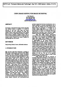
Our bodies are always making new cells, so we can grow, replace worn-out cells, or heal damaged cells after an injury. This process is controlled by certain genes. All cancers are caused by changes to these genes. Changes usually happen during our lifetime, although a small number of people inherit such a change from a parent. Normally, cells grow and multiply in an orderly way. However, changed genes can cause cells to behave abnormally. Lung cancer occurs for out-of-control cell growth in one or both lungs, tumors arising from cells lining the airways of the respiratory system and it will expand into airways. Cancer cells can be carried away from the lungs in blood or lymph fluid that surrounds lung tissue. Lymph flows through lymphatic vessels, which drain into lymph nodes located in the lungs and in the centre of the chest. Lung cancer often spreads toward the centre of the chest because the natural flow of lymph out of the lungs is toward the centre of the chest. Metastasis occurs when a cancer cell leaves the site where it began and moves into a lymph node or to another part of the body through the blood stream [1]. Cancer that starts in the lung is called primary lung cancer. There are several different types of lung cancer, and these are divided into two main groups: Small cell lung cancer and non-small cell lung cancer which has three subtypes: Carcinoma, Adenocarcinoma and Squamous cell carcinomas. Tumors can be classified as: Benign (non-cancerous), which the size of the tumor is less than 3mm. This is the starting level of cancer tumor, under this category is easily curable. Malignant (cancerous), which the size of the tumor is greater than 3mm. This is an uncontrollable level of cancer tumor, under this category is not curable, as shown in Fig. 1.
www.jmest.org JMESTN42350851
3613
Journal of Multidisciplinary Engineering Science and Technology (JMEST) ISSN: 3159-0040 Vol. 3 Issue 1, January - 2016
diaphragm, resulting in a large variation of contrast to the background. Nodules present a large variation in density – and hence visibility on a radiograph (some nodules are only slightly denser than the surrounding lung tissue, while the densest ones are calcified). There are many tissues that overlap each other on chest radiographs. The shadows of cancerous tumors seen on chest radiograph are usually vague.
Fig. 1. Types of tumors
Lung cancer is a major cause of mortality as demonstrated by the statistical numbers published every year by the American Lung Cancer Society. They indicate that the 5-year survival rate for patients with lung cancer can be increased up from an average of 14% to 50%, if the lungs cancer is diagnosed and treated at its early stage [2]. If the lung cancer is not treated in the earliest stage, this growth can spread beyond the lungs in a process called metastasis into nearby tissue cells and, eventually, into various parts of the body rapidly. The survival rate is significantly improved but there is need to increase this survival rate more than the current value. This should be done without opening the patient body. The task is performed after having inner view of the human body. The multiple methods are used to take the medical chest images from inside the body like X-rays, CT scans, MRI etc. These medical chest images are considered the most widely used technique for detection of the lung cancer. On the medical chest image of someone with lung cancer, there is usually a visible mass or nodule. This nodule will look like a white spot on the chest images taken of lungs that does not look like normal lung tissue or blood vessels while the lung itself will appear black. A nodule may also be called a lesion. The nodule may range in size. If the nodule is very small, for example, the size of a grain of rice, a marble, or even a walnut, then the surgery to remove the lobe in which the nodule was found may be the only treatment that is needed. On the other hand, if the nodule is larger than the size of a walnut, lemon, or even larger, it needs to have more treatment than just the surgery. The other treatments might include chemotherapy, radiation, or both of them [3]. The detection of lung cancer at an early stage is very important to cure it; however, there is a difficulty to diagnose the lung cancer on medical chest images because of the following reasons [4]: Nodules can appear anywhere in the lung field and vary widely in sizes. A nodule diameter can take any value between a few millimeters up to several centimeters. As nodules can appear anywhere in the lung field, they can be obscured by the ribs, blood vessels and other normal anatomic structures beneath the
The obtained medical chest images are of not good quality. There is a need of medical expert to give an opinion on the chest images. The medical experts with same expertise are not available at every place. There is a need of certain guidance for such medical experts. Even if medical experts are available, there are chances of human errors due to similarity of tissues, veins and small nodules presenting the image at the initial stage. Moreover, most of the techniques that diagnose lung cancer are expensive and time consuming; also most of them are detecting the lung cancer in its advanced stages, where the patients' chance of survival is very low. Therefore, there is a great need for a new technology to diagnose the lung cancer in its early stages, which lead to develop image mining techniques to provide a good quality tool for improving the manual analysis. B.
Image Mining
Data mining is used to extract hidden, previously unknown and potentially useful information about data. It is an increasingly popular field that uses statistical, visualization, machine learning, and other data manipulation and knowledge extraction techniques for decision making. Data mining can be used to help in predicting future patient behavior, disease diagnosis, and improving treatment programs [5]. Image mining is an extension of data mining technique. Image mining is a vital technique which is used to mine knowledge straightforwardly from image. Image mining technique deals with the implicit knowledge extraction, image data association and additional patterns, which are not clearly accumulated or stored in the images. Image mining is rapidly gaining attention among researchers in the field of data mining, information retrieval, and multimedia databases because of its potential in discovering useful image patterns that may push the various research fields to new frontiers. The fundamental challenge in image mining is to determine how low-level, pixel representation contained in a raw image or image sequence can be efficiently and effectively processed to identify high-level image objects and relationships. The image mining is highly specific because the image databases are non relational. In addition, many image attributes are not directly visible to the user.
www.jmest.org JMESTN42350851
3614
Journal of Multidisciplinary Engineering Science and Technology (JMEST) ISSN: 3159-0040 Vol. 3 Issue 1, January - 2016
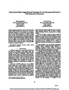
Image data are usually unstructured by nature. There are no well-defined fields of data with precise and nonambiguous meaning, and the data must be processed to arrive at fields that can provide content information about it. Another difficulty in mining of image data is its variability, heterogeneous nature, and the huge volume of data. All these characteristics of image data make image mining is demanding and interesting [6]. C. Image Mining Stages for Diagnosis of Lung Cancer There are a major stages for applying image mining techniques in the form of medical chest images for diagnosis of lung cancer in its early stages, these stages includes Data Preprocessing, Segmentation, Feature Extraction and Classification in order to classify the small lung nodules into benign (noncancerous) or malignant (cancerous), as shown in Fig. 2.
Segmentation Stage
The segmentation is the second stage which isolates the lung region from the medical chest images to locate objects and boundaries (lines, curves, etc.) in images through partitioning a digital image into multiple segments (sets of pixels). More precisely, image segmentation is the process of assigning a label to every pixel in the image such that pixels with the same label share certain visual characteristics. The goal of segmentation is to simplify or change the representation of the medical chest image into something that is more meaningful and easier to analyze. Segmentation divides the image into regions; region is group of the connected pixels that are similar with respect to some characteristic such as gray levels, colors, intensity. The result of image segmentation is a set of segments that collectively cover the entire image [8]. This stage is useful in checking each individual pixel to see whether it belongs to regions of interest or not (nodule detection).
Feature Extraction Stage
After extracting the regions of interest, we need to determine whether all the extracted regions are nodules or not. We have to analyze the features of each nodule to distinguish true nodules from the false positive nodules. The Image Features Extraction stage is very important in image processing, which extracts the most interesting features to decrease the complexity of image processing. These features will help to identify the small cancerous nodules (for example: regular and circular nodules are more likely to be benign, whereas irregular nodules are more likely to be cancerous). These features were organized in a database for using as an input for the classification process [2].
Fig. 2. Image mining stages for diagnosis of lung cancer
Data Preprocessing
Each medical chest image is scanned, and stored. During the scanning, the quality of medical chest image is affected due to non uniform intensity, variations, shift, and noises. Thus, there is a need to pre-processing process of the image, which aims at improving the quality of medical chest images to present them in an appropriate format, and cleaning the data by removing the unwanted noise present in the scanned medical chest images without affecting the details to improve the quality of the images, this stage plays a key role in the diagnostic and analysis process. Hence, image filtering [7] is an important step in preprocessing process, which improves the quality of the images through enhancing the contrast and reducing the noise that has corrupted image but leaving the rest of image.
Classification Stage
The final stage is classification stage, this stage aims at classify the small lung nodules into benign (starting level of cancer tumor) or malignant (an uncontrollable level of cancer tumor). Classification consists of predicting a certain outcome based on a given input. In order to predict the outcome, the algorithm processes a training set containing a set of attributes and the respective outcome. The algorithm tries to discover relationships between the attributes that would make it possible to predict the outcome. Next the algorithm is given a data set not seen before, which contains the same set of attributes, except for the prediction attribute – not yet known. The algorithm analyses the input and produces a prediction. Many advanced classification approaches, such as neural networks, fuzzy-sets, decision trees, SVM, and expert systems, have been widely applied for classification of the medical chest images. In most cases, image classification approaches are grouped as [9]: Supervised Classification, which is a method of using samples of known informational classes (training sets) to classify pixels of unknown identity. For
www.jmest.org JMESTN42350851
3615
Journal of Multidisciplinary Engineering Science and Technology (JMEST) ISSN: 3159-0040 Vol. 3 Issue 1, January - 2016
example: minimum distance to means algorithm, parallelepiped algorithm, maximum likelihood algorithm. Unsupervised Classification, which is a method examines a large number of unknown pixels and divides it into number of classes based on natural groupings present in the image values. Computer determines spectrally separable class and then defines their information value. No extensive prior knowledge is required. For example: K-means clustering algorithm. Early diagnosing lung cancer system makes the job of the radiologists easier and more accurate, but it won't replace what they do in. The output from the system would be used to alert the radiologists to the cancerous nodule locations, and the final diagnostic decision would then be made by the radiologists. The objective of this research is to present an overview of some existing technical articles for diagnosis of lung cancer by analyzing the medical chest images using the image mining techniques to enhance the lung cancer diagnosis. II.
LITERATURE REVIEW:
Numerous researches have been carried on applying image mining techniques for diagnosis of lung cancer. This section presents a brief description for some of these existing image mining techniques. M.Gomathi et al. (2010) [10] presented Computer Aided Diagnosing (CAD) system for automatic detection of lung cancer. The initial process was lung region detection by applying basic image processing techniques such as Bit-Plane Slicing, Erosion, Median Filter, Dilation, Outlining, Lung Border Extraction and Flood-Fill algorithms to the chest computer tomography (CT) scan images. After the lung region was detected, the segmentation algorithm was carried out with the help of Fuzzy Possibilistic C Mean (FPCM) clustering algorithm to identify the region of interest (ROIs) which helped in determining the cancer region from the extracted lung image. Then, the features were extracted and the diagnosis rules were generated. These rules were then used for learning with the help of Support Vector Machine (SVM). The experimentation was performed with 1000 images obtained from the reputed hospital.
from reputed hospital. The main objective of the project was to develop a CAD (Computer Aided Diagnosis) system for finding the lung fissures and lesions using the lung CT images and classify the lesions as Benign or Malignant. The accuracy of this system was 80% approximate the accuracy indicated by surgeons and radiologists for locating fissure regions during reading clinical CT images in 2.5–7.0 mm. Disha Sharma et al. (2011) [12] developed an automatic CAD system for detection of lung cancer by analysis of computed tomography images. This system developed cancer detection system based on texture features extracted from the slice of DICOM Lung CT images for the identification of cancerous nodules. In developing this system, it passed the available lung CT images and its database in basic three stages to achieve more accuracy: Firstly, a pre-processing stage involving some image enhancement techniques helped to solve the problem. This system preprocessed the images (by contrast enhancement, thresholding, filtering, and blob analysis) obtained after scanning the lung CT Images, and secondly, separated the suspected nodule areas (SNA) from the image by a segmentation process by using thresholding segmentation mechanism by Otsu thresholding algorithm and region growing techniques. Finally, it relied on texture features which helped to make a comparison between cancerous and non cancerous images. For accurate detection of cancerous nodules, it developed an artificial neural network to differentiate the cancerous nodules from the other suspected nodule areas in the CT images, and trained the neural network by the backpropagation algorithm and tested it with different images from a database of the DICOM CT Lung images of NIH/NCI Lung Image Database Consortium (LIDC) dataset. The accuracy of this system was 85% approximate the accuracy indicated by surgeons and radiologists for locating cancerous nodules during reading clinical CT images in 2.5–7.0 mm.
B.Magesh et al. (2011) [11] proposed a Computer Aided Diagnosing (CAD) system for detection of lung nodules from the computed tomography (CT) images. This system initially applied the image preprocessing techniques to remove the noise from the lung image by applying two steps: Denoising and Median Filter algorithms. Then, for lung regions extraction, Bit-Plane Slicing, Erosion, and Dilation algorithms was used to separate the lung structure from all other uninteresting structures.
Ms. Swati et al. (2012) [13] provided a Computer Aided Diagnosis System (CAD) for early detection of lung cancer nodules from the Chest Computer Tomography (CT) images. There were five main phases involved in this CAD system. They were image pre-processing, extraction of lung region from chest computer tomography images, segmentation of lung region, feature extraction from the segmented region, classification of lung cancer as benign or malignant. Initially, total variation based denoising was used for image denoising, and then segmentation was performed using optimal thresholding and morphological operations.
After the segmentation was performed on lung region, the features can be obtained from it and the diagnosis rule can be designed to exactly detect the cancer nodules in the lungs. For experimentation of the proposed technique, the CT images were collected
Textural features extracted from the lung nodules using gray level co-occurrence matrix (GLCM). For classification, SVM classifier was used. The main aim of this method was to develop a CAD (Computer Aided Diagnosis) system for finding the lung tumor using the
www.jmest.org JMESTN42350851
3616
Journal of Multidisciplinary Engineering Science and Technology (JMEST) ISSN: 3159-0040 Vol. 3 Issue 1, January - 2016
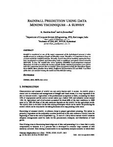
lung CT images and classify the tumor as Benign or Malignant. The lung tumor system is shown in Fig. 3. The accuracy of this system was 92.5%. However, this accuracy was evaluated based on applying the test process with only 15 diseased lung CT JPEG images of size 196x257.
Fig. 4. Sudha.V Tidke's block diagram of lung nodule
detection system
Fig. 3. Swati's lung tumor system
Sudha.V Tidke et al. (2012) [14] provided an automated lung nodule detection system. Initially, the lung region was extracted from the CT image by performing thresholding and morphological reconstruction. Then, segmentation was performed using global thresholding and morphological operation. The segmented nodules were used for feature extraction. By using these steps, the nodules were detected and segmented and some features were extracted. The extracted features were tabulated for classification. Genetic programming-based classifier (GPC) technique was used to classify the nodules as cancerous and non-cancerous nodules. This CAD system was evaluated using the LIDC database, a publicly available database from the National Biomedical Imaging Archive (NBIA), and its nodules have been fully annotated by multiple radiologists. The basic layout of this system is shown in Fig. 4.
Hiram Madero Orozco et al. (2013) [15] presented a computational alternative to classify lung nodules using computed tomography (CT) thorax images. The novelty of this method was the elimination of the segmentation stage. The contribution consisted of several steps. After image acquisition, eight texture features were extracted from the histogram and the gray level co-occurrence matrix for each CT image. The features were used to train a classifier called support vector machine (SVM), used to classify lung tissues into two classes: with lung nodules and without lung nodules. A total of 128 public clinical data set (ELCAP, NBIA) with different number of slices and diagnoses were used to train and evaluate the performance of the methodology presented. The results obtained were validated by a radiologist to finally obtain a reliability index of 84%. Kawsar Ahmed et al. (2013) [16] proposed a lung cancer risk prediction system for predicting the risk levels of the patient. It used k-means clustering algorithm for identifying relevant and non-relevant data with two clusters where one cluster contained relevant data to lung cancer and another contained remaining data that means non relevant data. Then, AprioriTid and decision tree algorithm was used to mine the frequent patterns and extracted the significant frequent patterns from the clustered dataset. S.Sivkumar et al. (2013) [17] developed lung nodule detection scheme. This work presented an image segmentation approach using standard Fuzzy C-Means (FCM), Fuzzy-Possibilistic C-Means (FPCM) and weighted Fuzzy-Possibilistic C-Means (WFPCM) algorithm on the Chest Computer Tomography (CT) images. Then, this work performed nodule segmentation through fuzzy based clustering models; classification by using a machine learning technique called Support Vector Machine (SVM). This methodology used three different types of kernels (linear, polynomial and RBF). RBF kernel gave the better class performance. Table 1 shows the accuracy, specificity and sensitivity for these three different
www.jmest.org JMESTN42350851
3617
Journal of Multidisciplinary Engineering Science and Technology (JMEST) ISSN: 3159-0040 Vol. 3 Issue 1, January - 2016
kernels of SVM classifier. The best accuracy of this system was 80.36%. TABLE I. RESULT FOR THREE DIFFERENT KERNELS OF SVM CLASSIFIER
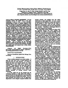
Varalakshmi.K (2013) [18] proposed a hybrid approach called neuro-fuzzy algorithm for automatic detection of lung nodules from the chest CT lung images and classifying these nodules into cancerous (Malignant) and non-cancerous (Benign) nodules, to reduce the false positive rate using Image processing techniques and Neural Network techniques. The segmentation was achieved by a series techniques including thresholding, median filtering, closing, and labeling. Lung region was extracted from the original CT image. From the lung region, the ROIs (regions of interest) were obtained. The nodules were evaluated based on features such as size of area, circularity, skewness, kurtosis and mean and then subjected for classification to classify the nodules. Neural fuzzy model was designed to extract suitable diagnosis rules, and classified the true nodules from the ROIs. The final results of experiments showed that the system can detect small lung nodule accurately as high as 89.3%. Meanwhile, the false positive per image was reduced as low as 0.3. The size of nodules that can be detected by this system is between 3mm and 5cm within lung field. This system was not able to detect nodules with size less than 3mm and also some 3mm nodules were missed. Mr.Vijay et al. (2014) [19] proposed a lung cancer detection system using image processing to classify the present of lung cancer in a CT- images. In this system, there were three main processes used; preprocessing, feature extraction and finally the classification process. MATLAB was used in every process made throughout the project. This system used convolution filters with Gaussian pulse to smooth the cell images. The contrast and color of the images were enhanced. Then, the nucleuses in the images were segmented by thresholding. After that, the system utilized morphological and colorimetric to extract feature from image of the nucleuses. The extracted morphologic features included the average intensity, area, perimeter and eccentricity of the nucleuses. On this basis, a lung cancer cell identification nodule was employed to analyze those features to judge whether cancer cells exist in the specimens or not. Moreover, if there were cancer cells, the cancer cell type was identified. The entire diagnosis process of this system is shown in Fig. 5.
Fig. 5. Vijay's lung cancer detection system
Raviprakash S. Shriwas et al. (2015) [20] proposed a lung cancer detection system. In the lung cancer detection system the contrast and color of the images were enhanced. After that, the nucleuses in the images were segmented by thresholding. After that, the system developed morphologic and colorimetric techniques to extract features from the images of the nucleuses. On this basis, neural network classifier was employed to analyze those features to judge whether cancer cells exist in the specimens or not. Similarly, if there were cancer cells, the cancer cell type is identified. Neural Network algorithm was implemented using open source and its performance was compared to other classification algorithms. It gave the 96.04% result as compare to other classifiers. Sheeraz Akram et al. (2015) [21] proposed a pulmonary nodule detection system using Artificial Neural Networks (ANN) based on hybrid features consisted of 2D and 3D Geometric and Intensity based statistical features. The lung volume was segmented using thresholding, 3D connected component labeling, contour correction and morphological operators. The candidate nodules were extracted and pruned based on the rules that were built using characteristics of nodules. The 2D and 3D Geometric features and Intensity Based Statistical features were extracted and used to train a Neural Network. This Computer-Aided Diagnostic (CAD) system was tested and validated using standard dataset of Lung Image Consortium Database (LIDC). The sensitivity of 96.95% was achieved with accuracy of 96.68%.The block diagram of this methodology is given in Fig. 6.
www.jmest.org JMESTN42350851
3618
Journal of Multidisciplinary Engineering Science and Technology (JMEST) ISSN: 3159-0040 Vol. 3 Issue 1, January - 2016
final problem is identification of affected nodules from all the candidate nodules. III. CONCLUSION: Lung cancer is the most dangerous disease and widespread in the world according to stage of discovery of the cancer cells in the lungs, this gives us indication that the process of early detection of this disease plays a very important and essential role to avoid serious stages and to reduce its percentage distribution in the world. In this research, different data mining techniques were presented which can be employed in automated lung cancer prediction system. Many existing techniques have been employed in recent years for the prediction of lung cancer. In this research an overview of different algorithms for segmentation and classification which are used in the field of lung cancer prediction was presented. The main focus is on using different classifiers combined with different segmentation algorithms for nodule detection in lung using image processing. REFERENCES [1] "Non-Small cell lung cancer", Available at: http://www.katemacintyrefoundation.org/pdf/nonsmall-cell.pdf, Adapted from National Cancer Institute (NCI) and Patients Living with Cancer (PLWC), 2007.
Fig. 6. Block diagram of the pulmonary nodule detection
system
From the mentioned researches, we conclude that the computer-aided diagnosis (CAD) system can be used for detection of lung cancer by analyzing the medical chest images. The main idea of developing a CAD system is not to delegate the diagnosis to a machine, but rather that a computer algorithm acts as a support to the radiologist and points out locations of the suspicious objects. CAD systems achieve four main objectives: Improving the quality and the accuracy of diagnosis. Increasing therapy success by early detection of cancer. Avoiding unnecessary biopsies examination of tissues taken from a living body).
(an
Reducing the radiologist interpretation time.
Also, we conclude that many CAD systems, regardless of implementation, share the same basic idea, which is that in order to produce a successful Computer Aided Diagnosis system, several problems has to be resolved. First problem is removing the noise present in the scanned medical chest images without affecting the details. Segmentation is the second problem to be considered which helps in generation of candidate region for detecting the cancer nodules. The
[2] Zakaria Suliman Zubi, and Rema Asheibani Saad, "Using some data mining techniques for early diagnosis of lung cancer", Recent Researches in Artificial Intelligence, Knowledge Engineering and Data Bases, Libya, 2007. [3]
http://lungcancer.about.com.
[4] Gomathi, and Thangaraj, "Computer aided medical diagnosis system for detection of lung cancer nodules a survey", the Free Library, pp. 3-12, 2012. [5] Divya Tomar, and Sonali Agarwal, "A survey on data mining approaches for healthcare", International Journal of Bio-Science and BioTechnology, vol. 5, no. 5, pp. 241-266, 2013. [6] L. Khan, and V. A. Petrushin, "Multimedia data mining and knowledge discovery", 2007. [7] Ramesh Jain, Rangachar Kasturi, and Brian G. Schunck, "Machine vision", McGraw-Hill, pp. 112139, 1995. [8] Salem Saleh Al-amri, N.V.Kalyankar, and Khamitkar S.D, "Image segmentation by using thresholding technique", Journal of Computing, vol. 2, no. 5, 2010. [9] Pooja Kamavisdar, Sonam Saluja, and Sonu Agrawal, "A Survey on image classification approaches and techniques", International Journal of Advanced Research in Computer and Communication Engineering, vol. 2, no. 1, 2013. [10] M. Gomathi, and P. Thangaraj, "A Computer aided diagnosis system for lung cancer detection",
www.jmest.org JMESTN42350851
3619
Journal of Multidisciplinary Engineering Science and Technology (JMEST) ISSN: 3159-0040 Vol. 3 Issue 1, January - 2016
American Journal of Applied Sciences, vol. 7, no. 12, pp. 1532-1538, 2010. [11] B. Magesh, Pgscholar, P. Vijayalakshmi, and M. Abirami, "Computer aided diagnosis system for the identification and classification of lessions in lungs", International Journal of Computer Trends and Technology, 2011. [12] Disha Sharma, and Gagandeep Jindal, "Computer aided diagnosis system for detection of lung cancer in CT scan images", International Journal of Computer and Electrical Engineering, vol. 3, no. 5, 2011. [13] Ms. Swati, P. Tidke, Prof. Vrishali, and A. Chakkarwar, "Classification of lung tumor using SVM", International Journal of Computational Engineering Research, vol. 2, no. 5, pp. 1254-1257, 2012. [14] Sudha.V, and Jayashree.P, "Lung nodule detection in CT images using thresholding and morphological operations", International Journal of Emerging Science and Engineering (IJESE), vol. 1, no. 2, 2012. [15] Hiram Madero Orozco, and Osslan Osiris Vergara Villegas, "Lung nodule classification in CT thorax images using support vector machines", 12th Mexican International Conference on Artificial Intelligence, pp. 277-283, 2013.
[17] S.Sivkumar, and Dr.C.Chandrasekar, "Lung nodule detection using fuzzy clustering and support vector machine", International Journal of Engineering and Technology, vol. 5, no. 1, 2013. [18] Varalakshmi.K, "Classification of lung cancer nodules using a hybrid approach", Journal of Emerging Trends in Computing and Information Sciences, vol. 4, no. 1, 2013. [19] Mr.Vijay, A.Gajdhane, and Prof. Deshpande L.M.,"Detection of lung cancer nodule on computed tomography images by using image processing", International Journal of Application or Innovation in Engineering & Management (IJAIEM), vol. 3, no. 7, 2014. [20] Raviprakash S. Shriwas, and Akshay D. Dikondawar, "Lung cancer detection and prediction by using neural network", IPASJ International Journal of Electronics & Communication (IIJEC), vol. 3, no. 1, 2015. [21] Sheeraz Akram, Muhammad Younus Javed, Usman Qamar, Aasia Khanum, and Ali Hassan, "Artificial neural network based classification of lungs nodule using hybrid features from computerized tomographic images", Applied Mathematics & Information Sciences, An International Journal, vol. 9, no. 1, 2015.
[16] Kawsar Ahmed, Abdullah-Al-Emran, Tasnuba Jesmin, Roushney Fatima Mukti, Md Zamilur Rahman, and Farzana Ahmed, "Early detection of lung cancer risk using data mining", Asian Pacific Journal of Cancer Prevention, vol. 14, no. 1, pp. 595598, 2013.
www.jmest.org JMESTN42350851
3620