Apr 3, 2016 - like a loaf of bread (bread-loaf technique) or variations thereof, allowing only a small sampling of all surgical margins to be analyzed, causing ...
6 – ORIGINAL ARTICLE MODELS, BIOLOGICAL
Validation of a new technique to freezing and embedding tissue in Mohs surgery, using an animal model1 Frederico Hassin SanchezI, Joaquim Ribeiro FilhoII, Asdrubal Cesar RussoIII DOI: http://dx.doi.org/10.1590/S0102-865020160080000006 Fellow Master degree, Postgraduate Program in Surgical Sciences, Medical School, Universidade Federal do Rio de Janeiro (UFRJ), Brazil. Conception and design of the study, acquisition of data, manuscript writing. II PhD, Associate Professor, Postgraduate Program in Surgical Sciences, Medical School, UFRJ, Rio de Janeiro-RJ, Brazil. Conception and design of the study, critical revision, final approval of the version to be published. III Full Professor, Graduation Program in medicine, Medical School, Universidade Federal Fronteira Sul (UFFS), Chapecó-SC, Brazil. Acquisition of data. I
ABSTRACT PURPOSE: To validate the innovative Dry Ice method, comparing it with two standard methods currently used for tissue processing in Mohs surgery, the Heat Sink method and the Miami Special. METHODS: Forty eight samples of pigs kin with the standard beveled Mohs technique were used, and randomly allocated into six groups. Each group was processed with one of the 3 methods and evaluated for: The freezing time, the depth required to cut into the block to obtain a complete section, and the quality of histological slides analyzed with a image software. The statistical analysis was performed with the software SAS® System. The inferential analysis was made by one-way ANOVA. RESULTS: The Miami Special showed a processing time significantly shorter than Dry Ice method and Heat Sink method. There was no significant difference in the depth required to cut into the blocks, and area of surgical margins visualized. CONCLUSION: The Dry Ice method was as efficient as the other two methods currently used in Mohs surgery, considering the individual advantages and disadvantages of each method. Key words: Mohs Surgery. Skin Neoplasms. Dermatology. Equipment and Supplies. Inventions. Swine.
Acta Cirúrgica Brasileira - Vol. 31 (8) 2016 - 533
Sanchez FH et al.
Introduction The Mohs micrographic surgery is a gold standard procedure to treat several types of skin cancer1. It is a meticulous procedure that includes a number of technical steps, from the tumor removal until the tissue preparation, mapping the surgical margins and the microscopic analysis. The tissue processing performed in Mohs surgery distinguishes from the conventional histological techniques. In conventional standard procedures, the tissues are frozen, and cut, like a loaf of bread (bread-loaf technique) or variations thereof, allowing only a small sampling of all surgical margins to be analyzed, causing false negatives, and increasing the chance of tumor recorrence2,3. Consequently in the conventional histologic techniques, generally 2 to 4 mm of tissue is left unexamined at each interval, and less than 1% of the excised margin is analysed2-4. In contrast, during Mohs surgery, the tissue is removed with a specific angulation of the scalpel, allowing flattening of the lateral and deep surgical margins in the same plane. This sectioning of flat tissue, allows the confection of histological slides containing all surgical margins in the same section, allowing the analysis of 100 % of all surgical margins2,5,6. The great rate of efficacy of Mohs surgery is attributed to the ability to view all surgical margins in a single en face section5,6. The high quality frozen sections are fundamental to allow the visualization of the peripheral and deep margins in the same plane7. Each successive step of the surgery demands time, and the patient needs to wait for the tissue processing and the microscopic analysis. When large tumors are removed and multiple samples need to be processed, a long waiting time can be necessary, and can increase the morbidity, the staff fatigue and technical errors8. Many innovative techniques have been developed to improve efficiency and speed of the tissue processing in Mohs surgery, keeping the high quality of histological slides8,9. A survey of Mohs surgeons published in 2003 showed a great variety of the devices and techniques used to flatten, to freeze, and to prepare the tissues in Mohs surgery, among them, the heat extractor flattening in the cryostat (22.6%), relaxing incisions (16.1%), heat extractor and relaxing incisions (14.2%), freezing on a glass slide (10.3%), relaxing incision and glass slide (9.4%), and the Miami Special technique (3.9%) are the most commonly used10. Other less used devices, such as the Davidson CryocupTM (Bradley Products Inc., Bloomington, MN), CryoHistTM (Cryo Histology, Inc., Shawnee, KS), the Cryo-EmbedderTM ( Salt Lake City, UT), the face-down cryoembedding of tissue using stainless stell embedding wells, and the use of dry ice have been described
534 - Acta Cirúrgica Brasileira - Vol. 31 (8) 2016
to optimize the tissue preparation process8,9,11,12. One of the most popular techniques used to flatten and freeze specimens is the heat extractor method, used for flattening and freezing the tissues inside the cryostat12,13. However, the warm weather and high temperatures, increase the thermal exchange between the cryostat chamber and the environment, and it can slow down the process, especially when many samples need to be processed7,12. In an attempt to minimize this problem, some surgeons have used over 30 years the Miami Special method, that uses a specific device to mechanical flattening with liquid nitrogen7. In 2014 a promising process for freezing and embedding tissue with dry ice was described, which did not require the use of cryostat or liquid nitrogen to freeze the tissue12. However despite the apparent cost-effectiveness advantages, there is a lack of comparative studies. The purpose of this article is to compare the innovative dry ice technique12 with two standard methods currently used for tissue processing in Mohs surgery, the Heat Sink method5,13 and the Miami Special7, using an animal model. Methods The research was approved by the Ethics Committee for the use on animal experimentation of the Universidade Federal do Rio de Janeiro (UFRJ) (150-13). Porcine skin from two newly slaughtered animals (Sus scrofa domestica) was used and donated by an official inspected slaughterhouse (Cooperativa Central Aurora Alimentos, ChapecóSC). A large area of pig belly skin was excised from the two animals, and immediately cooled until the use in the same day. The same author marked and excised circular samples 1.0 cm in diameter from the pig belly skin, using the standard beveled Mohs technique. Forty eight specimens were randomly allocated into six groups of samples. The group 1, group 2 and group 3, were composed of 10 circular samples each one. A cut in the middle of the circular samples was performed to promote the relaxation of the edges14,15. The remaining 18 samples were randomly allocated into three groups of 06 samples each one. The samples of the groups 4, group 5, and group 6, were bisected and processed as two separate pieces, resulting in 12 semi-circular samples in each group (Figure 1). The processing as two parts, is a laboratory technique commonly used in Mohs surgery, especially used when preparing a thicker skin sample, to facilitate the flattening and to allow better assessment of surgical margins (Figure 2)5,14. Each group of the not divided samples, and each group of bisected
Validation of a new technique to freezing and embedding tissue in Mohs surgery, using an animal model
samples were processed as one of the three methods of freezing and tissue processing (Table 1).
To eliminate technician variability in the results, a single and experienced histology technician with proficiency in the three methods worked on the project. For each method the same embedding medium was used in the preparation of all samples: The Optimal Cutting Temperature Compound (OCT) (TissueTek®, Sakura Finetek USA, inc., Torrence, CA). The tissue processing with each technique were the following: Heat extractor method (Heat Sink technique)
FIGURE 1 - Flowchart of the arrangement of samples in each group.
FIGURE 2 - A circular sample with a central relaxation cut (pacman), and a bisected semicircular sample.
In the heat extractor method, the sample is placed with surgical margin side down on a chilled heat extractor and the epidermal edge is gently laid flat on the cold heat sink surface to allow flattening the deep and peripheral margin in a single plane. The OCT is applied on the specimen on the heat extractor, covering it entirely and left to freeze until it gets an opaque appearance. OCT is applied to a cutting chuck to freeze inside the cryostat. The heat extractor with the tissue is immediately inverted onto the chuck containing OCT, and they freeze together as a sandwich. After freezing, the heat extractor is pulled away from the chuck containing the tissue. An additional drop of the OCT is applied on the frozen block surface to fill clefts at the tissue and then the heat extractor is pressed against to the block surface for few seconds, leaving the cutting chuck prepared for sectioning5 (Figure 3).
TABLE 1 - Characteristic of the groups. Groups Status of Number of Method of Samples samples ( n ) processing Group 1 Group 2
Circular Circular
10 10
Group 3 Group 4 Group 5
Circular Semicircular Semicircular
10 12 12
Group 6
Semicircular
12
Heat sink method Miami Special method Dry Ice method Heat Sink method Miami Special method Dry Ice method
The margins of the all specimens were inked like standard Mohs technique. Some authors recommend the application of en face ink to detect and define embedding errors in Mohs specimens16,17. The standard ink was used in the edge of the all samples, for easy viewing to the end point of sectioning by histotechnician.
FIGURE 3 - Heat Sink method. 1) The sample placed with surgical margin side down on a chilled heat extractor, and the OCT is applied on the specimen on the heat extractor, covering it entirely. 2) The heat extractor with the tissue is immediately inverted onto the chuck containing OCT, and they freeze together as a sandwich. 3 and 4) The heat extractor is pressed against to the block surface for few seconds, leaving the cutting chuck prepared for sectioning.
Acta Cirúrgica Brasileira - Vol. 31 (8) 2016 - 535
Sanchez FH et al.
Liquid nitrogen method (Miami Special technique) An adapted obstetric clamp has been used at the University of Miami for over 30 years, to facilitate the embedding tissue process in the hot and humid environment of Florida7 (Figure 4). The device is designed to hold a chuck and a portion of tissue together with parallel faces while being dipped in liquid nitrogen to rapidly freeze with tissue and OCT thus forming a block. A small amount of embedding medium is placed over the chuck from the cryostat and dipped for few seconds in liquid nitrogen until it acquires a gray translucent appearance8. The chuck is placed into the chuck holder of the Miami Special clamp. On the opposite side of the device, the tissue sample is gently placed and flattened on the plane surface the Miami Special clamp with deep and peripheral margins in the same flat plane. The Miami Special device is closed and the chuck is clamped into fixed position like a common hemostat, allowing the sandwiched tissue and chuck to be dipped into liquid nitrogen to freeze together18. The device is opened and an additional drop of OCT is applied on the tissue surface to fill any clefts, and then it is closed again to press the tissue surface against to the device flat surface and dipped for few seconds to complete the freezing process18. The chuck with the embedding tissue is removed from the device ready for sectioning (Figure 4).
FIGURE 4 - Miami Special method. 1) The sample is gently placed on the chuck. 2) The chuck is placed into the chuck holder of the Miami Special clamp. 3) The sandwiched tissue and chuck are dipped into liquid nitrogen to freeze together. 4) The device is opened and an additional drop of OCT is applied on the tissue surface to fill any clefts, and then it is closed again to press the tissue surface against to the device flat surface and dipped for few seconds to complete the freezing. 5) The chuck with the embedding tissue is removed from the device ready for sectioning.
536 - Acta Cirúrgica Brasileira - Vol. 31 (8) 2016
Dry ice method (Rio de Janeiro technique) This technique was developed to accelerate the embedding tissue process in the hot climate of Rio de Janeiro (Brazil) and uses an aluminum box containing 8-12 dry ice cubes, with a thermal insulation surrounding the side and bottom surfaces of the box (polyurethane support)12. The flat upper surface of the box, which we called the external freezing bar (EFB) works just like the internal freezing bar of the cryostat, with the advantage that it enables to handle the samples outside of the cryostat and allow to process several samples simultaneously. The specimen is placed on the surface of EFB with lower surgical margin facing down, and the peripheral margins are flattened on the bar surface. Gentle pressure with a finger is applied to the center of the sample to ensure that the deep and peripheral margins are in contact with the frozen surface. The OCT medium is applied directly over the specimen, and an additional OCT is applied on the chuck from the cryostat. The embedding medium on the metal surface and the chuck must be only partially frozen when sandwiched together so they will freeze together in a solid block. After few seconds the freezing block is separated from the EFB and a small additional portion of OCT is applied to the surface of the embedding tissue on the prepared cutting chuck and pressed against the EFB for approximately 10 seconds to flatten the surface. The prepared chuck is ready to be transferred to the cryostat for sectioning (Figure 5)12.
FIGURE 5 - a) Dry ice inside the device; b) Dry ice inside the device; c) The semicircular sample flattened on the plane surface of the device; d) The sample freezing with the OCT; e) Chucks against the freezing samples; f) The cutting chuck ready to sectioning, with the deep and lateral margins of the sample in the same flat plane.
Validation of a new technique to freezing and embedding tissue in Mohs surgery, using an animal model
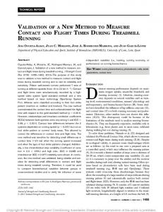
For each group, the time of the tissue processing and the mounting of the blocks was recorded, from the freezing, until ready for sectioning, inside the cryostat. Because all specimens were treated the same after this, the end point was the samples ready for sectioning inside the cryostat. In all samples, sections every 5 microns (μm) were performed, until it was possible to view the depth margin, and epidermal margin in a same plane. The depth required to cut into the block to obtain a complete section was recorded in microns. These samples were stained with hematoxylin and eosin, and then mounted with glass coverslips standardly. The same investigator evaluated only one slide of each sample, with the first complete section. Although the optimal session is defined by the complete representation of the epidermis, dermis and subcutaneous, many authors consider as a most important quality criterion of the slides, the visualization of more than 90% of epidermal margins5,8,11. The sections were microscopically analyzed by an experimented surgeon, and the complete sections were defined as having at least 90% of the epidermis and a complete representation of dermis and fat tissue, and the visualization of the inke in all surgical margins. It was the end point to stop the sectioning of the block. In a second moment, the slides were scanned using a slide scanner (Aperio ScanScope slide scannerTM – Leica Biosystems) and the software ImageJ (Rasband, W.S., ImageJ, U. S. National Institutes of Health, Bethesda, Maryland, USA.) was used to estimate the visualization of the margin in each slide (Figures 6 and 7).
FIGURE 7 - a) The entire semicircular sample; b) A detail of the peripheral margin with the epidermis; c) A detail of the deep margin delimited by the green ink.
Statistical analysis Statistical analysis was performed with the statistical software SAS® System, version 6.11 (SAS Institute, Inc., Cary, North Carolina). The inferential analysis was made by the statistical method one-way ANOVA, and the multiple comparison test of Tukey. The normality hypothesis was not rejected according to the Shapiro-Wilks test. The significance of determining the level adopted was 5%. The analytical design was defined by one-way ANOVA for each type of sample, rather than two-way ANOVA, with the main objective to analyze the influence of the each method separately for each sample type, circular (Groups 1, 2 and 3) or semi-circular (Groups 4, 5 and 6). The descriptive analysis presented in tables, and the observed data were expressed as mean and standard deviation in each group. Illustrative graphics were constructed expressed by the mean, and 95% confidence interval. Results The Tables 2 and 3 show the mean and standard deviation (SD) of the processing time, the depth of sectioning into the block, and the percentage of visualized margins, according to freezing
FIGURE 6 - a) The scanned histological slide, with entire deep and peripheral margin visualized; b) The area of the histological slide analyzed by the ImageJ software; c) A histological sample with lost of part of surgical margins; d) The estimate visualized area of the sample analyzed by the ImageJ software.
methods, and the corresponding descriptive level (p value) of the statistic test, of the circular samples and of the bisected semicircular samples, respectively.
Acta Cirúrgica Brasileira - Vol. 31 (8) 2016 - 537
Sanchez FH et al.
samples.
TABLE 2 – Result of the processing time, depth of the block sectioning, and margins visualized, in each group with circular
Object of analysis
Group / Method Group 1 (Heat Sink) Group 2 (Liquid Nitrogen) Group 3 (Dry Ice)
mean 168.8 73.1 124.2
± ± ± ±
SD 18.3 7.8 7.9
Depth of the block sectioning (µm)
Group 1 (Heat Sink) Group 2 (Liquid Nitrogen) Group 3 (Dry Ice)
525.0 622.5 484,5
± ± ±
94.3 138.5 85,5
Margins Visualized (%)
Group 1 (Heat Sink) Group 2 (Liquid Nitrogen) Group 3 (Dry Ice)
94.0 93.7 94.1
± ± ±
1.8 2.2 1.8
Processing Time (seg)
p value a
≠ significant b
< 0.0001
Group 1 ≠ Group 2 Group 1 ≠ Group 3 Group 2 ≠ Group 3
0.025
Group 2 ≠ Group 3
0.89
SD: Standard Deviation. a one-way ANOVA. b multiple comparison test of Tukey at 5%.
TABLE 3 – Result of the processing time, depth of the block sectioning, and margins visualized, in each group with semicircular samples. ≠ significant b p value a Object of Analysis Group (Method) mean ± SD Group 3 (Heat Sink) 129.1 ± 18.2 Group 3 ≠ Group 4 < 0.0001 Processing Time (sec) Group 4 (Liquid Nitrogen) 71.3 ± 6.0 Group 3 ≠ Group 5 Group 5 (Dry Ice) 115.3 ± 12.4 Group 4 ≠ Group 5
Depth of the Block Sectioning (µm)
Group 3 (Heat Sink) Group 4 (Liquid Nitrogen) Group 5 (Dry Ice)
339.6 364.6 352.1
± ± ±
45.1 58.8 54.2
Margins Visualized (%)
Group 3 (Heat Sink) Group 4 (Liquid Nitrogen) Group 5 (Dry Ice)
95.6 96.0 95.9
± ± ±
2.0 2.9 2.1
0.52
0.93
SD: Standard Deviation. a One-way ANOVA. b Multiple comparison test of Tukey at 5%.
For the groups containing circular samples (Groups 1, 2 and 3), according to one-way ANOVA analysis, there was a significant difference in processing time (p