Vascular patterning defects associated with expression of activated Notch4 in embryonic endothelium H. Uyttendaele*†, J. Ho†‡, J. Rossant‡, and J. Kitajewski*§ *Departments of Pathology and Obstetrics兾Gynecology, Columbia University, 630 West 168th Street, New York, NY 10032; and ‡Samuel Lunenfeld Research Institute, Mount Sinai Hospital, and Department of Molecular and Medical Genetics, University of Toronto, 600 University Avenue, Toronto, ON, Canada M5G 1X5
Notch 兩 Flk1 兩 Jagged1 兩 vasculature
F
ormation of the vascular system begins early in mammalian development, with the specification and aggregation of angiogenic precursors in the embryo and formation of blood islands in the visceral yolk sac. In the yolk sac, there is a close association between the primitive hematopoietic cells and the developing endothelium, suggestive of a common precursor, the so-called hemangioblast (1). Within the embryo itself, the first blood vessels develop in the absence of hematopoiesis, suggesting that these precursors are solely angiogenic (2). The early blood vessels of the embryo or the yolk sac develop by aggregation of angioblasts that form de novo into a primitive network of simple endothelial tubes—the process of vasculogenesis (3). Later development of the mature vessel system involves a complex process of remodeling and refining the initial pattern, with the proliferation and sprouting of new vessels from existing ones—the process of angiogenesis (3). Full elaboration of the vascular system is more complex than what is suggested by this division into two phases. As vessels begin to be remodeled into a functioning circulation system, they need to undergo localized proliferation and regression as well as programmed branching and migration. They must be specified into different calibers and types of vessels, including divisions into arteries, veins, and lymphatics, with separate subdivisions into large vessels, venules, arterioles, and capillaries. In addition, vessels must recruit supporting cells, smooth muscle cells, and pericytes (4) to ensure the stability of the vessels formed. A number of signaling pathways, such as VEGF兾VEGFR (vascular endothelial growth factor and its receptor) and Angiopoietin兾Tie, are involved in vascular development. VEGF signaling plays a major role in promoting the proliferation and differentiation of the endothelial lineage from the earliest stages of development, whereas the Angiopoietin兾Tie pathway acts slightly later to promote the recruitment of supporting cells and vessel stabilization (5). However, the action of these factors alone is not sufficient to explain the complex patterning of the developing vasculature. There is www.pnas.org兾cgi兾doi兾10.1073兾pnas.091584598
growing evidence for specific vascular roles for the components of other signaling pathways, including the Ephrin兾Eph, TGF- (transforming growth factor ), PDGF (platelet-derived growth factor), FGF (fibroblast growth factor), and Delta兾Notch pathways. The Delta兾Notch pathway was originally characterized in Drosophila (6). This pathway is conserved through evolution from Caenorhabditis elegans (7) to humans (8, 9). Notch signaling is often associated with the promotion of a stem cell vs. differentiated cell fate (10) as, for example, in primary neurogenesis (11) or during T cell development (12). However, it is also involved in patterning processes in development, including lateral inhibition of cell-fate choices at both a single-cell level and at a group-decision level. In other situations, Notch signaling acts in a more classically inductive manner to regulate cell-fate decisions. Several Notch receptors and ligands are expressed in the developing vasculature, and Notch4 is restricted in expression to this lineage in mouse embryos (9, 13). Notch4 expression has been detected as early as 7.5 days postcoitum (dpc) by reverse transcription–PCR (RT-PCR), and whole-mount in situ analysis has shown endothelial-specific expression of Notch4 throughout embryogenesis in a pattern similar to that observed for Flk1 (13). A Notch ligand, Delta-like 4 (Dll4), is expressed in arterial endothelium and can function as a ligand for both Notch1 and Notch4 (14). Genetic evidence supports a role for Notch signaling in vascular development in both mice and humans. Mouse mutations in Notch ligands, Delta1 and Jagged1, cause vascular problems—hemorrhaging in Dll1 mutants (15) and vascular-remodeling defects in Jagged1 mutants (16). Mutation of the presenilin 1 (PS1) gene, proposed to be involved in Notch signaling, produces a complex phenotype and shows extensive hemorrhaging (17, 18). A Notch1兾Notch4 double mutant has been generated and results in embryonic lethality with severe defects in angiogenic vascular remodeling (19); a similar phenotype is seen in Notch1 mutant embryos (19). Expression of activated Notch4 or Jagged1 can promote the differentiation of cultured endothelial cells into vessel-like structures (20). The most compelling evidence that Notch genes are involved in human vascular disorders comes from an analysis of a human disorder termed CADASIL (cerebral autosomal dominant arteriopathy with subcortical infarcts and leukoencephelopathy) which is manifested by stroke and vascular dementia and characterized by arteriopathy of cerebral arterioles. Mutations in human Notch3 have been characterized in CADASIL patients (21). It is not clear from mutants whether the Notch signaling pathway acts autonomously in the endothelium, because all of the mutated This paper was submitted directly (Track II) to the PNAS office. Abbreviations: dpc, days postcoitum; SMA, smooth muscle actin; HA, hemagglutinin epitope; RT-PCR, reverse transcription–PCR; ES, embryonic stem; EBs, embryoid bodies. See commentary on page 5377. †H.U. §To
and J.H. contributed equally to this work.
whom reprint requests should be addressed. E-mail:
[email protected].
The publication costs of this article were defrayed in part by page charge payment. This article must therefore be hereby marked “advertisement” in accordance with 18 U.S.C. §1734 solely to indicate this fact.
PNAS 兩 May 8, 2001 兩 vol. 98 兩 no. 10 兩 5643–5648
BIOLOGY
Notch proteins function as receptors for membrane-bound ligands (Jagged and Delta-like) to regulate cell-fate determination. We have investigated the role of Notch signaling in embryonic endothelium of the mouse by expressing an activated form of the Notch4 protein in vasculature under the regulation of the Flk1 (VEGFR) locus. Expression of activated Notch4 results in a growth and developmental delay and embryonic lethality at about 10 days postcoitum. The extent of the developing vasculature in mutant embryos was restricted, fewer small vessels were seen, and vascular networks were disorganized. The brain periphery of mutant embryos contained large dilated vessels with evidence of compromised vessel-wall integrity and large areas of necrosis; yolk-sac vasculature was abnormal. Expression of an activated form of Notch4 in embryonic vasculature leads to abnormal vessel structure and patterning, implicating the Notch pathway in phases of vascular development associated with vessel patterning and remodeling.
DEVELOPMENTAL
Edited by Richard O. Hynes, Massachusetts Institute of Technology, Cambridge, MA, and approved February 26, 2001 (received for review December 9, 2000)
genes show expression in many different cell types, nor is it clear what phase of vascular development depends on Notch signaling. Addressing these issues requires endothelial-specific manipulation of Notch function. In both flies and worms, Notch functions have been defined by the opposite phenotypes of loss-of-function and constitutively activated Notch mutations (22). Here, we investigate the role of Notch signaling in the mammalian endothelium by expressing an activated form of the Notch4 protein in endothelial cells under the regulation of the Flk1 (VEGFR) locus. This expression resulted in embryonic lethality, associated with abnormal vessel structure and vascular patterning, implicating the activity of the Notch pathway in the later phases of vascular development but not in the initial specification of the lineage. Materials and Methods Production of Flk1兾int-3HA Embryonic Stem (ES) Cells. A targeting
vector was constructed that inserts int-3HA [the activated Notch4, hemagglutinin epitope (HA)-tagged at the carboxyl terminus], with a simian virus 40 poly(A) signal, in place of lacZ sequences in the previously generated Flk1 targeting vector (23). The backbone of the vector is pPNTloxP, containing PGKneo (flanked by two loxP sites) for positive selection and PGK-TK for negative selection. int-3HA is inserted in the 5⬘ untranslated region of Flk1 and contains its own translational initiation signal. This construct was electroporated into Tie1⫹/⫺ ES cells, and correctly targeted clones (Flk1兾int-3) were positively selected for by using G418 and negatively selected for by using ganciclovir. Tie1⫹/⫺ ES cells in which one allele of the Tie1 gene is replaced by the lacZ gene were used. Thus, targeted ES-cell clones express Notch兾int-3 under control of the Flk1 promoter and express lacZ under control of the Tie1 promoter. The expression of -galactosidase was used as a marker for endothelial cells. Southern blot hybridization determined that ES-cell clones had one Flk1 allele replaced with Notch4兾int-3 by homologous recombination (Fig. 1). Generation of Flk1兾int-3 ES Embryos. ES-cell lines were used in
aggregations with tetraploid inner cell mass embryos. Two-cell diploid embryos were fused to become tetraploid one-cell embryos, cultured to the four-cell stage, and then aggregated with ES cells. Composite embryos were cultured overnight and then transferred to pseudopregnant recipients. Embryos that were entirely Flk1兾int-3 and control Tie1-lacZ-derived embryos were generated by using the technique described above. -Galactosidase Staining. Embryos were dissected at various stages,
fixed, stained for lacZ expression, and counterstained as previously described (23).
Whole-Mount RNA in Situ Hybridization and Antibody Staining.
Digoxigenin-labeled RNA probes were transcribed from linearized templates and whole-mount in situ hybridization was carried out as described (24). ␣-Smooth muscle actin (SMA) antibody (Clone 1A4) was obtained from Sigma, and the Tek兾Tie2 antibody was kindly provided by N. Takakura (Kumamoto University School of Medicine, Kumamoto, Japan) (25). Whole-mount antibody staining was performed as described (26). In Vitro Differentiation of ES Cells. ES cells were allowed to differentiate into cystic EBs in vitro (27). ES cells were passaged twice in the presence of lymphocyte inhibitory factor (LIF) on gelatincoated plates. On day 6, after the second passage, ES colonies were treated with dispase, washed, and grown in suspension in bacterial dishes in ES medium without LIF. On day 8 after the dispase treatment, -galactosidase activity was detected as described. RT-PCR. Poly(A)⫹ RNA from mouse kidney and EBs generated
from either control ID5 ES-cell line or the targeted ES-cell line
5644 兩 www.pnas.org兾cgi兾doi兾10.1073兾pnas.091584598
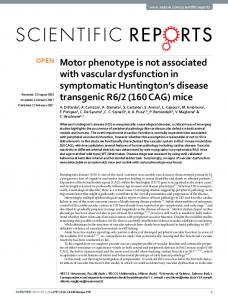
Fig. 1. Presentation of ES-cell generation and analysis of ES-cell clones. (A) Schematic representation of ES-targeting strategy. (B) Targeting construct. The targeting vector is designed to replace the exon of Flk1 that encodes the initiating methionine with a cassette containing int-3HA and the neomycin (neo) gene cassette. (C) Genotype analysis of ES clones by Southern blotting. The targeted locus places int-3HA under the gene-regulatory region of Flk1 and introduces a NotI (N) site at the junction of the int-3HA and neo genes. Genomic DNA was digested with NotI, and hybridization was done with an Flk1-specific probe. (D) Expression analysis for int-3HA transcripts in embryoid bodies (EBs) by RT-PCR. Analysis was conducted with control RNA from kidney, from parental line 1D5, and from a homologous recombinant line, 1A12.
1A12 was analyzed by RT-PCR. RT-PCR was performed with Robocycler (Stratagene). For the expression analysis of Notch4, Notch3, Tie1, and the int-3HA transgene, specific primers were used to amplify fragments by using equal amounts of cDNA. Primer sequences are available upon request. Northern Blot Analysis. Total RNA was isolated from EBs, and Northern blot analysis was performed with 20 g of total RNA. 32P-radiolabeled riboprobes were transcribed from Flk1, Jagged1, and -actin cDNAs. The hybridization was done in 60% (vol兾vol) Formamide兾5⫻ SSC兾5⫻ Denhardt’s solution (0.02% polyvinylpyrrolidone兾0.02% Ficoll兾0.02% BSA)兾1% SDS兾20 mM Uyttendaele et al.
NaH2PO4 (pH 6.8)兾0.1 mg/ml salmon sperm DNA兾100 mg/ml yeast tRNA兾10 mg/ml poly(A)⫹ mRNA兾7% (vol兾vol) dextran sulfate for 14 h at 65°C. Washes were performed at room temperature with 2⫻ SSC and 1% SDS at 50°C for 15 min each, followed by a 2-h wash at 80°C with 0.2⫻ SSC and 1% SDS. Hematopoietic Colony Assay. Individual yolk sacs from 9.5 dpc
Tie1-lacZ and Flk1兾int-3 embryos were treated with 0.5 ml of 0.1% collagenase兾0.45% dispase兾20% (vol兾vol) FCS in PBS for 1 h at 37°C as described (28). The cells were then plated in 2 ml of 1.0% methylcellulose in Iscove’s modified Dulbecco’s medium (IMDM) supplemented with 15% (vol兾vol) FCS兾100 M 2-mercaptoethanol兾2 mM L-glutamine兾1% BSA兾10 g/ml pancreatic insulin兾200 g/ml human transferrin兾3 units/ml erythropoietin兾10 ng/ml IL3兾10 ng/ml IL-6兾50 ng兾ml stem cell factor (Methocult GF M3434, StemCell Technologies, Vancouver). Colonies were incubated in a humidified CO2 atmosphere at 37°C and scored visually at day 10. Results
Flk1兾int-3 Embryos Die Between 9.5 and 10.5 dpc, and Display Restricted and Disorganized Vasculature. Flk1兾int-3 and control
Tie1-lacZ ES-cell lines were aggregated with tetraploid wild-type embryos (Fig. 1 A) and transferred into pseudopregnant recipients. The tetraploid cells failed to contribute to embryonic tissues such that aggregation of ES cells and a tetraploid embryo produced a conceptus in which the entire embryo, as well as the mesoderm of the yolk sac, was wholly ES-cell derived (31). The vasculature in Flk1兾int-3 and Tie1-lacZ embryos was visualized by -galactosidase staining in both whole mounts (Fig. 2) and histological sections of embryos (Fig. 3). At 8.5 dpc, Flk1兾int-3 embryos appeared morphologically normal, and the vascular plexus in embryos and yolk sacs appeared identical to control Tie1-lacZ embryos (data not shown). By 9.5 dpc, Flk1兾int-3 embryos were growth and developmentally delayed (Fig. 2 B and D) and died soon thereafter (between 9.5 and 10.5 dpc), presumably because of vascular problems. The extent of the developing vasculature in Flk1兾int-3 embryos was restricted, fewer small vessels were seen, and vascular networks were disorganized. Major blood vessels such as the dorsal Uyttendaele et al.
Fig. 2. Embryo whole-mounts composite. Control Tie1-lacZ (A and C) and Flk1兾int-3 (B and D) embryos at 9.5 dpc were processed for whole-mount -galactosidase staining to visualize the endothelial cells. At 9.5 dpc, vasculature in mutant embryos appears to be more restricted and disorganized. Yolk-sac vasculature is shown for Tie1-lacZ (E) and Flk1兾int-3 (F). Analysis of vessels in the umbilical cord (arrow) is shown for Tie1-lacZ (G) and Flk1兾int-3 (H).
aorta, cardinal veins, intersomitic arteries, and arteries in the brain were present in mutant embryos (Fig. 2 B and D), suggesting that the initial stages of vascular development occurred in Flk1兾int-3 embryos. However, the fine, tree-like vascular networks observed in the brains of control Tie1-lacZ embryos (Fig. 2 A and C) failed to form in Flk1兾int-3 embryos (Fig. 2 B and D). Instead, disorganized and dilated vessels were observed, and vessels in the brain periphery either did not develop or degenerated. Sagittal sections made through the heads of Flk1兾int-3 embryos confirmed the absence of small branching vessels and the persistence of large dilated vessels. The vessel wall integrity of these dilated vessels was often lost, and large areas of necrosis were seen in mutant embryos (Fig. 3B). The hearts in the Flk1兾int-3 embryos PNAS 兩 May 8, 2001 兩 vol. 98 兩 no. 10 兩 5645
BIOLOGY
of the extracellular domain of Notch4 results in a constitutively active Notch protein, which we will term Notch4(int-3) (29). By using a ‘‘knockin’’ strategy, we introduced Notch4(int-3) into the Flk1 locus in such a manner that the endogenous gene was mutated and replaced by Notch4(int-3). This replacement should have resulted in expression of Notch4(int-3) in an endothelial-specific manner. To provide a marker of endothelial development, targeting was performed in two different parental ES-cell lines (1D5 and 1D10) that are heterozygous for a targeted insertion of lacZ into the Tie1 locus (ref. 30; Fig. 1 A). Clones were isolated from parental ES-cell lines after selection and were genotyped for homologous recombination by Southern blotting (Fig. 1C). Of 243 clones, 9 were correctly targeted; for further analysis, 2 from each parental line were used (2G6 and 1A12 from 1D5; 5C9 and 5F1 from 1D10). The Notch4(int-3) cDNA encodes an HA epitope at the carboxyl terminus and has identical activity as nontagged Notch4(int-3)(29). To demonstrate that Notch4(int-3) transcripts are expressed in the ES-cell lines, RT-PCR using Notch4(int-3) and HA-specific primers was performed on ES-cell RNA and RNA from EBs. Expression of Notch4(int-3) transcripts was observed only in EBs derived from ES cells after homologous recombination and was not seen in control EBs (Fig. 1D). All further experiments were done by using four independently generated ES-cell lines (2G6, 1A12, 5C9, and 5F1) and two parental ES-cell lines (1D5 and 1D10), and resulted in consistent and reproducible phenotypes. Unless otherwise specified, we will refer to the four ES-cell lines that were generated by homologous recombination as Flk1兾int-3, and refer to the two parental ES-cell lines as Tie1-lacZ.
DEVELOPMENTAL
Generation of Flk1兾int-3 ES Cells. We have established that deletion
Fig. 3. Embryo sections composite. -Galactosidase staining of histological sections of Tie1-lacZ (A, C, and E) and Flk1兾int-3 embryos (B, D, and F) at 9 dpc. A and B show cross sections through the brain. C and D show cross sections through the spinal cord, cardinal vessels, and heart. E and F show cross sections through yolk sacs. In Flk1兾int-3 embryos, enlarged and collapsed vessels are observed, large areas of necrosis are seen, and fewer -galactosidase-staining cells are visible (B and D). (Bars ⫽ 1 mm.)
appeared relatively normal, although enlarged and normal heart looping and trabeculation were observed (Fig. 3D), suggesting that Flk1兾int-3 embryos do not die because of heart defects. The yolk-sac vasculature was abnormal in Flk1兾int-3 embryos. The honeycomb vascular plexus formed normally at 8.5 dpc, but was largely unchanged across most of the surface of the yolk sac at 9.5 dpc (Fig. 2F). The major vitelline vessels associated with the connection between the yolk sac and the embryo were formed, however, and were actually enlarged in caliber (Fig. 2H). As development proceeded, the vasculature of the yolk sacs of mutants appeared to degenerate in a gradient, beginning at the periphery and ending with the area where the major yolk-sac vessels connect the sac and the embryo (data not shown). This observation is similar to the observed degeneration in the brain vascular plexus. Is There a Vascular Remodeling Defect in Flk1兾int-3 Embryos? The phenotype observed in Flk1兾int-3 embryos is superficially similar to the vascular-remodeling defects observed in Tie2 mutants and in embryos overexpressing its negatively acting ligand, Ang2 (32). We investigated possible interactions between the Tie and Notch pathways in these embryos. Other reports show that mice that are singly heterozygous for either Flk1 (23) or Tie1 (30) are normal. The strategy used to generate the Flk1兾int-3 embryos involved knocking out one allele of Flk1 in a background heterozygous for a targeted mutation of Tie1. The embryos were thus doubly heterozygous for Flk1 and Tie1 mutations. To test whether the doubly heterozygous genotype alone resulted in a vascular phenotype, Flk1⫹/⫺ mice were crossed with 5646 兩 www.pnas.org兾cgi兾doi兾10.1073兾pnas.091584598
Fig. 4. Staining of embryo whole mounts and sections. Analysis of genes expressed in vasculature in Tie1-lacZ embryos (A, C, E, and G) and Flk1兾int-3 embryos (B, D, F, and H). Whole-mount embryos were stained with probes for Tie2 (A and B), with antibodies for ␣-SMA (C and D), and with probes for Jagged1 (G and H). Histological sections were stained with antibodies for ␣-SMA (E and F).
Tie1⫹/⫺ mice, and the offspring were genotyped. In the two litters (22 embryos) examined, the produced genotypes reflected the normal Mendelian ratios, showing that doubly heterozygous animals for Flk1 and Tie1 were normal. It is possible that Notch signaling directly affected expression of Tie genes, leading to the phenotypic effects. Clearly, this was not true for Tie1, because the Tie1-lacZ allele was normally expressed in the endothelium of the developing embryos. However, Tie1 seems to be involved with later development of the microvasculature and is not likely to be involved in the early events seen here. We then examined expression of Tie2 by whole-mount RNA in situ and found that its expression was unaffected (Fig. 4 A and B). This finding suggested that Notch signaling might be involved in vascular remodeling downstream of Tie2 signaling. The major defect seen in mutants in the Ang兾Tie2 pathway was a failure of recruitment of perivascular cells to the developing blood vessels. To test this supposition, we examined the expression of SMA, a marker of perivascular cells. SMA antibody staining revealed that there was some recruitment of accessory cells to the abnormally developing Uyttendaele et al.
Table 1. Hematopoietic colonies generated by Tie1-lacZ and Flk1兾int-3 9.5 dpc yolk sacs in methylcellulose colony assays Genotype
Erythroid colonies
Myeloid colonies
Total colonies
Tie1-lacZ Flk1兾int-3
59.4 ⫾ 16.9 47.2 ⫾ 17.5
34.6 ⫾ 8.5 39.7 ⫾ 8.0
94.0 ⫾ 23.7 86.9 ⫾ 22.6
Colonies were visually scored at day 10 of culture. The mean number of colonies was not significantly different between the Tie1-lacZ and Flk1兾int-3 yolk sacs (P ⬎ 0.2, P ⬎ 0.2, P ⬎ 0.5, respectively).
by whole-mount in situ hybridization of mutant embryos (Fig. 4 E and F). These data suggest that this phenotype is the result of an inherent defect in endothelial cells that express a constitutive activated Notch4 protein, and that the expression of activated Notch4 disrupts the development of vascular networks. Hematopoietic Development Is Not Affected in Flk1兾int-3 Embryos.
Flk1兾int-3 EBs Develop Abnormal Vascular Arrays. Examination of the phenotype of Flk1兾int-3 embryos suggested that the effect is somehow involved in the basic patterning rather than in the process of vessel stabilization during development. If this is true, we would expect to observe a phenotype even during differentiation of ES cells in vitro, where primitive endothelial tubes, but not mature blood vessels with associated cells, can develop. EBs were generated from the control Tie1-lacZ兾⫹ and Flk1兾int-3 ES-cell lines. The vascular networks observed in EBs (derived from Flk1兾int-3 ES cells) were disorganized and did not form the fine-caliber vessels observed in control EBs (Fig. 5A). Instead, the vessels were large and often clumped together, without the extensive branching seen in the controls. This defect was apparent despite the continued expression of several markers of endothelial development, including Tie1, Flk1, Notch3, Notch4, and Jagged1 (Fig. 5 B and C). Northern blot analysis of EBs did not reveal significant changes in Jagged1 expression (Fig. 5C) in contrast to Jagged1 levels analyzed Uyttendaele et al.
PNAS 兩 May 8, 2001 兩 vol. 98 兩 no. 10 兩 5647
BIOLOGY
vessels in the transgenic embryos, although the extent of SMA staining was reduced concomitantly with the reduction in the vasculature (Fig. 4 C and D). SMA antibody staining of embryo sections also showed staining of perivascular cells associated with both major vessels and the dorsal aorta (Fig. 4 E and F). The defects seen are also potentially similar to the phenotype of Jagged1 loss-of-function mutations. We examined expression of Jagged1 in the embryos and observed a down-regulation of expression (Fig. 4 G and H), suggesting that there may be a feedback between activation of Notch signaling and expression of appropriate ligands, as seen in several other situations where Notch signaling is active.
Discussion Vascular development is a complex process controlled by many different signaling pathways. By expressing an activated form of the Notch4 gene specifically in endothelial cells, we have demonstrated that this pathway is likely to play an intrinsic role in endothelial cells by regulating branching morphogenesis and patterning the developing vasculature. Expression of Notch4(int-3) under the regulatory elements of the Flk1 gene will lead to expression in the common precursors of the endothelial and hematopoietic lineages, the so-called hemangioblasts. We did not observe overt deficiencies of hematopoietic precursors in the Flk1兾int-3 embryos, and early development of the endothelium appeared normal. This observation suggests that Notch4 signaling does not regulate the early cell-fate decision to become hematopoietic or endothelial. This observation does not preclude a later role for Notch signaling in cell-fate decisions within the hematopoietic lineages. Indeed, there is good evidence that Notch signaling pathways are involved in cell-fate specification within the T cell lineage (12). Notch-family proteins can display distinct signaling activities (33); thus, it may be that activation of other Notch pathways may be important in regulating lineage decisions in the hemangioblast. Phenotypic effects were first observed in the vasculature of Flk1兾int-3 embryos around 9.5 dpc, at the time when vascular remodeling occurs (3). However, it does not seem that the defect is in the vessel stabilization pathway regulated by the Angiopoietin兾Tie pathway. Supporting cells that expressed SMA were recruited to the abnormal vessels of Flk1兾int-3 embryos, and both Tie receptors apparently were normally expressed in the endothelium. When ES cells differentiate in vitro into EBs, endothelial cells develop and undergo the initial stages of vasculogenesis, vessel formation, and patterning. However, these vessels do not undergo
DEVELOPMENTAL
Fig. 5. EBs composite. (A) EBs were generated from either Tie1-lacZ or Flk1兾int-3 (40⫻ or 100⫻ magnification, respectively). The endothelial cells in EBs were visualized by -galactosidase staining. (B) RT-PCR analyses for Tie1, Notch3, and Notch4 were carried out by using RNA derived from kidney (k), control EBs (1D5), and Flk1兾int-3 EBs (1A12). (C) Northern blot analysis was used to detect Flk1, Jagged1, and -actin transcripts in control EBs (1D5) or Flk1兾int-3 EBs (1A12).
Flk1 is expressed early enough in development to be a marker of the putative common precursor of the endothelial and the hematopoietic system, the hemangioblast. Thus, expression of activated Notch under the Flk1 promoter might affect allocation of the progeny of the hemangioblast to the endothelial vs. the hematopoietic lineages, if this decision is under Notch control. We examined the development of hematopoietic stem cells in the genetically altered embryos. Histological sections made through control and Flk1兾int-3 embryos revealed the presence of hematopoietic cells within the lumen of the blood vessels (Fig. 3 E and F), showing that hematopoiesis had not been completely suppressed. We then measured the extent of hematopoiesis by in vitro colony assay (Table 1) by using yolk-sac cells of 9.5 dpc embryos. Tie1-lacZ- and Flk1兾int-3-derived yolksac cells produced equivalent numbers of erythroid and macrophage colonies, suggesting that expression of Notch4(int-3) in early hematopoietic cells does not perturb their development.
vascular remodeling and stabilization. Because the blood vessels developing in Flk1兾int-3 EBs apparently show vascular defects similar to the embryos (namely, failure of fine branching morphogenesis and enlarged primary vessels), it seems that activation of the Notch4 pathway is acting intrinsically in the endothelial lineage to affect the primary patterning of the vessels. The defects in the Flk1兾int-3 embryos suggest that Notch4 may normally regulate branching morphogenesis in the developing vasculature. When activated ubiquitously in the endothelial lineage under the Flk1 promoter, Notch4 signaling leads to a major block in fine branching of the vasculature, suggesting that it regulates the branching phase of vascular development. Temporally and spatially localized activation of the Notch signaling pathway could thus play an important role in normal development by establishing the final pattern of vascular branching seen in both embryos and yolk sacs. That Notch signaling has a role in the regulation of branching morphogenesis is consistent with observations in other systems. The int-3 oncogene, the activated Notch used here, leads to mammary tumorigenesis. However, its initial effect is to block epithelial branching in the mammary gland (34). We have also shown that activated Notch blocks branching morphogenesis induced by hepatocyte growth factor in mammary epithelial cultures in vitro (29). Recent studies have implicated Notch in tracheal branching in Drosophila (35) and in neurite branching in the mammalian nervous system (36). Our interpretation of the Flk1兾int-3 phenotype would suggest that gain-of-function mutations of Notch proteins in the endothelial lineage would lead to loss of fine branching, whereas loss-offunction mutations would lead to excessive branching. The recent loss-of-function mutation in Jagged1 in mice does not necessarily seem to fit this hypothesis, because the images presented of the mutant embryos show an absence of smaller vessels similar to that reported here in the gain-of-function case. However, it is not known whether the vascular defects seen in Jagged1 mutants are caused by a primary or a secondary effect on the endothelium. There are
added complications in all these analyses that are brought about by the complex feedback loops that control expression of the genes of the Notch pathway. The expression of Jagged1 seems to be downregulated in the Flk1兾int-3 embryos, which could explain the similarity between the two phenotypes. Resolution of this complexity will require an analysis of mutants in different components of the pathway, especially those generated in a tissue-specific manner, where the influence of indirect effects of Notch signaling caused by its action in multiple lineages can be excluded. The strategy we used to activate Notch in embryonic vasculature may not directly address the normal role of Notch in development, but it proved to be informative. A recent analysis of Notch1 mutant embryos and Notch1兾Notch4 doubly mutant embryos showed that loss of Notch expression can also lead to vascular remodeling defects and abnormal vascular patterning (19). This phenotype is somewhat similar to that seen in Flk1兾int-3 mutant embryos. One point of contrast is the dilated vessels in the Flk1兾int-3 embryos as opposed to the collapsed vessels seen in Notch1兾Notch4 double mutants (19), again suggesting a role for Notch signaling in regulating vessel size, patterning, or integrity. Because activated Notch blocks vessel branching in the embryo, it will be important to determine whether the Notch pathway is also involved in neovascularization in the adult and in pathological settings, such as tumor angiogenesis.
1. Robb, L. & Elefanty, A. G. (1998) BioEssays 20, 611–614. 2. Pardanaud, L. & Dieterlen-Lievre, F. (1993) Anat. Embryol. 187, 107–114. 3. Risau, W. (1997) Nature (London) 386, 671–674. 4. Sims, D. E. (1986) Tissue Cell 18, 153–174. 5. Carmeliet, P. (2000) Nat. Med. 6, 389–395. 6. Artavanis-Tsakonas, S. & Simpson, P. (1991) Trends Genet. 7, 403–408. 7. Greenwald, I. S., Sternberg, P. W. & Horvitz, H. R. (1983) Cell 34, 435–444. 8. Ellisen, L. W., Bird, J., West, D. C., Soreng, A. L., Reynolds, T. C., Smith, S. D. & Sklar, J. (1991) Cell 66, 649–661. 9. Uyttendaele, H., Marazzi, G., Wu, G., Yan, Q., Sassoon, D. & Kitajewski, J. (1996) Development (Cambridge, U.K.) 122, 2251–2259. 10. Fortini, M. E. & Artavanis-Tsakonas, S. (1993) Cell 75, 1245–1247. 11. Chitnis, A., Henrique, D., Lewis, J., Ish-Horowicz, D. & Kintner, C. (1995) Nature (London) 375, 761–766. 12. Robey, E. (1999) Annu. Rev. Immunol. 17, 283–295. 13. Shirayoshi, Y., Yuasa, Y., Suzuki, T., Sugaya, K., Kawase, E., Ikemura, T. & Nakatsuji, N. (1997) Genes Cells 2, 213–224. 14. Shutter, J. R., Scully, S., Fan, W., Richards, W. G., Kitajewski, J., Deblandre, G. A., Kintner, C. R. & Stark, K. L. (2000) Genes Dev. 14, 1313–1318. 15. Hrabe de Angelis, M., McIntyre, J., II, & Gossler, A. (1997) Nature (London) 386, 717–721. 16. Xue, Y., Gao, X., Lindsell, C. E., Norton, C. R., Chang, B., Hicks, C., GendronMaguire, M., Rand, E. B., Weinmaster, G. & Gridley, T. (1999) Hum. Mol. Genet. 8, 723–730. 17. Shen, J., Bronson, R. T., Chen, D. F., Xia, W., Selkoe, D. J. & Tonegawa, S. (1997) Cell 89, 629–639. 18. Wong, P. C., Zheng, H., Chen, H., Becher, M. W., Sirinathsinghji, D. J., Trumbauer, M. E., Chen, H. Y., Price, D. L., Van der Ploeg, L. H. & Sisodia, S. S. (1997) Nature (London) 387, 288–292. 19. Krebs, L. T., Xue, Y., Norton, C. R., Shutter, J. R., Maguire, M., Sundberg, J. P., Gallahan, D., Closson, V., Kitajewski, J., Callahan, R., et al. (2000) Genes Dev. 14, 1343–1352.
20. Uyttendaele, H., Closson, V., Wu, G., Roux, F., Weinmaster, G. & Kitajewski, J. (2000) Microvasc. Res. 60, 91–103. 21. Joutel, A., Vahedi, K., Corpechot, C., Troesch, A., Chabriat, H., Vayssiere, C., Cruaud, C., Maciazek, J., Weissenbach, J., Bousser, M. G., et al. (1997) Lancet 350, 1511–1515. 22. Greenwald, I. (1998) Genes Dev. 12, 1751–1762. 23. Shalaby, F., Rossant, J., Yamaguchi, T. P., Gertsenstein, M., Wu, X. F., Breitman, M. L. & Schuh, A. C. (1995) Nature (London) 376, 62–66. 24. Decimo, D., Georges-Labouesse, E. & Dolle, P. (1995) Gene Probes: A Practical Approach Book (Oxford Univ. Press, London) pp. 46–53. 25. Takakura, N., Huang, X. L., Naruse, T., Hamaguchi, I., Dumont, D. J., Yancopoulos, G. D. & Suda, T. (1998) Immunity 9, 677–686. 26. Conlon, R. A., Reaume, A. G. & Rossant, J. (1995) Development (Cambridge, U.K.) 121, 1533–1545. 27. Bautch, V. L., Stanford, W. L., Rapoport, R., Russell, S., Byrum, R. S. & Futch, T. A. (1996) Dev. Dyn. 205, 1–12. 28. Wong, P. M., Chung, S. W., Chui, D. H. & Eaves, C. J. (1986) Proc. Natl. Acad. Sci. USA 83, 3851–3854. 29. Uyttendaele, H., Soriano, J. V., Montesano, R. & Kitajewski, J. (1998) Dev. Biol. 196, 204–217. 30. Puri, M. C., Rossant, J., Alitalo, K., Bernstein, A. & Partanen, J. (1995) EMBO J. 14, 5884–5891. 31. Nagy, A., Gocza, E., Diaz, E. M., Prideaux, V. R., Ivanyi, E., Markkula, M. & Rossant, J. (1990) Development (Cambridge, U.K.) 110, 815–821. 32. Maisonpierre, P. C., Suri, C., Jones, P. F., Bartunkova, S., Wiegand, S. J., Radziejewski, C., Compton, D., McClain, J., Aldrich, T. H., Papadopoulos, N., et al. (1997) Science 277, 55–60. 33. Beatus, P., Lundkvist, J., Oberg, C. & Lendahl, U. (1999) Development (Cambridge, U.K.) 126, 3925–3935. 34. Jhappan, C., Gallahan, D., Stahle, C., Chu, E., Smith, G. H., Merline, G. & Callahan, R. (1992) Genes Dev. 6, 345–355. 35. Llimargas, M. (1999) Development (Cambridge, U.K.) 126, 2355–2364. 36. Sestan, N., Artavanis-Tsakonas, S. & Rakic, P. (1999) Science 286, 741–746.
5648 兩 www.pnas.org兾cgi兾doi兾10.1073兾pnas.091584598
We thank Violaine Closson, Carolina Mailhos, and Indy Das for comments. We acknowledge Ken Harpal for histology and Colleen Craig for assistance. This work was supported by the National Institutes of Health Grant RO1 HL62454 (to J.K.), the Marilyn Bokemeier Sperry Fund, an Established Investigator Award 9940177N from the American Heart Association, a predoctoral fellowship from the United States Army Medical Research and Materiel Command (to H.U.) under Grant DAMD17–94-J-4153, and a grant from the National Cancer Institute of Canada (to J.R.). J.R. is a Medical Research Council Distinguished Scientist and a Howard Hughes Medical Institute International Scholar.
Uyttendaele et al.