Himalayas, surrounded by India on 3 sides and China to ... Nepal and India, social, cultural, and economic activities in ... Kathmandu or the Armed Forces Re-.
LETTERS
10.
Block C, Roitman M, Bogokowsky B, Meizlin S, Slater PE. Forty years of meningococcal disease in Israel: 1951– 1990. Clin Infect Dis. 1993;17:126–32.
Address for correspondence: Colin Block, Department of Clinical Microbiology and Infectious Diseases, Hadassah-Hebrew University Medical Center, PO Box 12000, Jerusalem 91120, Israel; email: colinb@ekmd. huji.ac.il
Identification of All Dengue Serotypes in Nepal To the Editor: Nepal is situated on the southern slopes of the Himalayas, surrounded by India on 3 sides and China to the north. Nepal’s altitude ranges from 8,848 m in the Himalayas to 90 m in the Terai, the southern, low, flatland bordering India. Nepal is a disease-endemic area for many vector-borne diseases, including malaria, kala-azar, Japanese encephalitis, and lymphatic filariasis. Because of the porous border between Nepal and India, social, cultural, and economic activities in cross-border areas are common. Dengue is an emerging disease in Nepal; presumably transmission is moving north from India into the Terai (1–5). The first report of dengue virus isolation or RNA (serotype 2 with nucleotide homology closest to a dengue virus type 2 isolate from India) was in 2008 involving a Japanese patient returning from Nepal in October 2004 (5). Entomologic investigations from the 1980s showed Aedes albopictus in the Terai plains, but Ae. aegypti has not been previously reported. After Indian outbreaks now known to include all 4 dengue serotypes (6), a team from the Epidemiology and Disease Control Division,
Kathmandu, investigated suspected cases of dengue fever during September–October 2006 in Banke, the district bordering Uttar Pradesh, India. The team collected blood samples from persons in Banke and, subsequently, from persons in a number of other districts and sent them to the National Public Health Laboratory in Kathmandu or the Armed Forces Research Institute of Medical Sciences, Bangkok, Thailand for analysis with ELISA, reverse transcription–PCR, (RT-PCR), or both. Case definitions for dengue fever were adopted based on World Health Organization guidelines (7). Blood samples were obtained from patients with an acute febrile illness of 2–7 days’ duration and with >2 of the following manifestations: headache, retro-orbital pain, muscular or joint pain, and rash. If laboratory tests were positive, cases were confirmed. Results were confirmed by ELISA performed at the Armed Forces Research Institute of Medical Sciences as previously described (8). Positive results were immunoglobulin (Ig) M >40 units or IgG >100 units. RT-PCR was performed by extracting RNA from 140 μL of each serum sample using QIAGEN Viral RNA Extraction Kit per manufacturer’s instructions (QIAGEN, Germantown, MD, USA). RTPCR and nested PCR were conducted according to the Lanciotti protocol (9) with the following modifications. Reverse transcriptase from avian myeloblastosis virus (Promega, Madison, WI, USA) was used in the first round RT-PCR. The concentrations of the primers used in the RT-PCR and nested reactions were reduced from 50 pmol to 12.5 pmol per reaction, and the number of nested PCR amplification cycles was increased to 25. Serum specimens were obtained from 70 suspected case-patients from 16 districts from October 13 through December 3, 2006; 25 confirmed cases (13 by ELISA, 10 by RT-PCR, and 2 by both tests) came from 9 districts
(Table). The average age was 29 years (range 5–65 years); 80% of the casepatients were men. Three patients had a history of travel to India, but clusters of dengue fever cases reported in October (Banke and Dang districts) indicated local transmission was occurring among patients with no travel history. The Terai districts accounted for 80% of cases. Entomologic collections done indoors and outside at 5 different sites reporting suspected cases identified Ae. albopictus and Ae. aegypti in all 5 districts. These clinical and laboratory test results confirmed the presence of all 4 dengue serotypes. Notably, patients from the Dang district had no travel history outside the Dang valley. Because Aedes spp. have been identified in Dang, the data strongly suggest the existence of an endemic cycle of dengue. Underreporting is expected in the absence of diagnostic facilities at the field level. It is unclear whether the predominance of male patients is indicative of greater outdoor as opposed to indoor transmission. Of note, Ae. albopictus has been found in the country since the 1980s; in this study, we found Ae. aegypti in Nepal. Men typically wear short-sleeved clothes due to hot and humid conditions and, therefore, are frequently exposed to mosquito bites. However, men may also access the healthcare system more frequently. The ages of case-patients point to a relative lack of dengue immunity among the older population, and this finding is consistent with a new introduction of dengue. Because dengue hemorrhagic fever appears when >1 serotype becomes endemic to an area (10), the presence of all 4 serotypes portends the emergence of more severe dengue disease in Nepal. Acknowledgments We thank laboratory personnel at the National Public Health Laboratory and the Armed Forces Research Institute of Medical Sciences for their expertise.
Emerging Infectious Diseases • www.cdc.gov/eid • Vol. 14, No. 10, October 2008
1669
LETTERS
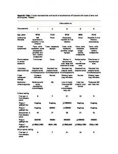
Table. Dengue laboratory test results, National Public Health Laboratory, Nepal, and AFRIMS, Bangkok, 2006* Patient no. Age, y Gender Residence Travel history ELISA 1 39 M Kathmandu Unknown Positive 2 48 M Banke Yes Negative 3 18 M Banke No Negative 4 20 M Banke No Negative 5 22 M Banke No Negative 6 25 M Banke No Negative 7 25 M Kathmandu Unknown Negative 8 26 F Kathmandu Unknown Positive 9 38 M Parsa No Negative 10 16 M Dhading Unknown Negative 11 25 M Jhapa No Positive 12 37 F Parsa Unknown Positive 13 38 M Dhading No Positive 14 24 M Banke No Positive 15 36 M Banke No Positive 16 22 M Parsa Unknown Positive 17 5 F Rupandehi No Positive 18 13 M Dang No Positive 19 35 F Parsa No Positive 20 20 M Kathmandu Yes Positive 40 M Kapilbastu No Positive 21 22 20 F Rupandehi No Positive 23 42 M Dang No Positive 24 65 M Banke Yes Positive 25 28 M Dang No Positive
RT-PCR DEN-3 DEN-3 DEN-3 DEN-3 DEN-3 DEN-3 DEN-1 DEN-3 DEN-4 DEN-2 ND ND ND ND ND ND ND ND ND ND ND ND ND ND ND
*AFRIMS, Armed Forces Research Institute of Medical Sciences; RT-PCR, reverse transcription–PCR; DEN, dengue; ND, not done.
Sarala Malla, Garib D. Thakur, Sanjaya K. Shrestha, Manas K. Banjeree, Laxmi B. Thapa, Gyanendra Gongal, Prakash Ghimire, Bishnu P. Upadhyay, Purosotam Gautam, Shyam Khanal, Ananda Nisaluk, Richard G. Jarman, and Robert V. Gibbons Author affiliations: National Public Health Laboratory, Kathmandu, Nepal (S. Malla, B.P. Upadhyay, S. Khanal); Epidemiology and Disease Control Division, Kathmandu (G.D. Thakur, M.K. Banjeree, L.B. Thapa, P. Gautam); Walter Reed/Armed Forces Research Institute of Medical Sciences, Kathmandu (S.K. Shrestha); WHO Regional Office for South-East Asia, New Delhi, India (G. Gongal); Tribhuvan University, Tribhuvan, Nepal (P. Ghimire); and United States Army Medical Command–Armed Forces Research Institute of Medical Sciences, Bangkok, Thailand (A. Nisaluk, R.G. Jarman, R.V. Gibbons) DOI: 10.3201/eid1410.080432
1670
References Pandey BD, Rai SK, Morita K, Kurane I. First case of dengue virus infection in Nepal. Nepal Med College J. 2004;6:157–9. 2. Pandey BD, Morita K, Khanal SR, Takasaki T, Miyazaki I, Ogawa T, et al. Dengue virus, Nepal. Emerg Infect Dis. 2008;14:514–5. 3. Sherchand JB, Pandey BD, Haruki K, Jimba M. Serodiagnosis of Japanese encephalitis and dengue virus infection from clinically suspected patients of Nepal. J Inst Med. 2001;23:25–31. 4. Malla S, Ghimire P, Dumre S, Khanal SP, Subedi BK, Wierzba TF. A first report of dengue fever cases in Nepal. In: Health action in Nepal: health newsletter [cited 2008 Aug 27]. Available from http://www. who.int/hac/crises/npl/sitreps/2005/Nepal_Health_Action_Issue_III.pdf 5. Takasaki T, Kotaki A, Nishimura K, Sato Y, Tokuda A, Lim CK, et al. Dengue virus type 2 isolated from an imported dengue patient in Japan: first isolation of dengue virus from Nepal. J Travel Med. 2008;15:46–9. 6. Bharaj P, Chahar HS, Pandey A, Diddi K, Dar L, Guleria R, et al. Concurrent infections by all four dengue virus serotypes during an outbreak of dengue in 2006 in Delhi, India. Virol J. 2008;5:1. DOI: 10.1186/1743-422X-5-1
7.
1.
8.
9.
10.
World Health Organization. Dengue haemorrhagic fever: diagnosis, treatment, prevention and control. 2nd ed. Geneva: The Organization; 1997. Innis BL, Nisalak A, Kusalerdchariya S, Chongswasdi V, Suntayakorn S, Puttisiri P, et al. An enzyme-linked immunosorbent assay to characterize dengue infections where dengue and Japanese encephalitis co-circulate. Am J Trop Med Hyg. 1989;40:418–27. Lanciotti RS, Calisher CH, Gubler DJ, Chang GJ, Vorndam AV. Rapid detection and typing of dengue viruses from clinical samples by using reverse transcriptase– polymerase chain reaction. J Clin Microbiol. 1992;30:545–51. Halstead SB, Nimmannitya S, Cohen SN. Observations related to the pathogenesis of dengue hemorrhagic fever. IV. Relation of disease severity to antibody response and virus recovered. Yale J Biol Med. 1970;42:311–28.
Address for correspondence: Robert V. Gibbons, United States Army Medical Command–Armed Forces Research Institute of Medical Sciences, APO AP 96546, USA; email: robert.gibbons@ afrims.org
Emerging Infectious Diseases • www.cdc.gov/eid • Vol. 14, No. 10, October 2008