kind of within-object spatial analysis was processed by this area whatever the goal ..... responding anatomical regions and Brodmann areas (BA). Matching Task.
Visual Pathways for Object-Oriented Action and Object Recognition: Functional Anatomy with PET
Isabelle Faillenot, Ivan Toni, Jean Decety, Marie-Claude Grégoire1 and Marc Jeannerod
The purpose of this study was to identify the functional anatomy of the mechanisms involved in visually guided prehension and in object recognition in humans. The cerebral blood flow of seven subjects was investigated by positron emission tomography. Three conditions were performed using the same set of stimuli. In the ‘grasping’ condition, subjects were instructed to accurately grasp the objects. In the ‘matching’ condition, subjects were requested to compare the shape of the presented object with that of the previous one. In the ‘pointing’ condition (control), subjects pointed towards the objects. The comparison between grasping and pointing showed a regional cerebral blood flow (rCBF) increase in the anterior part of the inferior parietal cortex and part of the posterior parietal cortex. The comparison between grasping and matching showed an rCBF increase in the cerebellum, the left frontal cortex around the central sulcus, the mesial frontal cortex and the left inferior parietal cortex. Finally, the comparison between matching and pointing showed an rCBF increase in the right temporal cortex and the right posterior parietal cortex. Thus object-oriented action and object recognition activate a common posterior parietal area, suggesting that some kind of within-object spatial analysis was processed by this area whatever the goal of the task.
experimental data have suggested that parietal cortex might also be involved in some aspects of shape discrimination. Electrophysiological studies in the monkey have shown the existence, in the anterior intraparietal area (AIP), of neurons which fire in anticipation of manipulative hand movements directed at specific objects (Taira et al., 1990; Sakata and Taira, 1994). Transient inactivation of this area abolishes the preshaping of the hand during prehension (Gallese et al., 1994). Neuropsychological observations also point to a similar organization of visual cortical pathways in man. Parietal lesions produce spatial disorientation, especially when located in the right hemisphere. A subset of these lesions centered in the intraparietal sulcus (IPS) produce a specific visuomotor disorder known as ‘optic ataxia’. Patients with optic ataxia are unable to appropriately orient their hand when reaching out towards target objects (Perenin and Vighetto, 1988), or to correctly calibrate their finger grip to the size of these objects (Jeannerod, 1986). Conversely, whereas parietal patients have no difficulty in discriminating between the same objects, patients with bilateral occipito-temporal lesions present object agnosia (see review in Farah, 1992). Goodale et al. (1991) reported the case of one such patient who was unable to visually identify objects but had normal performance in grasping them. These observations have prompted a re-evaluation of the functional distinction between the two cortical visual streams (e.g. Goodale and Milner, 1992). Both streams would process visual information about object orientation and shape, but each stream would use it in order to achieve different goals. The ventral stream plays a critical role in the visual perception of objects while the dorsal stream mediates the sensorimotor transformations required for visually guided action directed towards them. The present experiment was designed to identify the neural network involved in object analysis when it is performed with the purpose of either recognizing the object or generating an action towards it. Subjects were thus instructed either to make a shape judgement on non-verbalizable objects of different shapes and sizes, or to grasp and manipulate them. These two tasks are not equivalent, however, because making a shape judgement — a perceptual task — is devoid of any motor component. For this reason, in order to compare the two tasks, we used pointing as a third condition for substracting from grasping as much of its motor component as possible.
A large body of anatomical data indicate that connections within the monkey visual cortex are organized along two main streams of projections. Both streams originate from the primary visual areas: the dorsal stream goes through extrastriate visual areas and reaches the associative parietal areas, whereas the ventral stream reaches the inferotemporal cortex (Mishkin et al., 1983; Boussaoud et al., 1990). Although these pathways can be seen as two separate routes for processing visual input, they are also interconnected (Morel and Bullier, 1990). The point of whether these two visual pathways correspond to distinct and complementary visual functions has been the subject of many physiological studies. The inferotemporal cortex has been known for a long time to be involved in shape discrimination (Gross et al., 1972; Pohl, 1973). Neuronal populations in these areas respond more to complex visual patterns than to simple bars or spots (Tanaka et al., 1990; Tanaka, 1993). Many cells exhibit remarkable specificity for particular stimuli, like faces, without being sensitive to more elementary attributes of these stimuli, such as spatial position, size, luminance, color or spatial frequency (Perrett et al., 1982, 1987). This property is a strong argument for the claim that inferotemporal areas are functionally involved in processing invariant aspects of visual stimuli and thus represent the final stage of an object identification mechanism. Posterior parietal cortex, by contrast, has been assigned a role in processing spatial relationships between objects and between the body and other objects. Lesions of this region lead to disturbances in discriminating the respective positions of objects and in orienting movement in extrapersonal space (Pohl, 1973; Faugier-Grimaud et al., 1978). More recently, however,
Vision et Motricité, INSERM U 94, 16 Av du Doyen Lépine, 69500 Bron and 1CERMEP, 59 bld Pinel, 69003 Lyon, France
Materials and Methods Subjects Seven healthy male volunteers (age 24 ± 1 years) were studied. All subjects were right-handed according to the Edinburgh Handedness Inventory (Oldfield, 1971). None of them had any history of neurological disease. Subjects gave their informed consent for the experimental procedures and were paid for their participation. The experiments were Cerebral Cortex Jan/Feb 1997;7:77–85; 1047-3211/97/$4.00
carried out in accordance with the Declaration of Helsinski and with approval from the local Ethical Committee. Experimental Design and Apparatus The subjects were studied during three different activation tasks repeated twice in a pseudo-randomized order. The following specific instructions concerning each task were given immediately prior to each scan:
. . .
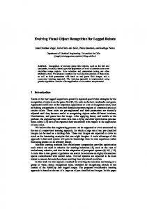
Point with the right index finger towards the center of the object and come back to the starting position (pointing task). Take the object with the right hand and place it on the table (grasping task). Subjects were asked to grasp the objects between their fingertips (precision grip) and to place at least one finger in the notch (see Fig. 1) in order to adapt their hand shape and orientation to the object. Observe the objects and press a mouse button each time the shapes of two consecutive objects are identical, irrespective of size and orientation (matching task). Each subject’s right hand was placed in contact with the mouse for the duration of the task.
The same set of stimuli was presented at the same rate (0.3 Hz) and in the same pseudo-randomized order in the three conditions. The stimuli consisted of white wooden blocks (1.0–2.5 mm thick) with five different shapes and three different sizes (Fig. 1). Each object was presented three times (45 presentations). The sequence was arranged in such a way as to involve 15 occurrences of consecutive identical shapes. Objects were designed in order to elicit different patterns of grasp movements and were sufficiently similar to require careful visual analysis to discriminate between each other. The object presentation device was positioned on the camera-bed. It consisted of two planes, an inferior horizontal table and a superior oblique board with a circular hole (13 cm diameter). The subject’s right forearm was placed on the horizontal table, above the subject’s chest. The superior board was positioned in such a way that the hole was at reaching distance and perpendicular to the subject’s gaze axis. Objects were fixed on a disk (14 cm diameter) by Velcro and presented one at a time through the hole. In this way, subjects could only see the stimulus upon a black background. The object notch was always presented on the right side, but as objects were manually fixed on the disk, small variations in the orientation of the object could occur. This is why the subjects were instructed not to pay attention to the orientation. The movements performed by the subjects towards the objects consisted of rotation of the arm at the shoulder joint. Data Acquisition Regional cerebral blood f low (rCBF) was measured by recording the distribution of cerebral radioactivity following injections of H215O. Any increase in rCBF entails an increase in the amount of radioactivity observed in that region (Raichle, 1987). Each subject received six i.v. bolus injections of 50 mCi with a 15 min inter-scan interval. Head movements were restricted with a molded-foam head holder, which was individually made for each subject and used for both magnetic resonance imaging (MRI) and positron emission tomography (PET) scans. Anatomical images (FLASH 3D, T1) were obtained with a Siemens 1.5 Tesla scanner. Using MRI references, PET data were acquired parallel to the intercommissural (AC–PC) plane. PET scans were performed on a TTV03 time-of-f light camera (LETI). This scanner provides images with seven slices (Mazoyer et al., 1990). Since the axial field of view of this camera (84 mm) is smaller than the whole brain size (∼110 mm), two acquisitions were required for each condition. This was performed by moving the bed of the scanner in order to cover the whole cerebrum. Radiation attenuation phenomenon was corrected by transmission scans using an external positron emitting source (68Ge) rotating around the subject’s head. Bolus injection, data acquisition and object presentation started at the same time. Data were acquired in list mode and a dynamic series was generated to plot activity as a function of time. Off-line reconstruction started when 20% of maximal activity was reached, on average 18 s after the injection. Emission data were reconstructed for 90 s using a Hanning filter with a cut-off frequency of 0.15 mm–1. Initial images were in a 128 ×
78 Cerebral Regions Involvement in Grasping and Shape Matching • Faillenot et al.
Figure 1. Object shapes used in the tasks. Each shape was built in three sizes.
128 × 7 pixel format with a 2 × 2 × 12 mm voxel size. MRI data were converted in a 128 × 128 format with cubic voxel of 2 mm3. Image Transformations Image calculations and manipulations were carried out on Sun SPARK workstation using the ANALYZE image display software and SPM package running on MATLAB. PET scans were normalized for global f low for each subject. Possible head movement between scans was corrected by aligning all scans on the first one, using Automated Image Registration (AIR) software (Woods et al., 1993). For each subject, the two sets of PET images were aligned on MRI data in order to be parallel to the AC–PC plane. After merging, a unique set of PET images representing the whole brain was available for each condition. PET and MR images were transformed into a standard space (Talairach and Tournoux, 1988) using a stereotactic normalization developed by Friston et al. (1989). This normalizing spatial transformation matches each scan to a template image that already conforms to the standard space. The procedure involves an affine (linear) and quadratic (nonlinear) three-dimensional transformation. This is followed by a two-dimensional piecewise (transverse slices) nonlinear matching. MR images were transformed in the same manner and were averaged across subjects. The blurring in the mean MRI scan ref lects the variability in position of anatomic structures for our group. Subsequently, the PET images were filtered with a low-pass Gaussian filter (FWHM of 12 mm) for smoothing the data in three dimensions. This allowed the suppression of noise and effects due to residual differences in functional and gyral anatomy during intersubject averaging. Statistical Parametric Mapping The task and subject effects were estimated according to the general linear model at each and ever y voxel. To test the hypothesis about regional specific task effects the estimates were compared using linear contrasts. The resulting set of voxel values for each contrast provided a statistical parametric map [SPM (Z)]. In this study, the resulting foci were then characterized in terms of peak height (u). The significance of each region was estimated in terms of the probability that the peak height observed could have occurred by chance [P (Zmax > u)] over the entire volume. Stimulus-induced changes in rCBF were considered to be significant at a threshold of P < 0.05 (Z-score > 4.2) after correcting for multiple comparisons. Significant rCBF increases were superimposed on averaged MRI. This procedure enabled us to report activated foci in terms of Talairach and Tournoux coordinates as well as by reference to anatomical structures. Singular Value Decomposition (SVD) In addition to the inferential statistical analysis described above, a descriptive statistical analysis concerning the pattern of correlated activity of PET data was also performed. Principal components of the covariance matrix were extracted and maps of functional networks corresponding to significant components were analysed (Friston, 1994). SVD reveals the spatial and/or temporal patterns that explain most of the variance of the data.
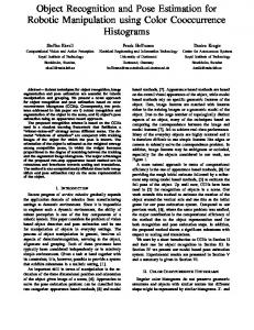
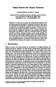
Results Behavioral Observations In the pointing task, the subject’s hand was placed with the palmar surface resting on the table. During the pointing movement, subjects extended their index finger and f lexed the other fingers. In the grasping task, subjects effectively adapted their hand posture to the different objects by changing wrist orientation, and adjusting grip size and the number of fingers involved. In the matching task, although all subjects reported some difficulty in sustaining attention, the mean error rate was only 6%. Subjects’ strategy for matching objects was based primarily on global shape and secondarily on shape details. Two subjects mentioned that they had tried to name the objects. None of them reported having used mental rotation to match the shapes. Statistical Parametric Mapping The Talairach and Tournoux atlas coordinates of all significant rCBF increases are shown in Table 1, together with the corresponding anatomical regions and Brodmann areas (BA). Matching Task When the matching task was compared with the pointing task, three foci of rCBF increases appeared in the right hemisphere (Fig. 2, upper part): one in the temporal lobe and two in the parietal lobe. In the right temporal lobe a discrete but highly significant rCBF increase was located just above the temporo-occipital sulcus in the inferior temporal cortex (BA 37). In the right parietal lobe the rCBF increases were both located in the dorsal cortex around the intraparietal sulcus (IPS). The lower region was in the posterior part of the superior parietal lobule (between BA 19 and 7). The upper region was in the IPS (between BA 40 and 7). When the matching task was compared with the grasping task (Fig. 2, lower part), two rCBF increases appeared. The right temporal activation observed in the previous subtraction was still present. The other rCBF increase was in the left middle frontal cortex (BA 10). The dorsal parietal activations observed in the previous subtraction were not significant. Grasping Task Comparing the grasping task with the pointing task revealed only one region of activation in the whole brain. The Talairach and Tournoux atlas coordinates of this rCBF increase corresponds to BA 40 in the left hemisphere (contralateral to the grasping hand) extending from AC–PC +16 to +32 mm (Fig. 3). Careful inspection of the averaged and individual MR images revealed that this area was located in the inferior part of the post-central sulcus (BA 2/40). The comparison between grasping and matching shows the ensemble of regions involved in the grasping movement, as illustrated in Figure 4. Planes below AC–PC –8 mm demonstrate activation in the vermis and in the right lateral cerebellum. The planes +40 to +48 mm show activation in the left frontal cortex around the central sulcus (BA 1–4), which extends to the pre-central sulcus (BA 6). This region covers the sensorimotor hand area (AC–PC +40 to +48 mm) and the lateral premotor cortex. The planes +48 and +52 mm show an rCBF increase in the mesial frontal cortex (BA 6), corresponding to SMA. Moreover, there are activations in the left inferior post-central sulcus (BA 2/40) (AC–PC +20 to +32 mm), in the left SII (AC–PC +8 to +16 mm), in the left insula (AC–PC +0 to +8 mm) and in the
Table 1 Anatomical regions that showed significant rCBF increase Subtraction Anatomical region
Brodmann area Coordinates x
Matching vs R intraparietal sulcus pointing R occipito-parietal cortex R inf. temporal gyrus Matching vs L mid. frontal gyrus grasping R inf. temporal gyrus Grasping vs L inf. postcentral sulcus * pointing
Grasping vs vermis matching R cerebellum L inf. postcentral sulcus* L parietal operculum* (SII) L post. insula * L post-central gyrus L central gyrus L mesial frontal cortex L cuneus R post-central sulcus
y
Z-score z
40/7
34
–52
44
4.80
19/7 37
24 44
–72 –46
40 –8
4.78 4.25
10
–26
60
8
4.81
37
44
–46
–8
4.60
–48
–18
16
4.64
–50
–28
28
4.43
0
–66
–16
5.37
12 –48 –42 –36 –32 –30 0 –16 38
–56 –22 –22 –28 –32 –22 –2 –76 –26
–16 24 8 4 48 48 48 16 32
4.58 5.13 4.47 4.41 4.71 4.57 4.55 4.49 4.42
2/40
2/40
1. 2. 3 4 6 18 2/40
Coordinates are in millimeters and correspond to the atlas of Talairach and Tournoux (1988). Brodmann areas are defined according to the same atlas. Z-score > 4.2 and > 4.6 correspond to P < 0.05 and < 0.01 respectively. Post, inf, mid, R and L indicate posterior, inferior, middle, right and left respectively. *Peaks of activity localized according to their position on averaged MRI (see figures). The coordinates do not correspond to these anatomical regions in the atlas.
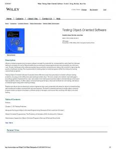
left cuneus (BA 18) (AC–PC +16 to +20 mm). In addition, an activated area was found in the fundus of the ipsilateral (right) post-central sulcus (BA 2/40) (AC–PC +32 mm). The limited zones in the occipito-parietal cortex and in the IPS that appeared to be activated when the matching task and the pointing task were compared (Fig. 2) did not reach the 0.05 level of significance when comparing the matching task to the grasping task and the grasping task to the pointing task. In fact, the maximal Z-scores of these areas was just below the significance level. IPS showed an rCBF increase in the grasping versus pointing comparison (Z-score = 4.13, P = 0.06, coordinates: 30, –52, 44) whereas the occipito-parietal cortex showed an rCBF increase in the matching versus grasping comparison (Z = 4.09, P = 0.07, coordinates: 24, –72, 40). Because the SPM analyses are inherently conservative, these results suggest that the IPS is likely to be involved in both matching and grasping, whereas the occipito-parietal cortex is more distinctive of object identification. Functional Networks The SVD analysis showed that the main factor accounts for 77.5% of the observed variance–covariance structure (Fig. 5A). Figure 5B presents the score for this factor, revealing that the main effect is present in the grasping and matching conditions. In other words, the PET activity observed in the present experiment was distributed between two poles corresponding to our two main experimental conditions. Therefore, the pointing task can be considered as a control condition for the other two tasks. The eigenimages corresponding to the main factor are shown in Figure 5C and D. The positive eigenimage (Fig. 5C) represents
Cerebral Cortex Jan/Feb 1997, V 7 N 1 79
Figure 2. Cerebral regions activated during the matching task compared with the pointing task, and during the matching task compared with the grasping task. These regions are superimposed on sections of the averaged MRI. Under horizontal slices, numbers indicate the distance (in mm) from the AC–PC plane. The red and yellow areas indicate pixels with Z-score > 3.1 and > 4.2 (P corrected < 0.05) respectively. The right hemisphere is on the right. VAC is the vertical traversing the anterior commissure.
80 Cerebral Regions Involvement in Grasping and Shape Matching • Faillenot et al.
Figure 3. Cerebral regions activated during the grasping task compared with the pointing task. All conventions are as for Figure 2.
the functional network in the grasping task. This network includes principally the cerebellum and the left sensorimotor cortex. It also extends to the SM A, premotor cortex, cingulate cortex, ipsilateral sensorimotor cortex, left inferior parietal cortex (SI, SII, BA 40), bilateral insular cortex and parietooccipital sulci. The negative eigenimage (Fig. 5D) shows the functional network involved in the matching task. The pattern shows a greater co-activation of the right hemisphere. Its highest loadings are in the right inferior and middle temporal gyri and in the medial part of the superior frontal gyrus (BA 8/9). All the prefrontal cortex and the right IPS seems to be involved in the matching task.
Discussion The aim of this study was to determine the neural substrate of object processing according to the task in which the subject is
involved. Cortical rCBF was measured during a task involving visuomotor transformation (grasping objects) and a task involving perceptual visual judgement (matching object shapes with each other). As the same set of stimuli was used in both conditions, only the task differed. These two tasks both require analysis of object properties, but for different purposes. In the case of grasping, object size, volume and shape are analyzed for programming and executing the prehension movement (see Jeannerod, 1981). In the case of matching, the analysis of object shape is made for the purpose of memorizing and comparing. The results of this experiment were twofold. First, the activation of some cortical areas was task-specific: whereas the inferior parietal cortex in the contralateral hemisphere was involved in the network performing the visuomotor transformation, perceptual matching involved the temporal cortex on the right side. Second, and most importantly, a limited cortical area in the right intraparietal sulcus was common to both tasks.
Cerebral Cortex Jan/Feb 1997, V 7 N 1 81
Figure 4. Cerebral regions activated during the grasping task compared with the matching task. All conventions are as for Figure 2.
Temporal Cortex The significant rCBF increase in the temporal cortex during the matching task was limited to a restricted region in the Brodmann area 37 (Fig. 2). This local increase in blood f low, however, was included in a widespread functional network, as shown in Figure 5D. This network extends over the occipito-temporal junction, the middle and inferior temporal gyri, a large fraction of the prefrontal lobe and the posterior parietal cortex. Similar results (except for the parietal activation discussed below) were obtained in previous studies dealing with visual processing of objects or faces both in humans (Haxby et al., 1991; Sergent et
82 Cerebral Regions Involvement in Grasping and Shape Matching • Faillenot et al.
al., 1992; Kosslyn et al., 1994) and non-human primates (Mishkin et al., 1983; Bachevalier and Mishkin, 1986; Tanaka, 1993). A recent study on shape matching using unknown abstract objects found a restricted activation in the posterior inferior temporal gyrus (BA 37) (Smith et al., 1995), i.e. lateral with respect to the area activated during the processing of unknown faces which involved mainly the fusiform gyrus. The fact that the right hemisphere was more involved than the left one in our matching task is also consistent with previous reports by Horwitz et al. (1992) and by McIntosh et al. (1994). In these studies, the object vision condition was similar to ours: it
Figure 5. Results of principal components extraction. (A) The SVD analysis showed two factors. Factor 1 accounts for 77.5% of variance of all the data. (B) The main factor is related to the difference between the grasping task (G) and the matching task (M). (C) The positive eigenimage corresponds mainly to the functional network involved in the grasping task. (D) The negative image shows the network involved in the matching task. The eigenimages are displayed as a maximum intensity projection in standard SPM format. The color scale is arbitrary and each image is scaled to its maximum. The numbers indicate the size in mm of the stereotactic box. R indicates the right hemisphere. VAC and VPC are the verticals traversing the anterior and posterior commissures.
required that the attributes of objects (faces) be analyzed for the purpose of comparison. Sergent et al. (1992) also reported an activation at the right occipito-temporal junction (BA 19, 37) during face identification, whereas the left occipito-temporal cortex (BA 19, 20, 21) was activated during object categorization. This observation is concordant with behavioral data suggesting the existence of two independent visual-form systems (e.g. Marsolek, 1995). One of them, which operates in the right hemisphere, stores local details for distinguishing specific instances of a given form. The other one, in the left hemisphere, stores invariant information for discriminating different form
categories. Thus, the activation of cortical areas in the right hemisphere during our matching task would be consistent with this model. Anterior Parietal Cortex During the grasping–matching subtraction, areas involved in somatosensory processing became visible (Fig. 4). This pattern of activation also appears on the positive eigenimage of correlated activity analysis (Fig. 5C). In the parietal lobe, the rCBF increased in primary somatosensory areas (BA 1, 2 and 3) at the level of the hand representation (Z > +40 mm; see Matelli
Cerebral Cortex Jan/Feb 1997, V 7 N 1 83
et al., 1993; Grafton et al., 1994; Sadato et al., 1996). A distinct focus of activity was located in the left inferior postcentral sulcus (BA 2/40) and extended towards the secondary sensory area (SII) and the posterior insular cortex. The latter two regions, which are anatomically linked, have often been shown to be coactivated (e.g. Seitz and Roland, 1992; Burton et al., 1993; Stephan et al., 1995). Thus the parietal operculum was involved in both the pointing and the grasping tasks, but the ventral part of the postcentral sulcus was activated by the grasping task only (see also Grafton et al., 1996). The finding of a specific activation in the post-central sulcus during the grasping task is surprising. Based on the localization of lesions affecting visuomotor transformation (Perenin and Vighetto, 1988), we had predicted that grasping would involve a more posterior region in the parietal lobe. Instead, we found activation in an anterior region (BA 2/40, Z < +32 mm) when the pointing task was subtracted from grasping. Activation of the inferior part of the postcentral sulcus was reported in a number of different tasks, such as stimulation by moving visual stimuli (Dupont et al., 1994), tactile discrimination (O’Sullivan et al., 1994) and mental imagery of grasping movements (Decety et al., 1994). In addition, Grafton et al. (1996) found an area in the lateral parietal operculum involved in the grasping task but not in the pointing task. Its anatomical location as well as its Talairach coordinates closely correspond to the ventral part of the area activated in the present study. These results are consistent with Deiber et al. (1996), who showed that the anterior parietal cortex (BA 40) close to the postcentral sulcus plays a critical role in the use of visual instructions for motor preparation. The Talairach coordinates of this area suggest that it overlaps the dorsal part of the area involved in the present grasping task. Considering its function and its anatomical location, the most anterior part of the human IPL bears some homology to the inferior parietal area 7b in the monkey (Hyvärinen, 1981). This latter area, which is connected to other parietal areas such as SII, A IP and VIP and to the ventral premotor cortex, is part of a network for producing motor responses to visual and somatosensory stimuli (Graziano et al., 1994; Graziano and Gross, 1993). Occipito-parietal Cortex The shape matching task activated a region in the right occipito-parietal cortex which was found not to be concerned in either the grasping or the pointing task. This region is at the border between Brodmann areas 19 and 7. Haxby et al. (1994), who found that this area was activated in a location matching task, concluded that it was involved in the processing of spatial vision. This can hardly be the case in the present experiment, since our two tasks both carried spatial location analysis. Another possible interpretation (Kosslyn et al., 1994, 1995) is that this region is active during object identification tasks because it is involved in shifting attention to the location of an expected object part or property. Since the stimuli used in our study were abstract and unknown, devoid of semantic or functional cues, shape comparison depended exclusively on perceptual information. It is likely that in these conditions, the attentional component was more important in the matching task than in the other tasks (as the subjects themselves reported). Because the matching task relied, at least in part, on localizing the area of the object where the critical properties were located, subjects had to orient their local attention. Whatever the mechanism involved (perceptual or attentional), it is logical to
84 Cerebral Regions Involvement in Grasping and Shape Matching • Faillenot et al.
submit that this part of the occipito-parietal cortex is involved in the local analysis of object properties. One cannot exclude that the predominent right hemisphere activation in the matching condition was due, at least partly, to the subtraction methodology: when the grasping (or the pointing) condition was subtracted from matching, a predominant activation in the left fronto-parietal cortex was removed. Intraparietal Sulcus Both the grasping and, to a greater extent, the matching tasks were associated with an rCBF increase in the IPS (BA 40/7). This area is thus clearly associated with the visual analysis of objects. Indeed, both tasks required visual processing in order to analyze the size, volume and orientation of the object irrespective of its spatial location. During the grasping task, the object size and volume determined the grip size and the adequate number of fingers that were necessary to grasp it; during the matching task size and volume had to be processed in order to extract the invariant shape properties that were used for the comparison. A possible interpretation is that this area in the IPS plays a role in integrating the three-dimensional properties of objects, based on the finding that visual IPS neurons in the monkey respond to three-dimensional object properties (axis or surface orientation, thickness, shape) from binocular disparity signals (Ohtsuka et al., 1995). Further studies are needed to determine the specific role of IPS in visual object analysis.
Conclusion The present results do not fully support the dissociation between visual pathways specialized for ‘perception’ (the occipito-temporal pathway) and for ‘action’ (the occipitoparietal pathway) (e.g. Goodale and Milner, 1992), which would have predicted that a specific region in the posterior parietal cortex is distinctly involved in grasping as compared with matching. This region was not found in the present study. Rather, an area in the IPS, common to the two tasks, was activated. This finding underlines that recognition and grasping are not independent processes using strictly parallel pathways. Rather, our results support the idea that object-oriented action and shape comparison both involve a form of visual object analysis which is processed by the same posterior parietal area. This mechanism would be distinct from the analysis of shape mediated by the temporal cortex, and its exclusion by a lesion would not prevent object recognition.
Notes The authors are grateful to M. T. Perenin and Y. Rossetti for their advice on experimental design. I.F. was supported by a grant from the Human Science Frontier Program. I.T. was supported by the Fondation Fyssen (Paris). Correspondence should be adressed to Dr Jean Decety, Vision et Motricité, INSERM U.94, 16 Av du Doyen Lépine, F-69500 Bron, France.
References Bachevalier J, Mishkin M (1986) Visual recognition impairment follows ventromedial prefrontal lesions in monkeys. Behav Brain Res 20:249–261. Boussaoud D, Ungerleider LG, Desimone R (1990) Pathways for motion analysis: cortical connections of the medial superior temporal and fundus of the superior temporal visual areas in the macaque. J Comp Neurol 296:462–495. Burton H, Videen TO, Raichle ME (1993) Tactile-vibration-activated foci in insular and parietal-opercular cortex with PET: mapping the second somatosensory area in humans. Somatosens Motor Res 10:297–308. Decety J, Perani D, Jeannerod M, Bettinardi V, Tadary B, Mazziotta JC,
Woods R, Fazio F (1994) Mapping motor representations with positron emission tomography. Nature 371:600–602. Deiber MP, Ibanez V, Sadato N, Hallett M (1996) Cerebral structures participating in motor preparation in humans: a positron emission tomography study. J Neurophysiol 75:233–247. Dupont P, Orban GA, Bruyn Bd, Verbruggen A, Mortelmans L (1994) Many areas in the human brain respond to visual motion. J Neurophysiol 72:1420–1424. Farah MJ (1992) Agnosia. Curr Opin Neurobiol 2:162–164. Faugier-Grimaud S, Frenois C, Stein DG (1978) Effects of posterior parietal lesions on visually guided behavior in monkeys. Neuropsychologia 16:151–168. Friston KJ (1994) Functional and effective connectivity in neuroimaging: a synthesis. Hum Brain Mapping 2:56–78. Friston KJ, Passingham RE, Nutt JG, Heather JD, Sawle GV, Frackowiak RSJ (1989) Localisation in PET images: direct fitting of the intercommissural (AC–PC) line. J Cereb Blood Flow Metab 9:690–695. Gallese V, Murata A, Kaseda M, Niki N, Sakata H (1994) Deficit of hand preshaping after muscimol injection in monkey parietal cortex. NeuroReport 5:1525–1529. Goodale MA, Milner AD (1992) Separate visual pathways for perception and action. Trends Neurosci 15:20–25. Goodale MA, Milner AD, Jacobson LS, Carey DP (1991) A neurological dissociation between perceiving objects and grasping them. Nature 349:154–156. Grafton ST, Fagg AH, Woods RP, Arbib MA (1996) Functional anatomy of pointing and grasping in humans. Cereb Cortex 6:226–237. Grafton ST, Wood RP, Tyszka M (1994) Functional imaging of procedural motor learning: relating cerebral blood f low with individual subject performance. Hum Brain Mapping 1:221–234. Graziano MSA, Gross CG (1993) A bimodal map of space: somatosensory receptive fields in the macaque putamen with corresponding visual receptive fields. Exp Brain Res 97:96–109. Graziano SA, Yap GS, Gross CG (1994) Coding of visual space by premotor neurons. Science 266:1054–1056. Gross CG, Rocha-Miranda CE, Bender DB (1972) Visual properties of neurons in the inferotemporal cortex of the macaque. J Neurophysiol 35:96–111. Haxby JV, Grady CL, Horwitz B, Ungerleider LG, Mishkin M, Carson RE, Herscovitch P, Schapiro MB, Rapoport SI (1991) Dissociation of object and spatial visual processing pathways in human extrastriate cortex. Proc Natl Acad Sci USA 88:1621–1625. Haxby JV, Horwitz B, Ungerleider LG, Maisog JM, Pietrini P, Grady CL (1994) The functional organization of human extrastriate cortex: a PET-rCBF study of selective attention to faces and locations. J Neurosci 14:6336–6353. Horwitz B, Grady CL, Haxby JV, Schapiro MB, Rapoport SI, Ungerleider LG, Mishkin M (1992) Functional associations among human posterior extratriate brain regions during object and spatial vision. J Cogn Neurosci 4:311–322. Hy värinen J (1981) Regional distribution of functions in parietal association area 7 of the monkey. Brain Res 206:287–303. Jeannerod M (1981) Intersegmental coordination during reaching and natural visual objects. In: Attention and performance (Long J, Baddeley A, eds), pp 153–168. Hillsdale, NJ: Lawrence Erlbaum. Jeannerod M (1986) The formation of finger grip during prehension, a cortically mediated visuomotor pattern. Behav Brain Res 19:99–116. Kosslyn SM, Alpert NM, Thompson WL, Rauch SL, Anderson AK (1994) Identifying objects seen from different viewpoints: PET investigation. Brain 117: 1055–1071. Kosslyn SM, Alpert NM, Thompson WL (1995) Identifying objects at different levels of hierarchy: a PET study. Hum Brain Mapping 3:107–132. Marsolek CJ (1995) Abstract visual-form representations in the left cerebral hemisphere. J Exp Psychol Hum Percept Perf 21:375–386. Matelli M, R izzolatti G, Bettinardi V, Gilardi MC (1993) Activation of
precentral and mesial motor areas during the execution of elementary proximal and distal arm movements: a PET study. NeuroReport 4:1295–1298. Mazoyer B, Trebossen R, Shoukroun C, Verrey B, Syrota A, Vacher J, Lemasson P (1990) Physical caracteristics of TTV03, a new hight spatial resolution time-of-f light positron tomograph. IEEE Trans Nucl Sci 37:778–782. McIntosh AR, Grady CR, Ungerleider LG, Haxby JV, Rapoport SI, Horwitz B (1994) Network analysis of cortical visual pathways mapped with PET. J Neurosci 14:655–666. Mishkin M, Ungerleider LG, Macko K A (1983) Object vision and spatial vision: two cortical pathways. Trends Neurosci 6:414–417. Morel A, Bullier J (1990) Anatomical segregation of two cortical visual pathways in the macaque monkey. Vis Neurosci 4:555–578. O’Sullivan BT, Roland PE, Kawashima R (1994) A PET study of somatosensor y discrimination. Microgeometry versus macrogeometry. Eur J Neurosci 6:137–148. Ohtsuka H, Tanaka Y, Kusunoki M, Sakata H (1995) Neurons in monkey parietal association cortex sensitive to axis orientation. J Jpn Ophthalmol Soc 99:59–67. Oldfield RC (1971) The assessment and analysis of handedness: the Edinburgh inventory. Neuropsychologia 9:97–113. Perenin MT, Vighetto A (1988) Optic ataxia: a specific disruption in visuomotor mechanisms. Brain 111:643–674. Perrett DI, Rolls ET, Caan W (1982) Visual neurons responsive to faces in the monkey temporal cortex. Exp Brain Res 47:329–342. Perrett DI, Mistlin AJ, Chitty AJ (1987) Visual cells responsive to faces. Trends Neurosci 10:358–364. Pohl W (1973) Dissociation of spatial discrimination deficits following temporal and parietal lesions in monkeys. J Comp Physiol Psychol 82:227–239. Raichle ME (1987) Circulatory and metabolic correlates of brain function in normal humans. In: Handbook of physiology, the nervous system, part 2 (Mountcastle VB, Plum F, Geiger SR, eds), pp 643–674. Bethesda, MD: Waverly Press. Sadato N, Ibanez V, Deiber M-P, Campbell G, Leonardo M, Hallett M (1996) Frequency-dependent changes of regional cerebral blood f low during finger movements. J Cereb Blood Flow Metab 16:23–33. Sakata H, Taira M (1994) Parietal control of hand action. Curr Opin Neurobiol 4:847–856. Seitz RJ, Roland PE (1992) Vibratory stimulation increases and decreases the regional cerebral blood f low and oxidative metabolism: a PET study. Act Neurol Scand 86:60–67. Sergent J, Ohta S, Macdonald B (1992) Functional neuroanatomy of face and object processing — a PET study. Brain 115:15–36. Smith EE, Jonides J, Koeppe R A, Awh E, Schumacher EH, Minoshima S (1995) Spatial versus object working memor y: PET investigation. J Cogn Neurosci 7:337–356. Stephan KM, Fink GR, Passingham RE, Silbersweig D, Ceballos-Baumann AO, Frith CD, Frackowiack RSJ (1995) Functional anatomy of the mental representation of upper extremity movements in healthy subjects. J Neurophysiol 73:373–386. Taira M, Mine S, Georgopoulos AP, Murata A, Sakata H (1990) Parietal cortex neurons related to the visual guidance of hand movement. Exp Brain Res 83:29–36. Talairach J, Tournoux P (1988) Co-planar stereotaxic atlas of the human brain. George W. Thieme: Stuttgart. Tanaka K (1993) Neuronal mechanisms of objets recognition. Science 262:685–688. Tanaka K, Saito H, Fukada Y, Moriya M (1990) Integration of form, texture and color information in the inferotemporal cortex of the macaque. In: Vision memory and the temporal lobe (Iwai H, Misitkin M, eds), pp 101–109. New York: Elsevier. Woods RP, Mazziotta JC, Cherry SR (1993) MRI–PET registration with automated algorithm. J Comput Assist Tomogr 17:536–546.
Cerebral Cortex Jan/Feb 1997, V 7 N 1 85