Apr 28, 2011 - April 28, 2011 11:47 WSPC/S0219-4775 167-FNL 00046 ... A successful pre-processing stage based on wavelet denoising ..... 7 (2009). 1â12. [12] C. J. De Luca, Surface Electromyography: ... [25] D. L. Donoho, Wavelet analysis and WVD: A ten minute tour, ... Wavelets and Applications, France (1992).
April 28, 2011 11:47 WSPC/S0219-4775 167-FNL 00046
Fluctuation and Noise Letters Vol. 10, No. 2 (2011) 157–167 c World Scientific Publishing Company � DOI: 10.1142/S0219477511000466
WAVELET-BASED DENOISING ALGORITHM FOR ROBUST EMG PATTERN RECOGNITION
ANGKOON PHINYOMARK, PORNCHAI PHUKPATTARANONT and CHUSAK LIMSAKUL Department of Electrical Engineering, Faculty of Engineering Prince of Songkla University, 15 Kanjanavanich Road Kho Hong, Hat Yai, Songkhla, 90112, Thailand Received 15 May 2010 Accepted 29 November 2010 Communicated by Hidetoshi Konno A successful pre-processing stage based on wavelet denoising algorithm for electromyography (EMG) signal recognition is proposed. From the limitation of traditional universal wavelet denoising, the optimal weighted parameter is assigned for universal thresholding method. The optimal weight for increasing EMG recognition accuracy is 50–60% of traditional universal threshold with hard transformation. Experimental results show that it improved approximately from 2 to 50% of recognition accuracy for EMG with signal-to-noise ratio (SNR) in the range of 20 to 0 dB compared to a baseline system (without pre-processing stage) and traditional universal wavelet denoising. The results are evaluated through a large EMG dataset with seven kinds of hand movements and eight types of muscle positions. Keywords: Electromyography (EMG) signal; white Gaussian noise; wavelet transform; wavelet denoising; robust recognition.
1. Introduction The EMG signal is one of the useful electrophysiological signals. It is measured by surface electrodes that are placed on the skin superimposed on the muscle. A compound of the whole motor unit action potentials (MUAPs) occurred in the muscles subjacent to the skin. The MUAPs are useful information in a number of medical and engineering applications. For instance, EMG signals are used for the diagnosis of neuromuscular and neurological problems in the clinic [1, 2] and also are used in the control of assistive devices and rehabilitation systems such as a prosthesis, electric-powered wheelchair, and exoskeleton robotic arm in engineering [3, 4]. Normally, in order to use the EMG signal as a diagnosis tool or a control signal, a feature is often extracted before performing the classification stage because a lot of information is obtained from the raw EMG data [5]. However, EMG signals that originate in various muscles and activities are also contaminated by various 157
April 28, 2011 11:47 WSPC/S0219-4775 167-FNL 00046
158
A. Phinyomark, P. Phukpattaranont & C. Limsakul
kinds of noises or interferences [6, 7]. This becomes a main problem to extract certain features and thus the reach to high accurate recognition. Hence, in the last decade, many researchers have been interested in developing better algorithms and improving the existing methods to reduce noises and to estimate the useful EMG information [8–10]. Generally, noise contaminated in EMG signals can be categorized into four main types: ambient noise, motion artifact, inherent instability of the EMG signal and inherence in electronic components in the detection and recording equipment [7, 11, 12]. The first three types have specific frequency bands and do not fall in the energy bands of the EMG signal. For example, motion artifact, cable motion artifact and instability in nature of EMG signal have most of their energy in the frequency range of 0 to 20 Hz, or power-line interference has the frequency component at 50 Hz (or 60 Hz). Generally, usage of conventional filters, i.e., band-pass filter and band-stop filter can reduce noises in this group [13]. However, noise of the last type is a main concern in the analysis of EMG signal. It is an inherent noise that is generated by electronic equipment. The frequency components of this noise are random in nature and range in the usable energy of EMG frequency band from 0 to several thousand Hz. It causes difficulty in elimination using the conventional filters. Moreover, using high-quality electronic components, intelligent circuit design and construction techniques, noises can be only reduced but it cannot be entirely eliminated [7, 12]. Hence, it may cause a problem in extracting the robust features. Ordinarily, white Gaussian noise (WGN) is used as a representative random noise in the last type in EMG signal analysis [14–17]. In this paper, different levels of WGN were used in the preparation of noisy environment. Adaptive filter or wavelet denoising algorithm, both advanced digital filters, is one of the modern methods that are commonly used as a powerful tool to remove random noise. However, the performance of adaptive filter is limited. Its performance depends on a reference input signal which is difficult to apply in real-world applications. On the other hand, wavelet denoising method does not require any reference signal. The pre-processing stage based on wavelet denoising algorithm for EMG upper- and lower-limbs movement recognition was successful in the last few years [8, 9, 11, 18–23]. In order to achieve the best performance in the wavelet denoising algorithm, four issues need to be addressed. The first and the second issues are the selection of the wavelet function and number of decomposition levels. We had presented in our previous works [18, 34] that the second order Daubechies wavelet (db2) and the fourth decomposition level provided a minimum mean square error value. Similar suggestions are also given in other literatures [8, 19]. The third issue is the selection of the wavelet threshold estimation method, which is the main focus of interest in this paper. Khezri and Jahed [20] proposed a useful wavelet threshold to estimate the denoised EMG signal, viz. Stein unbiased risk (SURE) method. It improved the accuracy of EMG recognition compared to the one without denoising the pre-processing stage and adaptive Bayes method. Following that,
April 28, 2011 11:47 WSPC/S0219-4775 167-FNL 00046
Wavelet-Based Denoising Algorithm for Robust EMG Pattern Recognition
159
in one of our recent works [18], we compared the SURE method with three other classical thresholding techniques: universal (UNI), hybrid between UNI and SURE, and minimax. Results showed that the UNI method yields better denoising performance than others including the SURE method. Moreover, Hussain et al. [8, 19] suggested that the pre-processing stage using the UNI method and hard transformation was able to improve the recognition system of the lower- and upper-limb motions. Successively, a modification of the UNI method based on level dependence was evaluated [21]. However, the former modified methods were not designed for the specific EMG system but were rather based on electrocardiography (ECG) signal, image, or simulation data [24–28]. In these literatures, some rescaled parameters such as the length of window data, logarithm of scale level, or two to the power of scale level, have been used to reduce a larger threshold value that was estimated from a fixed form of traditional UNI method [24–28, 35]. For instance, in an ECG signal, an optimal weighted parameter is found through the experimental results and a better performance of wavelet denoising is obtained [29]. Hence, in this paper, we are proposing the weighted parameter or pre-factor to modify traditional UNI method for improving the recognition of the EMG system. The fourth issue is the selection of thresholding process. After suitable threshold values are determined, the thresholding process can be done using two classical threshold transformations, namely hard and soft thresholding (HAD and SOF) [8, 9, 18–24]. 2. Wavelet-Based Denoising Algorithm The main idea of the wavelet denoising algorithm is to suppress the noise part of the signal s(n) by discarding the WGN e(n) and to recover the signal of interest f (n). The basic model is expressed as: s(n) = f (n) + e(n).
(1)
The procedure of the wavelet denoising algorithm consists of three steps. Firstly, the raw EMG signal is decomposed by the discrete wavelet transform (DWT) in order to obtain the detail and approximation coefficients (cD and cA). The cDs contain high frequency components from the high-pass filter (H) and cA contains low frequency components from the low-pass filter (L). The decomposition tree of DWT in this study is shown in Fig. 1. For wavelet signal denoising, noise parts are usually fallen in the cD bands. After that, the threshold value (THR) is calculated based on the noise variance and then is applied to the cDs using only a linear or non-linear transform. Finally, the denoised EMG signal is reconstructed based on the modified cDs and the retaining cA. From the introduction above, the traditional UNI method is better than the other classical thresholding methods. However, the THR that is estimated from the traditional UNI method is too large, which is not suitable for the EMG signal especially in the recognition point of view [21]. It not only removes noise part but
April 28, 2011 11:47 WSPC/S0219-4775 167-FNL 00046
160
A. Phinyomark, P. Phukpattaranont & C. Limsakul
Fig. 1. A signal’s wavelet decomposition tree by the DWT analysis to a 4-level decomposition.
Fig. 2. Responses of HAD and SOF with 0.4 THR value. The diagonal dashed line indicates the input signal and the solid line indicates the output signal through shrink function.
it also plausibly removes some important part of EMG signal. The traditional UNI method uses a fixed form threshold [24], which can be expressed as: � THR UNI = σ 2 log(N ), (2) where N is the length of EMG signal samples and σ is the standard deviation of noise that can be estimated using a median parameter. It can thus be calculated: σ=
median(|cDj |) , 0.6745
(3)
April 28, 2011 11:47 WSPC/S0219-4775 167-FNL 00046
Wavelet-Based Denoising Algorithm for Robust EMG Pattern Recognition
161
where cDj is the cD at scale level j. In this paper, the pre-factor or weight parameter (w) is assigned into the traditional UNI method in order to obtain the suitable factor for denoising the EMG signal, especially in the recognition viewpoint. In the experiments, the w range is from 0.05 to 0.95 and the incremental step is 0.05. The new threshold estimation can be defined as: � (4) THRUNI(W ) = wσj 2 log(N ). In addition, rescaling of the estimated threshold can improve the recognition performance in the EMG signal recognition where σj is the estimated σ for every decomposition level [21]. After suitable threshold values are defined, thresholding can be done using wavelet shrinkage or transformation function. In addition, the wavelet threshold transformation, HAD and SOF, can be respectively expressed as shown in Eqs. (5) and (6): � cDj , if |cDj | > THR j , (5) cDj = 0, otherwise � sgn(cDj )(cDj − THR j ), if |cDj | > THR j , (6) cDj = 0, otherwise where sgn() is a sign function that extracts the sign of a real number cDj . HAD is an easy shrinkage function. All the wavelet’s detail coefficients whose absolute values are lower than the threshold are set to zero and the other wavelet’s detail coefficients are kept. SOF is an expanded version of HAD [24], first zeroing all wavelet’s detail coefficients whose absolute values are lower than the threshold similar to that done with HAD, but shrinking the non-zero coefficients towards zero. 3. Experiments and Recognition System The procedure of the EMG recognition system consisting of three steps is shown in Fig. 3. Firstly, the EMG signals were recorded from surface electrodes to obtain raw EMG data. In this step, the pre-processing of EMG signal to avoid the variety of noises was conducted. As a result, raw EMG signals with very low noise were obtained as the training data. In order to evaluate the denoising performance,
Fig. 3. Block diagram of the proposed system.
April 28, 2011 11:47 WSPC/S0219-4775 167-FNL 00046
162
A. Phinyomark, P. Phukpattaranont & C. Limsakul
(a)
(b)
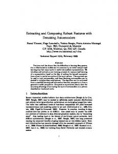
Fig. 4. (a) Seven upper-limb motions. (b) Eight channels of muscle on the right arm of subject 1 [19].
WGNs at different levels (20-0 dB SNR) were added into the raw EMG signals to prepare the noisy EMG signals as testing data in the experiments. In this paper, EMG data were collected from 30 subjects. EMG signals were recorded from eight electrode positions on the upper-limb and seven daily-life motions as shown in Figs. 4(a) and 4(b). In addition, EMG signals were collected from seven positions on the forearm and one position on the biceps brachii muscle using Duo-trode Ag-AgCl electrodes (Myotronics, 6140). A common ground reference was placed on the wrist using Ag-AgCl Red-Dot electrode (3M, 2237). The seven commonly used upper-limb movements are selected: wrist flexion, wrist extension, hand close, hand open, pronation, supination, and rest [31, 33]. Each motion is performed four times throughout a trial. For every subject, there are four sessions and six trials in each session. In all, there are 96 datasets. Each motion is subjected to a duration of three seconds. To guarantee the best weighted parameter optimized for EMG signal, WGNs were added to prepare the noisy EMG signals for five times in each dataset. A band-pass filter of 1–1000 Hz bandwidth and an amplifier with 60 dB gains (Grass Telefactor, Model 15) were set for the system. These signals were sampled at 3 kHz (National Instruments, PCI-6071E). In order to reduce time in recognition, EMG data were down-sampled to 1 kHz. More details of experiments and data acquisition are described in [30]. Secondly, we tested the performance of the denoising system as described previously in the last section. For the baseline system (BS), however, the second step is skipped and proceeds directly to the third step, i.e., without the denoising algorithm. Thirdly, features of the estimated EMG
April 28, 2011 11:47 WSPC/S0219-4775 167-FNL 00046
Wavelet-Based Denoising Algorithm for Robust EMG Pattern Recognition
163
signals are extracted in time domain including root mean square, waveform length, and coefficients of the fourth order of auto-regressive model [21]. As a result, a feature vector was created and fed into the classifier to recognize the control class. From the successful achievement of linear discriminant analysis [31], it was selected as a classifier in this paper. The window size and the window slide were defined as 256 and 128 ms for real-time constraint [31], respectively. In this study, the classification rate is used to evaluate the quality of the recognition system with the estimated EMG signal that is denoised by using different weighted UNI method. The recognition accuracy was calculated from the testing data for each subject training data with a leave-one-out cross validation. In order to confirm the optimal w value, the average of the classification rate of 30 subjects was performed and reported in the latter section. The optimal w value in the algorithms is selected when the recognition accuracy is closer to 100%. It means that useful information in EMG signal is maintained and undesirable parts of EMG signal are removed. In addition, the classification rate from the BS system was compared with that from the denoising system. 4. Results and Discussion We have found that the recognition performance of using HAD is always better than SOF in all cases, as shown in Figs. 5(a) and 5(b). Consequently, HAD is chosen in the thresholding process. Figure 5(a) shows the recognition accuracy using HAD of many different w values and noise levels. When w is between 0.5 and 0.6, its recognition performance is always superior or equal to the others including w at 1.00, which is the traditional UNI. Moreover, the proposed method performs significantly better than the standard UNI method with p-value less than 0.001. In this paper, the center of suitable w range is selected, i.e., 0.55, as a representation of weight in the validation of recognition accuracy. Figure 6 shows a comparison of the average recognition accuracy resulting from the denoising algorithm at weight 0.55 and the BS system. It can be seen that the improvement of 2 to 55% over the BS depending on the level of noise can be obtained by the denoising algorithm at weight 0.55. Moreover, we can confirm that the result of the optimal weight w is similar from each subject. Hence, this pre-factor is not dependent on the subject. According to the experimental results, a new wavelet threshold estimation equation can be written as follows: � (7) THR = 0.55σj 2 log(N ). The improvement of the recognition performance using wavelet denoising method in this paper succeeds better than the other recognition systems, be it speech recognition [32] or others. However, several other recognition systems are still employing the traditional UNI method that is not suitable for those systems. Hence, the idea in designation of the optimal weighted parameter to the traditional UNI method could be a better solution to improve the recognition performance of
April 28, 2011 11:47 WSPC/S0219-4775 167-FNL 00046
164
A. Phinyomark, P. Phukpattaranont & C. Limsakul
(a)
(b)
noisy EMG signal at 0 dB SNR noisy EMG signal at 5 dB SNR noisy EMG signal at 10 dB SNR
noisy EMG signal at 15 dB SNR noisy EMG signal at 20 dB SNR clean EMG signal (∞ dB SNR)
Fig. 5. Comparisons of average recognition accuracy from 30 subjects at various weighted parameters, and (a) hard thresholding, and (b) soft thresholding with.
specific recognition systems. Furthermore, our proposed weighted parameter in this paper can be widely used to improve both the denoising and the recognition performances in many EMG applications. Not only for controlling a prosthetic hand [3, 5, 14, 15, 18–21, 28, 30, 31, 33], it can also be used in other engineering and medical
April 28, 2011 11:47 WSPC/S0219-4775 167-FNL 00046
Wavelet-Based Denoising Algorithm for Robust EMG Pattern Recognition
Without denoising
165
With w at 0.55 and hard thresholding
Fig. 6. Comparisons, from 30 subjects, of the average recognition accuracy of the BS system, and the denoising system at various noise levels. Block diagram of the proposed system.
applications such as muscle fatigue detection [4], lower-limb prosthesis control or gait analysis [8], and MUAP detection [9, 16, 17].
5. Conclusion We have presented a pre-processing stage based on an optimal weighted parameter or pre-factor of the wavelet denoising algorithm for the EMG recognition system. It is apparent from the derived experimental results that this proposed method can be successfully employed for the extraction of robust features in noisy environments. Apart from a very small decrease in the recognition accuracy of a clean EMG signal, the improvement in the EMG recognition performance compared with the baseline system at all SNR is demonstrated. The weighted UNI method (w = 0.55) with hard thresholding is an effective tool at the pre-processing stage of an EMG recognition system in random noises in a useful EMG frequency band.
Acknowledgments This work was supported in part by the Thailand Research Fund (TRF) through the Royal Golden Jubilee Ph.D. Program (Grant No. PHD/0110/2550), and in part by NECTEC-PSU Center of Excellence for Rehabilitation Engineering, Faculty of Engineering, Prince of Songkla University. The authors also gratefully acknowledge the support of Dr. Adrian D. C. Chan from the Carleton University, Canada, for providing EMG data.
April 28, 2011 11:47 WSPC/S0219-4775 167-FNL 00046
166
A. Phinyomark, P. Phukpattaranont & C. Limsakul
References [1] M. H. Silber, Clinical utility of surface EMG, Neurology 55 (2000) 171–177. [2] S. Ko¸cer, Classification of EMG signals using neuro-fuzzy system and diagnosis of neuromuscular diseases, J. Med. Syst. 34 (2010) 321–329. [3] Y.-C. Dua, C.-H. Linb, L. -Y. Shyuc and T. Chena, Portable hand motion classifier for multi-channel surface electromyography recognition using grey relational analysis, Expert Syst. Appl. 37 (2010) 4283–4291. [4] D. K. Kumar, N. D. Pah and A. Bradley, Wavelet analysis of surface electromyography to determine muscle fatigue, IEEE Trans. Neural Syst. Rehabil. Eng. 11 (2003) 400–406. [5] M. Zecca, S. Micera, M. C. Carrozza and P. Dario, Control of multifunctional prosthetic hands by processing the electromyographic signal, Crit. Rev. Biomed. Eng. 30 (2002) 459–485. [6] E. A. Clancy, E. L. Morin and R. Merletti, Sampling, noise-reduction and amplitude estimation issues in surface electromyography, J. Electromyography Kinesiol. 12 (2002) 1–16. [7] M. B. I. Reaz, M. S. Hussain and F. Mohd-Yasin, Techniques of EMG signal analysis: Detection, processing, classification and applications, Biol. Proced. Online 8 (2006) 11–35. [8] M. S. Hussain, M. B. I. Reaz, F. Mohd-Yasin and M. I. Ibrahimy, Electromyography signal analysis using wavelet transform and higher order statistics to determine muscle contraction, Expert Syst. 26 (2009) 35–48. [9] X. Ren, X. Hu, Z. Wang and Z. Yan, MUAP extraction and classification based on wavelet transform and ICA for EMG decomposition, Med. Biol. Eng. Comput. 44 (2006) 371–382. [10] A. Phinyomark, C. Limsakul and P. Phukpattaranont, A novel feature extraction for robust EMG pattern recognition, J. Comput. 1 (2009) 71–80. [11] S. N. Kale and S. V. Dudul, Intelligent noise removal from EMG signal using focused time-lagged recurrent neural network, Appl. Comput. Intell. Soft Comput. 7 (2009) 1–12. [12] C. J. De Luca, Surface Electromyography: Detection and Recording (DelSys Incorporated, 2002). [13] C. J. De Luca, L. D. Gilmore, M. Kuznetsov and S.H. Roy, Filtering the surface EMG signal: Movement artifact and baseline noise contamination, J. Biomech. 43 (2010) 1573–1579. [14] M. Zardoshti-Kermani, B. C. Wheeler, K. Badie and R. M. Hashemi, EMG feature evaluation for movement control of upper extremity prostheses, IEEE Trans. Rehabil. Eng. 3 (1995) 324–333. [15] R. Boostani and M. H. Moradi, Evaluation of the forearm EMG signal features for the control of a prosthetic hand, Physiol. Meas. 24 (2003) 309–319. [16] F. Laterza and G. Olmo, Analysis of EMG signals by means of the matched wavelet transform, Electron. Lett. 33 (1997) 357–359. [17] P. Wellig and G. S. Moschytz, Analysis of wavelet features for myoelectric signal classification, Proc. IEEE Int. Conf. Electronics, Circuits and System, Lisbon, Portugal (1998), pp. 109–112. [18] A. Phinyomark, C. Limsakul and P. Phukpattaranont, A comparative study of wavelet denoising for multifunction myoelectric control, Proc. Int. Conf. on Computer and Automation Engineering, Bangkok, Thailand (2009), pp. 21–25. [19] M. S. Hussain, M. B. I. Reaz, M. I. Ibrahimy, A. F. Ismail and F. Mohd-Yasin, Wavelet based noise removal from EMG signals, Inform. MIDEM 37 (2007) 94–97.
April 28, 2011 11:47 WSPC/S0219-4775 167-FNL 00046
Wavelet-Based Denoising Algorithm for Robust EMG Pattern Recognition
167
[20] M. Khezri and M. Jahed, Surface Electromyogram signal estimation based on wavelet thresholding technique, Proc. 30th Ann. Int. IEEE EMBS Conf., Vancouver, British Columbia, Canada (2008), pp. 4752–4755. [21] A. Phinyomark, C. Limsakul and P. Phukpattaranont, EMG denoising estimation based on adaptive wavelet thresholding for multifunction myoelectric control, Proc. Conf. Innovative Technologies in Intelligent Systems and Industrial Applications, Monash University, Sunway campus, Malaysia (2009), pp. 171–176. [22] Z. Liu and Z. Luo, EMG de-noising by multi-scale product coefficient hard thresholding, J. Huazhong Univ. Sci. Nat. Sci. 36 (2008) 134–136. [23] Z. -Z. Luo, Q.-J. Zhang and J. -P. Jiang, Improving method of surface electromyography denoising based on wavelet transform, J. Zhejiang Univ. (Eng. Sci.) 41 (2007), pp. 213–216, 220. [24] D. L. Donoho, De-noising by soft-thresholding, IEEE Trans. Inf. Theory 41 (1995) 613–627. [25] D. L. Donoho, Wavelet analysis and WVD: A ten minute tour, Proc. Int. Conf. Wavelets and Applications, France (1992). [26] S. Zhong and V. Cherkassky, Image denoising using wavelet thresholding and model selection, Proc. Int. Conf. Image Processing, Vancouver, British Columbia, Canada (2000), pp. 262–265. [27] G. Song and R. Zhao, Three novel models of threshold estimator for wavelet coefficients, Proc. 2nd Int. Conf. Wavelet Analysis and Its Applications, Hong Kong (2001), pp. 145–150. [28] Z. Qingju and L. Zhizeng, Wavelet de-noising of electromyography, Proc. IEEE Int. Conf. Mechatronics and Automation, Luoyang, China (2006), pp. 1553–1558. [29] Y. Ying and W. Yusen, New threshold and shrinkage function for ECG signal denoising based on wavelet transform, Proc. 3rd Int. Conf. Bioinformatics and Biomedical Engineering, Beijing, China (2009), pp. 1–4. [30] A. D. C. Chan and G. C. Green, Myoelectric control development toolbox, Proc. 30th Conf. Canadian Medical and Biological Engineering Society, Toronto, Canada (2007), p. M0100. [31] K. Englehart, B. Hudgin and P. A. Parker, A wavelet-based continuous classification scheme for multifunction myoelectric control, IEEE Trans. Biomed. Eng. 48 (2001) 302–311. [32] O. Farooq and S. Datta, Wavelet-based denoising for robust feature extraction for speech recognition, Electron. Lett. 39 (2003) 163–165. [33] M. A. Oskoei and H. Hu, Myoelectric control systems — a survey, Biomed. Signal Process. Control 2 (2007) 275–294. [34] A. Phinyomark, C. Limsakul and P. Phukpattaranont, Optimal wavelet functions in wavelet denoising for multifunction myoelectric control, ECTI Trans. Elec. Eng. Electron. Commun. 8 (2010) 43–52. [35] S. Poornachandra, Wavelet-based denoising using subband dependent threshold for ECG signals, Digital Signal Process. 18 (2008) 49–55.