A Synchronization Algorithm of MRI Denoising and Contrast Enhancement Based on PM-CLAHE Model Di Jia, Fangfang Han, Jinzhu Yang, Yifei Zhang, Dazhe Zhao, Ge Yu
A Synchronization Algorithm of MRI Denoising and Contrast Enhancement Based on PM-CLAHE Model 1,2
Di Jia, 1Fangfang Han, 1Jinzhu Yang, 2Yifei Zhang, 1Dazhe Zhao,2Ge Yu Key Laboratory of Medical Image Computing of Ministry of Education, Northeastern University, LiaoNing, China,
[email protected] 2 College of Information Science and Engineering, Northeastern University, LiaoNing, China,
[email protected] 1
doi: 10.4156/jdcta.vol4.issue6.17
Abstract On clinical applications, many magnetic resonance (MR) Images obtained directly by the instruments are not satisfied by doctors. For example, there are some noises and poor contrast in the MR images. These shortcomings would affect the efficiency of feature extraction or recognition later. To solve this problem, the paper proposes a synchronization algorithm of denoising and contrast enhancement. By constructing a differential equation of limited adaptive histogram equalization, the edge denoising and enhancement are completed with the improved PM model. The algorithm draws on the advantages of the two original models, and the practicality and accuracy are proved by the results of the experiments.
Keywords: Partial differential equation, Level-set, Image denoising, Image enhancement, Histogram equalization
1. Introduction During the development of computer technology in the medical field, medical image processing and analysis are becoming more and more important. In the course of digital image processing, imaging equipment, physical methods and other factors often make the quality down. In general, medical images are accompanied by organizational boundaries blur, low contrast and noises. These factors not only have a great impact on the diagnosis, but also affect the accuracies of segmentation, registration and other image processing methods. The image denoising includes macro filter and micro filter. Macro filter is classic filter, such as wavelet-based denoising[1,2]. The advantages of these methods are that the noise and image can be easily separated through a high pass filter, and the drawback is not applicable to medical images. It is because that there are many low-frequency details in the medical image, and the noise and details will be removed together. A remedial strategy is the resumption of high-frequency signals, but the method is too complex to be applied in clinical cases. Micro filter is based on the differential equation such as P-M model[3] etc. Using this algorithm, the image can be smoothed through curve energy and the diffusion equation iteration. Macro filter is a special case of micro filter, such as the relationship between median filter and diffusion. The methods of image enhancement include histogram equalization[3], adaptive histogram equalization[4] etc. Caselles V raised a method of differential histogram equalization[6] in 1999, and it can implement the enhancement in micro area. At present, there are many types of image denoising and enhancement algorithms, but none of them could apply to medical image directly. A major reason is the contradiction between image denoising and enhancement. Removing noise followed by enhancement often make the edges weaker. If the removal of noise is not enough, there will be some issues such as the enhancement of noise etc. Enhancement followed by removing noise will make the noise amplified, so the denoising effect could be poor. The paper proposes a new model combined with P-M model and differential contrast limited adaptive histogram equalization (CLAHE). In equations the first formula is tangential force, and the second one is constituted by the differential CLAHE. By adjusting the two parameters, the result can be better.
144
International Journal of Digital Content Technology and its Applications Volume 4, Number 6, September 2010
2. Basic Model 2.1. PM denoising model PM model is a partial differential equation based on nonlinear diffusion. The diffusion is similar to the proliferation of physics. Impurities migrate from high concentration regions to low regions in the uneven medium. Two-dimensional nonlinear diffusion equation is shown in formula (1).
∂I ( x, y, t ) ∂2 I ∂2 I = div(∇I ) = 2 + 2 ∂t ∂x ∂y
(1)
I ( x, y, t ) is the evolutional matrix of image, ∇I is the gradient of Image, div(∇I ) is the gradient divergence. In order to denosing synchronize with protecting the edge, PM model introduces a coefficient transmission of the constraint function. In the flat regions, it can speed up the velocity of conduction. At the edge of the image, the velocity of conduction will decrease automatically. PM model of partial differential equation is constructed as follows: ∂I ( x, y, t ) = div[ g (| ∇I |)∇I ] (2) ∂t 1 , p = 1, 2 g (r ) = (3) 1 + (r / K ) p
g (| ∇I |) is the conductivity in formula (2). In formula (3), g(r) is a monotonic declined function, and the parameter is the value of gradient, so smoothing is able to combine with edge detection at the same time.
2.2. The differential model of histogram equalization(HE) The theory of HE model is to transform the non-uniform distribution to uniform distribution histogram. The purpose is to make visual sense more richness. Histogram equalization is generally expressed by the following equation: DY = f ( Dx ) = ( Dy max − Dy min ) ∫
D
Dx min
hx (τ )dτ + Dy min
DY is enhanced image, Dx is input image, f(*) is gray-scale transformation function.
(4)
∫
D Dx min
hx (τ )dτ
is cumulative histogram of the input image. The solving of traditional histogram equalization is as follows: (1) Statistics of the input image’s histogram, and calculate the cumulative histogram. (2) Use formula (4) to calculate the one-dimensional array. (3) The original image is enhanced with the array as a reference. The partial differential equation which Caselles V etc raised can be used to achieve histogram equalization. The form of equation is shown in formula (5). I n +1 = I n + Δt{[( Dy max − Dx min ) H ( I n )] − I n }
(5)
H(*) is cumulative histogram. The histogram equalization can be got from many times of iteration.
3. The Model of Denoising and Enhancement 3.1. The Differential model of CLAHE In general the image contrast can be improved by HE model, but the details would be poor. As special nature of medical images (edge blur, poor contrast), the algorithm is not suitable for such
145
A Synchronization Algorithm of MRI Denoising and Contrast Enhancement Based on PM-CLAHE Model Di Jia, Fangfang Han, Jinzhu Yang, Yifei Zhang, Dazhe Zhao, Ge Yu
categories. R. Cromartie propose a solution to the problem of the detail enhancement, it is CLAHE[7,8]. The principle is to block the original image, then process each sub-block combined with HE, at last reconstruct the entire image according to result. Reference with the HE differential equations, the differential model of CLAHE is raised as follows: ∂I ( x, y, t ) L (i ) (i ) (i ) = ∪ ((Dmax − Dmin )F( I (i ) ( x, y, t )) + Dmin ) − I ( x, y, t ) ∂t i =1
(6)
L
I n +1 = I n + Δt[∪ [( D(i ) max − D(i ) min ) F ( I (i ) n ) ]− In]
(7)
F ( I (i ) n ) = α H w ( I (i ) n ) + (1 − α ) H B ( I (i ) n )
(8)
i =1
F(*) is the function of the integration of sub-block histogram equalization, H w (*) is the normalized histogram inside the block, and H B (*) is the normalized histogram outside the block. i is the block number, D(i ) max is the maximum gray value of block i, D(i ) min is the minimum gray value of block i. Using gradient descent method, the solving function is as formula (7), L is the number of windows. When Δt =1, the ceremony will be transformed into the classic model of CLAHE.
3.2. The Model of Denoising and Contrast Enhancement In order to achieve the expected result, this paper will use the synchronized method of denoising and contrast Enhancement. The new expression is transformed from PM equation as the following formula: ∂I ∂I ∂I ∂I ∂I [ g (| ∇I |) ]+ [ g (| ∇I |) ] = ∂t ∂α ∂α ∂β ∂β = g (| ∇I |) Iαα + [ g (| ∇I |) + g ' (| ∇I |) | ∇I |]I ββ
= g (| ∇I |) Iαα + ψ (| ∇I |) I ββ
(9)
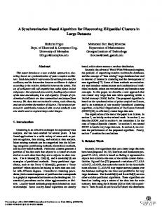
Figure 1. reference frame (α, β)
α is the tangential vector unit, β is the normal vector unit. From the formula (9), the equation of PM can be broken into two forms. The first form is constituted by the tangential vector, so the image will diffuse according this direction. This will make the edges more smoothly. The second form is constituted by the normal vector, and another part will spread in this direction. The differential equation can be completed by step iteration, so the additive combination of PM and CLAHE can remove the noise, smooth the edges, make enhancement within the unit step. Construction of the new equation is as follows: ∂I ( x, y , t ) (i ) (i ) (i ) = div[ g (| ∇I |)∇I ] + (( Dmax − Dmin ) F ( I ( i ) ( x, y , t )) + Dmin ) − I ( x, y , t ) ∂t
(10)
The first form is from PM model, the second and third form is from differential CLAHE. The result is solved through Gradient descent method, and explicit numerical calculations are as follows: L
I n +1 = I n + Δt{( P n + Q n ) + [∪ [( D(i ) max − D(i ) min ) F ( I (i ) n ] − I n ]} i =1
146
(11)
International Journal of Digital Content Technology and its Applications Volume 4, Number 6, September 2010
Supposing the size of moving window is 3×3, size of image is M×N, and L is (M-2)×(N-2). P n + Q n is the explicit expression of PM equation, the latter form is adopted from the gradient descent solution of CLAHE. Three adjustable parameters of extended equation are introduced into it, these can make the image denoising and enhancement corresponding with adjustment experience, and better results could be got through this image processing. The extended equation is shown as formula (10). L
I n +1 = I n + Δt{Cα P n + Cβ Q n + Cγ [∪[( D(i )max − D(i ) min ) F ( I (i )n ] − I n ]}
(12)
i =1
Cα is used to adjust the proportion of tangential smooth force. Cβ is used to adjust the proportion of normal smooth force. Cγ is used to enhance the scale factor. The better effect will be got through the regulation of these three parameters. As normal diffusion can make the edge of image more blurry, so we remove the second form, the final equation is as follows: L
I n +1 = I n + Δt{Cα P n + Cβ [∪ [( D(i ) max − D(i ) min ) F ( I (i ) n ] − I n ]}
(13)
i =1
Pn =
[ Dxx I n ( Dy I n ) 2 + Dyy I n ( Dx I n ) 2 − 2 Dx I n Dy I n Dxy I n ]
(14)
( Dx I n ) 2 + ( Dy I n ) 2 + ε
D* is the finite difference value of partial derivatives. It could be preceding difference, latter difference, or central difference. D** is approximation of the second order partial derivative of finite difference.
3.3. Algorithm Flow I in is original input image, and I out is output image, n is times of iterations. Cα , Cβ are tangential smoothing coefficient and enhanced coefficient, Δt is the step length and n is setting times of iteration. Algorithm implementation process is as follows: (1) I out = I in , set sliding window size. (2) The input image is processed through CLAHE, get the result R1. (3) Subtracting R1 and I in , multiply with Cβ to adopt I clahe . (4) Calculate I out = I out + Δt ( I clahe + I pm ) . (5) Cumulate number of iterations m, if (m < n) turn (2). (6) Output the final result I out .
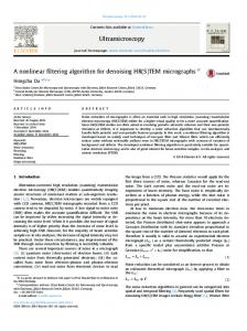
4. Experimental Results and Analysis The experimental CPU is 2.4GH and memory is 3.37GB. We use two brain MR images of 512×512 with noise to do experiment. One image contains the base of skull, another is cranial vault image. For the first image, experimental parameters are chosen as Δt = 5 , n = 100 (iteration times), α = 1, β = 0.005. For the second image, experimental parameters are chosen as Δt = 0.5 , n = 50, α = 1, β = 0.0025.
(a)
(b)
(c)
(d)
(e)
Figure 2. Comparison of experimental results. (a) is the original image. (b) is the image of enhance after denosing. (c) is the image of denosing after enhance. (d) is the image of PM. (e) is the image of PM-CLAHE.
147
A Synchronization Algorithm of MRI Denoising and Contrast Enhancement Based on PM-CLAHE Model Di Jia, Fangfang Han, Jinzhu Yang, Yifei Zhang, Dazhe Zhao, Ge Yu
(a)
(b)
Figure 3. Histogram comparison. (a) is the histogram of original image. (b) is the histogram of processed image using PM-CLAHE.
(a)
(b)
(c)
(d)
(e)
Figure 4. Comparison of experimental results. (a) is the original image. (b) is the image of enhance after denosing. (c) is the image of denosing after enhance. (d) is the image of PM. (e) is the image of PM-CLAHE.
(a)
(b)
Figure 5. Histogram comparison. (a) is the histogram of original image. (b) is the histogram of processed image using PM-CLAHE.
Experiment Number
Table 1. Experiment of time consuming Enhance after Denoising after PM denoising enhance
PM-CLAHE
Test 1
11.313 s
11.282 s
11.203 s
21.063 s
Test 2
5.719 s
5.656 s
5.593 s
10.844 s
Fig.2 (b)-(e) are the effect of 100 times of iterations. If the noise is not completely removed, it will lead to be amplified in the enhanced algorithm using enhancement after denoising, And Fig.2 show the effect. If the iterative times is increased much more, it will lead to weaker edges are smoothed, and this can affect the processing precision. Fig.2 (c) shows the results of denoiseing after enhancing. Noise is enhanced through enlarge phase, the smoothed image is superimposed with amplified noise. Fig.2 (d) is the effect of classic P-M algorithm. Comparing with Fig.2 (b)-(d), Fig.2 (e) of the PM-CLAHE algorithm not only can maintain edges, but also removed noise and enhanced contrast obviously. Fig.3 (a) and Fig.3 (b) are respectively histogram of the original image and PM-CLAHE image. It is clear that the improved algorithm makes more uniform gray. Fig.3 and Fig.4 are another group of comparison of experimental results. Table 1 shows the time comparison of these methods. The consuming of PM-CLAHE is nearly double times to the other methods, this is because CLAHE should be done each iterations.
148
International Journal of Digital Content Technology and its Applications Volume 4, Number 6, September 2010
5. Conclution The paper proposes a differential equation for MR image preprocessing. As the edge of the image can be protected better during the denosing phase through tangential smoothing, the new equation which removed normal smoothing has better effect on preventing edges diffusion. CLAHE can be good at keeping local details during the enhancement. Through the removal of normal diffusion and adding proportional coefficient, the equation combining with constructed differential CLAHE and PM denoising algorithm has advantages of the two algorithms.
6. References [1] Wang Wei, Wu Xiu-qing, Cheng Lei etc. “Image denoising based on stationary wavelet-based anisotropic diffusion”. Computer Engineering and Applications, vol.46, pp.180-182, 2010. [2] Li Feng, Lv Hui. “MRI Medical image denoising based on BEMD and wavelet thresholding”. Journal of Image and Graphics, vol.14, pp.1972-1977, 2009. [3] Perona P, Malik J. “Scale space and edge detection using anisotropic diffusion. IEEE PAMI, vol.12, pp.629-639, 1990. [4] Richard E etc. “Digital image processing using matlab”. Publishing House of Electronics Industry, pp.50-51, 2004. [5] Pizer S. “Adaptive histogram equalization and its variations”. Computer Vision Graphics and Image Processing, vol.39, pp.355-368, 1987. [6] Caselles V etc. “Shape-preserving local contrast enhancement”. IEEE IP, vol.8, pp.220-230, 1999. [7] Daxing Zhang, Shiming Liang etc. “An image authentication scheme based on correlation”. International Journal of Digital Content Technology and Its Applications. vol.4, pp.89-94, 2010. [8] Louis AJ, Belward J. “A variational approach to the radiomeric enhancement of digtial imagery”. IEEE Transaction Image Process, vol.4, pp.845-849,1995. [9] Syed A, Srinivasan V, K. Lal. “CT image denoising technique using GA aided window-based multivavelet transformation and thresholding with the incorporation of an effective quality enhancement method”. International Journal of Digital Content Technology and Its Applications, vol.4, pp.75-87, 2010. [10] H.Zhu, FHY Chan, FK Lam. “Image contrst enhancement by constrained local histogram equalization”. Comput Vision Image Understanding, vol.73, pp.181-190, 1999. [11] C.Chesneau, J. Fadili, J.L. Starck. “Stein block thresholding for image denoising”. Applied and Computational Harmonic Analysis. Vol.28, pp.67-88, 2010. [12] Dongwook Cho, Tien D.Bui. “Multivarite statistical modeling for image denoising using wavelet transforms”. Signal Processing: Image Communication, vol.20, pp.77-89, 2005. [13] Kazubek, M. “Wavelet domain image denoising by thresholding and winener filtering”. Signal Processing Letters, IEEE, vol.10, pp.324-326, 2003.
149