REVIEW
411
Development 135, 411-424 (2008) doi:10.1242/dev.000505
Wnt/Notch signalling and information processing during development The Wnt and Notch signalling pathways represent two major channels of communication used by animal cells to control their identities and behaviour during development. A number of reports indicate that their activities are closely intertwined during embryonic development. Here, we review the evidence for this relationship and suggest that Wnt and Notch (‘Wntch’) signalling act as components of an integrated device that, rather than defining the fate of a cell, determines the probability that a cell will adopt that fate.
Introduction The development of a multicellular organism requires the coordination in space and time of three events: cellular proliferation, the assignment of different fates to an ensemble of cells, and their organization into tissues and organs. Here, we shall focus on the second process, which is often referred to as ‘cell fate specification’, and on its driving force, a combination of complex gene regulatory networks (GRNs) (Davidson, 2006). These networks determine the phenotype of individual cells by providing diverse patterns of gene expression through the activity of intrinsic and extrinsic factors. The intrinsic factors are the transcription factors that a cell expresses at a given time that provide the coordinates that define a cell’s state (Alon, 2006). The extrinsic factors principally comprise signals that regulate and coordinate those states and the transitions that occur between them in groups of, and in individual, cells. Understanding the process of cell fate assignment requires us to describe the integration of both intrinsic and extrinsic factors in GRNs, as well as the mechanisms that underlie their precise and reproducible operation (Davidson, 2006). Multicellular organisms combine some intrinsic and extrinsic factors into a sophisticated biochemical kit, the ‘signal transduction’ machinery, that establishes, develops and coordinates fates within cell populations. The notion of signal transduction was introduced by Rodbell to explain how extracellular signals affect cell behaviour and was inspired by Cybernetics and Information Theory (reviewed by Rodbell, 1995). Originally, the theory proposed the existence of a signal, a transducer and an effector, together with an amplification step associated with the transduction event (Fig. 1). This scheme has provided a universal and useful framework for the analysis of signalling molecules in plant and animals for the last 30 years. Surprisingly, it never dealt with the issue of noise, fluctuations in the transduction process that affect and corrupt the outcome of the signalling event, which is central to Information Theory (Fig. 1). Perhaps this is because, at the time, little was known about the quantitative behaviour of signalling pathways, or, more likely, the issue of noise was not a consideration in biological systems. Recent detailed analysis of transcription in single-cell organisms has revealed that noise is a significant variable in the operation of Department of Genetics, University of Cambridge, Cambridge CB2 3EH, UK. *Author for correspondence (e-mail:
[email protected])
transcriptional networks, and it has been suggested that it might play an important role in development (Maamar et al., 2007; Suel et al., 2006; Suel et al., 2007). Transcriptional networks are integrated into the fabric of signal transduction, and putting noise back into this scheme will lead to important considerations about their organization and operation. Work over the last 20 years has established that there are six major and universal signal transduction devices in the cell (reviewed by Martinez Arias and Stewart, 2002): Hedgehog (Hh), Bone morphogenetic proteins (BMPs), Wnt (Wingless/Int1), Steroid hormone receptor, Notch and Receptor tyrosine kinase (RTK). Each of these pathways can be fitted into the signal transduction concept introduced by Rodbell, with effectors represented by pathwaydedicated transcription factors. Together, these pathways provide the basic machinery for cell fate transitions and assignments that underlie embryonic development. At present, signal transduction pathways are deemed to act as parallel information-processing channels that converge onto the enhancers of particular genes to create cell type-specific combinations of transcription factors that determine cells states and cell behaviour (Barolo and Posakony, 2002; Martinez Arias and Stewart, 2002). In this view, all pathways have an equal weight and a similar function. There is some truth in this view, which in model organisms, such as C. elegans and Drosophila, has led to a good understanding of some aspects of cell fate specification, e.g. muscle founders in the embryo (Carmena et al., 1998; Halfon et al., 2000) and photoreceptors (Silver and Rebay, 2005; Voas and Rebay, 2004) in Drosophila, as well as the specification of blastomeres in the eight-cell embryo of C. elegans (Newman-Smith and Rothman, 1998; Platzer and Meinzer, 2004; Rose and Kemphues, 1998). In these cases, a particular cell is defined by the combined activity of transcription factors, with precise spatial and temporal coordinates defined through the iterative activities of GRNs. However, genetic analyses of how some of these pathways affect the specification of cell types suggest that other activities and interactions between elements of the pathways also play an important role and remain to be explored (Brennan et al., 1999a; Carmena et al., 2006; Strutt et al., 2002; Tomlinson and Struhl, 2001). The nature and function of these interactions should be an important focus of research. Here, we look into how cross-regulatory interactions between elements of different signalling pathways affect the process of cell fate assignment. We do this in the context of Wnt signalling, and review the increasing evidence that an intricate functional relationship exists between Wnt and Notch signalling during the assignment of cells to particular fates. The roots of our analysis lie in the genetics of Drosophila and in our recent proposal that Wnt signalling is involved in regulating the probability that a cell adopts a particular fate during development (Martinez Arias and Hayward, 2006). We speculate that Notch signalling is intimately involved with Wnt in this process and that the interaction of both pathways results in greater accuracy and reliability of cell fate transitions, i.e. they act together to filter the noise intrinsic to this process. Our view
DEVELOPMENT
Penelope Hayward, Tibor Kalmar and Alfonso Martinez Arias*
REVIEW
Development 135 (3)
Noise
Source
Transmitter
CHANNEL
Receiver
Destination
TRANSDUCTION CHAIN
EFFECTOR
TARGETS
SIGNAL RECEPTOR
Fig. 1. Information and signal transduction. The term ‘signal transduction’, as used in cell biology, initially arose as an analogy with the transmission of information in telecommunications (Rodbell, 1995). In telecommunications, a channel is used to transduce information from a source to a specific destination. A transmitter places the information into the channel and a receiver picks it up and delivers it to the destination. In biological systems, the signal is the source, and the targets the destination. The signal transduction system is the channel that is accessed through a receptor. The system’s effector acts as the receiver and delivers the signal to the targets. In telecommunications, noise is an inherent property of the transduction process. Noise exists in the source and destination, but most significantly in the channel, which has to pass the information between the transmitter and the receiver. It is highly unlikely that biological signal transduction is immune to noise.
contrasts with a widely held one, whereby Wnt and Notch signalling simply provide individual elements of the complex combinations of signalling molecules and transcription factors that define the many different cell types of an organism. The ins and outs of Wnt signalling Wnt proteins are secreted glycoproteins that elicit cellular responses through their assembly of a membrane receptor complex that includes the Frizzled and members of the Low density lipoproteinrelated receptor (LRP) protein families (Fig. 2). This complex triggers a number of intracellular events that are represented by three modalities: (1) -catenin-mediated Wnt signalling, dedicated to the modulation of transcriptional activity and cell fates (Logan and Nusse, 2004); (2) planar cell polarity (Seifert and Mlodzik, 2007; Strutt and Strutt, 2005), which controls the activity of the cytoskeleton; and (3) Ca+2-related signalling, which targets adhesion and other processes (Kohn and Moon, 2005). Here, we will concentrate on -catenin mediated signalling. The effector of Wnt signalling in the nucleus is -catenin (Tolwinski and Wieschaus, 2004a). This protein was first identified as a linker between Cadherin and the cytoskeleton (Ozawa et al., 1989), but genetic studies in Drosophila (McCrea et al., 1991) and the analyses of colorectal tumours in humans and mice (Korinek et al., 1997; Morin et al., 1997; Munemitsu et al., 1995) revealed that a cytoplasmic pool of -catenin exists, the concentration, location and activity of which are modulated by Wnt signalling. In the absence of Wnt signalling, cytoplasmic -catenin is recruited to a complex that assembles around the scaffolding protein Axin (Behrens et al., 1998; Fagotto et al., 1999; Hart et al., 1998; Kishida et al., 1999), where it is phosphorylated at its N-terminus by Glycogen synthase 3 (GSK) (Ikeda et al., 1998) (see Fig. 2). Nterminus phosphorylated -catenin is targeted for degradation via the proteasome (Aberle et al., 1997; Jiang and Struhl, 1998; Marikawa and Elinson, 1998), which keeps the concentration of this cytoplasmic pool low. Upon Wnt signalling, a fraction of the soluble pool is stabilised (Riggleman et al., 1990), probably modified, and allowed to enter the nucleus (Tolwinski and Wieschaus, 2004a), where it interacts with members of the TCF (T cell factor) family of transcriptional regulators to modulate gene expression (Behrens et
al., 1996; Molenaar et al., 1996) (see Fig. 2). How the interaction of Wnt with its receptors leads to -catenin stabilization is not understood. As in other aspects of Wnt signalling, Dishevelled (Dsh in Drosophila, Dvl in vertebrates) plays an important role, which, in this case, is the modulation of the activity of the Axin-based destruction complex (Fagotto et al., 1999; Kishida et al., 1999). There is a good correlation between rises in the concentration of ‘soluble’ -catenin and its activity in the nucleus (Funayama et al., 1995; Korinek et al., 1997; Pai et al., 1997), but there is also evidence that the rise in -catenin concentration, per se, is not the only factor that determines its activity (Brennan et al., 2004; Guger and Gumbiner, 2000; Lawrence et al., 2001; Staal et al., 2002; Tolwinski et al., 2003). In particular, increases in cytoplasmic catenin concentration do not result in Wnt signalling activity (Brennan et al., 2004; Guger and Gumbiner, 2000; Staal et al., 2002). Furthermore, genetic analysis in Drosophila has shown that Axin has a second function in controlling the activity of Armadillo (Drosophila -catenin) (Tolwinski et al., 2003; Tolwinski and Wieschaus, 2004b), supporting the notion that the activity of catenin is regulated not only through changes to its cytoplasmic concentration. Recent reports have implicated endocytosis and membrane trafficking in the regulation of the Wnt signalling event (Blitzer and Nusse, 2006; DasGupta et al., 2005; Marois et al., 2006; Piddini et al., 2005; Rives et al., 2006; Seto and Bellen, 2006), but much remains to be done to link this information into a coherent argument as to how the Wnt signal is transferred to -catenin. The number of elements and the complexity of their interactions suggest that the formulation of kinetic models of the signalling event (Lee et al., 2003) will provide novel insights into the mechanism that underlies this process. Beyond signalling: a function for Wnt The lack of a detailed mechanism for Wnt signalling should not deter us from tackling aspects of its function. The current picture of Wnt/-catenin signalling offers two striking observations. First, many of the elements of this pathway are used in other signalling pathways or participate in a variety of cellular activities. For example, -catenin has a well characterised function in cell-cell adhesion (Ozawa et al., 1989); GSK3 and the various Casein kinases that participate in the signalling event play multiple roles in other signal transduction pathways and in cellular metabolism (Doble and Woodgett, 2003; Harwood, 2001; Polakis, 2002; Price, 2006); and Adenomatous polyposis coli (APC) has a central role in the biology of the cell through its interactions with microtubules (Nathke, 2006; Polakis, 1997). In addition, the list of Dshinteracting proteins increases continuously and some of these proteins are not easily linked to Wnt signalling, raising questions about whether Dishevelled is a core element of the pathway or a component of the basic biology of the cell that is used by Wnt signalling (Malbon and Wang, 2006; Wallingford and Habas, 2005; Wharton, 2003). In some ways, the multiple interactions and functions of each of these components of Wnt signalling link the signalling event to different processes, and thus place Wnt signalling at the heart of an integrated protein network. Two proteins that appear to escape these multifarious interactions are Axin and TCF [with its associated proteins, Legless (Lgs) and Pygopus (Pygo)], which seem to be dedicated to Wnt signalling (Logan and Nusse, 2004). The second striking feature of Wnt--catenin signalling is its ability to cooperate with transcription factors and effectors of other signalling pathways (reviewed by Martinez Arias and Hayward, 2006). In fact, it is often the case that the effects of Wnt become
DEVELOPMENT
412
Development 135 (3)
REVIEW
Wnt
Axin
Ub Ubiquitin-mediated proteolysis
Dsh
Ub
t
 i
Wnt
Dsh A x
d
i n
Axin
e
c
-catenin
-catenin
n
No signal CKI γ CK2
DSH
GBP
Soluble -catenin
With signal
CKI ε CKI α
CKI ε GBP CK2 CKI ␣
P
P
CKI γ
Axin
LRP5/6
C
DSH
-
e n c
-catenin
Axin-based destruction complex
-catenin
LRP5/6
a
Axin-based destruction complex
Axin-based destruction complex
Wnt
Dsh
Intracellular -TrCP
b
LRP5/6
LRP5/6
Frizzled
LRP5/6
Frizzled
LRP5/6
Plasma membrane
Wnt
Wnt
Wnt
a
Extracellular
Frizzled
Wnt Wnt Wnt
Frizzled
B
A
413
P
Groucho CtBP
GSK 3
PP2A
GSK 3
APC
CKI ε
Axin-based destruction complex
Lgs/BCL9
P
-catenin Free -catenin
APC
-catenin
Axin
CKI ε
CK2
Pygo
TCF LEF
Pol II
f
Nucleus
PP2A
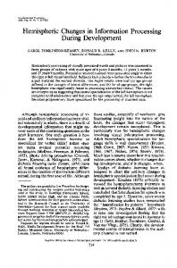
Fig. 2. The spatial and functional organization of the core mechanism of Wnt/-catenin signalling. (A) In the absence of ligand, a destruction complex assembles around the scaffolding protein Axin (see C for details), which binds and then labels -catenin for proteolysis via phosphorylation of specific residues at its N-terminus. (B) Wnt binds to Frizzled (a) and triggers signalling by initiating a chain of events, which at a biochemical level are not well characterized. At the cell surface, the Wnt-Frizzled complex forms a trimeric complex with LRP5/6 (Arrow in Drosophila) and this triggers the activity of Dishevelled (Dsh), which promotes the association of the destruction complex with the LRP5/6-Frizzled complex (b,c). A chain of events (detailed in C) leads to the LRP5/6-mediated degradation of Axin (d) and the release of -catenin from Axin (e). Free hypophosphorylated -catenin can then enter the nucleus (f), where it nucleates a transcription modulator complex around TCF. (C) Structure and function of the Axin-based destruction complex. In the absence of Wnt, the complex is assembled and can bind -catenin, which is then phosphorylated by GSK3. Phosphorylated -catenin is targeted for ubiquitination and proteasomal degradation. In the presence of Wnt, the destruction complex is recruited to the cell surface, where a series of phosphorylation events results in the degradation of Axin and the release of -catenin. For further details, see text and published literature (Logan and Nusse, 2004; Tolwinski and Wieschaus, 2004b). APC, Adenomatous polyposis coli; -TrCP, -transducin repeat-containing protein (also known as Ebi); CKI, Casein kinase; DSH, Dishevelled; CtBP, C-terminal-binding protein; GSK3, Glycogen synthase kinase 3; Lgs, Legless; LRP, Low density lipoprotein receptor-related protein; Pol II, RNA polymerase II; PP2A, Protein phosphatase 2A; TCF, T cell factor (also known as Pangolin); Ub, ubiquitination.
dampen fluctuations in gene expression over cell populations, that is, it acts as a noise filter (Martinez Arias, 2003; Martinez Arias and Hayward, 2006). Wnt and other signalling pathways: Notch In the light of the above observations, it is not surprising that Wnt signalling exhibits interactions with other signalling pathways, such as BMP, Hh and Ras/RTK (Hoppler and Moon, 1998; Janssen et al., 2006; Nusse, 2003; Sansom et al., 2006; Wilson et al., 2001; Zorn et al., 1999). Some of these interactions involve transcriptional effectors (Edlund et al., 2005; Labbe et al., 2000; Nishita et al., 2000), but others probably involve elements at different levels in the transduction chain (Carmena et al., 2006; Jeon et al., 2007; Luo et al., 2003). The consequences of many of these interactions remain to be analysed. However, there is one pathway with which Wnt signalling seems to have a recurrent and consistent interaction: Notch signalling (Hurlbut et al., 2007; Martinez Arias and Hayward, 2006).
DEVELOPMENT
obvious only in the context of other transcriptional effectors, to the point that its function appears to be to modulate their effects and activities (Baylies et al., 1995; Collins and Treisman, 2000; Cox and Baylies, 2005; Lowry et al., 2005; McGrew et al., 1997; Megason and McMahon, 2002; Wan et al., 2000) (reviewed by Martinez Arias and Hayward, 2006). This observation has led us to suggest that rather than acting as an instructive process, Wnt signalling acts in the stabilization of transcriptional events that are initiated by other factors and mechanisms. In this view, cell fate assignments might be divided into two separable steps: (1) an inducing step, which sets up an unstable multidimensional transcriptional state; and (2) the stabilization of this state, which is separate and requires Wnt signalling. Elements of this proposal have been drawn from an extrapolation of modern notions of ‘transcriptional noise’ to developmental systems, as well from considerations of emerging information about variability of expression at the level of single cells and single genes, in E. coli and S. cerevisiae. Our hypothesis concludes that Wnt functions to
414
REVIEW
Development 135 (3)
Signalling cell
Receiving cell
Co-repressors
A
NICD ECN
CBP CSL Nucleus
B b ADAM/S2
a Serrate/ Jagged/ Delta
ECN
␥-secretase/S3
MAML
e
rs so es pr e -r Co
f MAML
NICD
c
d
CBP
Pol II
CSL
NICD Nucleus
Notch belongs to a family of single-transmembrane-domain receptors that have an extracellular domain made up of EGF (Epidermal growth factor)-like repeats and an intracellular domain, the main structural feature of which is seven ANK (Ankyrin) repeats (Ehebauer et al., 2005; Nam et al., 2003; Nam et al., 2006; Zweifel et al., 2003). In contrast to the complexity of Wnt signalling, the mechanism of Notch signalling is apparently simple: the intracellular domain of Notch (NICD) acts as a membrane-bound transcription factor (Kopan, 2002), which is released by an interaction between Notch and its ligands, Delta and Serrate (Fig. 3). Free NICD translocates into the nucleus, where it interacts with CSL {for CBF in vertebrates, Suppressor of Hairless [Su(H)] in Drosophila and LAG-1 in C. elegans}, to drive the transcription of target genes (Bray, 2006; Ehebauer et al., 2006; Le Borgne, 2006). Recent work indicates that behind this basic biochemical mechanism, there is a certain degree of complexity at the level of the cleavage event, which seems to require endocytic trafficking or the localization of Notch to a specific endocytic compartment (Jaekel and Klein, 2006; Moberg et al., 2005; Thompson et al., 2005; Vaccari and Bilder, 2005) (reviewed by Bray, 2006; Le Borgne, 2006). Notch signalling was first identified in the context of lateral inhibition during the development of the peripheral nervous system (PNS) of insects (Simpson, 1990). During PNS
development, a group of ectodermal cells is endowed with the potential to be neural, but only one or two cells within the group adopt this fate and become a sensory organ precursor (SOP). In the process, SOPs suppress the neural potential of the surrounding cells through lateral inhibition mediated by Notch signalling (Hartenstein and Posakony, 1990; Heitzler and Simpson, 1991). Similar events have been described in the central nervous system (Campos-Ortega and Hartenstein, 1997) and in the muscles (Rushton et al., 1995) of Drosophila, as well as in the immune system (Radtke et al., 2004; Radtke et al., 1999), intestine (Crosnier et al., 2005; van Es et al., 2005; Zecchini et al., 2005) and nervous system (Henrique et al., 1997; Lutolf et al., 2002) of vertebrates. In Drosophila, the initial event that sets up the SOP fate depends on Wingless signalling (Couso et al., 1994; Phillips and Whittle, 1993) and is followed by lateral inhibition (Hartenstein and Posakony, 1990; Heitzler and Simpson, 1991). This leads to a simple and clear definition of the roles of Wnt and Notch, with Wnt mediating prepatterning and Notch mediating the inhibitory process (Martinez Arias, 2002). In this sequential and conditional relationship, lateral inhibition requires prepatterning, already suggesting that some sort of functional relationship exists between the two signalling systems.
DEVELOPMENT
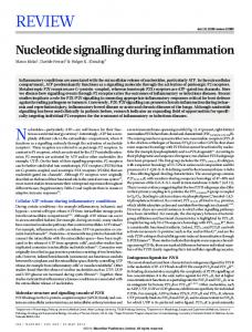
Fig. 3. Structure and functional organization of Notch signalling. Notch and its ligands (members of the DSL family) are transmembrane proteins. (A) In the absence of ligand, the full-length Notch protein is present at the cell surface as a heterodimer of the extracellular (ECN) and the transmembrane-intracellular domains. (B) (a) Binding of a Notch ligand (Serrate/Jagged/Delta) to specific EGF-like repeats (blue) triggers (b) a proteolytic cleavage, S2, in the extracellular juxtamembrane region (grey) of Notch by members of the ADAM tumour necrosis factor converting enzyme (TACE) proteases. This event primes (c) a second ligand-independent cleavage, S3, within the transmembrane domain (purple) of Notch, which is catalysed by the Presenilin–␥-secretase complex. As a result of S3 cleavage, (d) the intracellular domain of Notch (NICD) enters the nucleus, where it (e) interacts with CSL, displaces co-repressors and through Mastermind (MAML) recruits co-activators to (f) promote the transcription of target genes. For further details, see text and published literature (Bray, 2006; Ehebauer et al., 2006; Kopan, 2002; Le Borgne, 2006). ADAM, a disintegrin and metalloprotease; CBP, CREB binding protein (also known as Crebbp); CSL, CBF/Suppressor of Hairless/LAG-1; DSL, Delta/Serrate/LAG-2; ECN, Extracellular Notch; Pol II, RNA polymerase II; TACE, TNF␣-converting enzyme.
Development 135 (3)
A
REVIEW
415
D
Wingless Delta/Serrate
P
A V
B wg
wm
C Ser
D Ser & Dl
Ser & Dl
Ser & Dl
Ser & Dl
Dl
Wingless
E
Wingless
F
G
Senseless Achaete/Scute
Achaete/Scute
N/Dl lat. inhib.
Wingless
Interactions between Notch and Wnt signalling Interactions between Wnt and Notch signalling were first uncovered in the context of the development and patterning of the wing of Drosophila (Couso and Martinez Arias, 1994; Hing et al., 1994). Loss-of-function mutations in wingless, the Drosophila orthologue of Wnt1, and Notch synergise in a manner that indicates a close functional relationship between the two pathways. Since then, a detailed genetic analysis of wing development has provided explanations for some, but not all, of the observed interactions (Brennan et al., 1999b; Klein and Martinez Arias, 1999; Zecca and Struhl, 2007). In this section, we review these results and their interpretation and relate them to comparable situations in vertebrates. In Drosophila, Wingless and Notch signalling are used iteratively to drive the development and patterning of the wing (Klein and Martinez Arias, 1999; Zecca and Struhl, 2007) (Fig. 4). First, they synergise in the establishment and initial growth of the wing primordium (Couso and Martinez Arias, 1994; Klein and Martinez Arias, 1998). Later, Notch signalling promotes wingless expression at the future wing margin (Diaz-Benjumea and Cohen, 1995;
Neumann and Cohen, 1996). In turn, Wingless, after refining its expression (Micchelli et al., 1997), promotes the expression of the Notch ligands Delta and Serrate on either side of its expression domain to create a positive-feedback loop that maintains Notch signalling and Wingless expression and that patterns the wing margin (Fig. 4) (de Celis and Bray, 1997; Klein and Martinez Arias, 1998; Micchelli et al., 1997). This sequence of events can account for some of the genetic interactions that have been observed between Notch and Wingless in Drosophila, in particular, the extreme sensitivity of the wing margin to the dosage of Notch and wingless. Since loss of one copy of Notch produces a wing phenotype, it is not surprising that, given their close association during wing development, alterations in wingless gene dosage also have dramatic effects on wing development when they occur in a background of compromised Notch signalling (Couso and Martinez Arias, 1994; Langdon et al., 2006). In agreement with this, the haploinsufficient phenotype of Notch is a particularly sensitive assay for identifying proteins that interact genetically with Notch (Go and ArtavanisTsakonas, 1998; Hall et al., 2004; Mahoney et al., 2006; Verheyen et al., 1996). So, the mutual enhancement of mutations in wingless
DEVELOPMENT
Fig. 4. Functional networking of Wnt and Notch signalling during Drosophila wing development. During wing primordium patterning and sensory organ precursor (SOP) specification, the Wingless and Notch pathways act in a dynamic signalling landscape that endows cells with identity and orientation. The images to the left and right show wingless (wglacZ) expression in the epidermis of two wing discs at very early (left) and mid-third (right) instar. The white line straddling the middle of the Wingless stain represents the primordium of the wing margin (wm). Throughout the figure, ventral (V) is orientated down, dorsal (D) up, posterior (P) to the left and anterior (A) to the right. (A) Schematic of a wing disc between the first and second larval instars. Wing development is initiated at the intersection of the AP and DV boundaries by the joint activity of Notch and Wingless signalling. (B) During the transition from second to third instar, the DV boundary is established through the activity of Notch signalling triggered, initially, by the asymmetrically localized Notch ligands Serrate (Ser, dorsal, yellow) and Delta (Dl, ventral, red) that lead to the activation of wingless expression (blue) in a wide domain with a peak in the middle. This domain becomes progressively restricted to the DV boundary through an autoinihibitory effect of Wingless on its own expression. (C) Serrate and Delta are targets of Notch signalling, and Wingless signalling can contribute to this activation (arrows). As a result, a pattern emerges (D) of symmetric expression of Delta and Serrate (orange) and of Wingless (Wg, blue) at the DV boundary. This combination of events (B-D) leads to the formation and definition of the DV compartment boundary and its maintenance through a feedback loop in which Wingless maintains the expression of Serrate and Delta, which in turn maintain the expression of Wg. (E-G) As the third instar develops, the peripheral nervous system emerges (E). At the wing margin, it appears around the DV boundary within the domain of Delta and Serrate expression. In the second half of the third instar, high levels of Wingless signalling (blue) lead to the expression of proneural genes, such as senseless and members of the Achaete/Scute family (pink in F), which promote the appearance of SOPs (red in G). Notch-mediated lateral inhibition generates the spaced precursors that express high levels of AS-C and that will develop into sensory organs.
REVIEW
and Notch has a simple explanation in the GRNs that drive wing development. Similar, mutually dependent interactions between Notch and Wnt signalling have been observed in early germ layer specification in the sea urchin (Angerer and Angerer, 2003; McClay et al., 2000; Sherwood and McClay, 2001; Sweet et al., 2002) and in vertebrates, for example, during the development of skin precursors (Estrach et al., 2006), the patterning of the rhombomeres (Cheng et al., 2004) and during somitogenesis (Aulehla and Herrmann, 2004; Aulehla et al., 2003; Dequeant et al., 2006; Pourquie, 2003). In all of these cases, Wnt signalling activates the expression of Notch ligands. The recurrence of this regulatory relationship suggests that it acts in a manner similar to a network motif – a defined set of transcriptional interactions between two or more network elements that are repeated or used in different developmental or physiological contexts and that appear more often than they should in probabilistic terms (Alon, 2006). However, this is not the only way in which Wnt and Notch signalling interact. Genetic analysis of pattern formation in Drosophila has uncovered instances of interactions that cannot be accounted for by the modular transcription network described above (Brennan et al., 1997; Heitzler and Simpson, 1991; Langdon et al., 2006). The most significant of these is associated with the Abruptex (Ax) and Microchaete defective (Mcd) classes of Notch alleles (Brennan et al., 1999c; Couso and Martinez Arias, 1994; Heitzler and Simpson, 1993; Martinez Arias, 2002). The proteins encoded by these mutants behave as gain-of-function Notch receptors (Brennan et al., 1999c; de Celis and Bray, 2000; Heitzler and Simpson, 1993; Ramain et al., 2001). These mutants enable Su(H)independent Notch activity in the PNS during the prepatterning phase of development (Brennan et al., 1999c), but during lateral inhibition and during wing patterning they provide a Su(H)dependent activity (de Celis and Bray, 2000; Heitzler and Simpson, 1993). Furthermore, the Ax and Mcd alleles synergise with loss-offunction of wingless and their phenotype can be rescued, in part, by gain-of-function Wingless signalling (Couso and Martinez Arias, 1994). These observations are not easily reconciled with the welldescribed synergistic interactions that occur between loss-offunction mutations in components of both pathways. They suggest that Wingless and Notch signalling have an additional means of interacting with each other; this time, in an antagonistic manner. Although it could be argued that the interactions reflect targets that are repressed by one pathway and antagonized by the other, this is difficult to reconcile with the fact that often both modes of regulation act on the same target at the same time, creating a conflict, most significantly, with the allele-specific nature of these interactions. The possibility that there is a level of interaction that bypasses the transcriptional network is supported by epistasis analyses (Martinez Arias and Stewart, 2002; Suzuki and Griffiths, 1976), which were carried out on PNS and muscle precursor specification in Drosophila (Brennan et al., 1999a; Brennan et al., 1999b) (Fig. 5). These studies are grounded in the argument described above that Wingless signalling establishes a prepattern by creating equivalence groups through positional information, and that Notch signalling acts on these groups to restrict the neural potential to one or two cells through lateral inhibition (reviewed by Martinez Arias, 2002) (Fig. 5). Thus, the absence of Wingless leads to the absence of prepattern and, therefore, to no PNS specification for Notch to act on. The analysis of epistasis allows us to establish linear functional relationships between two mutations. Therefore, in a double mutant for Notch and wingless, the absence of Wingless should be dominant over the absence of Notch: no prepattern, no need for lateral inhibition. In this way, the phenotype of a Notch, wingless double
Development 135 (3) A
Initiating signal(s)
B
Wingless-mediated prepatterning
Notch
? Deltex
? Deltex
-catenin AS-C
Axin
Frizzled/Arrow Dishevelled
-catenin AS-C
Notch lateral inhibition Delta
Wingless
Notch
C
NICD Su(H)
Axin AS-C
Fig. 5. Integration of prepatterning and lateral inhibition functions via Wnt and Notch signalling in Drosophila. In the epidermis of the imaginal disc of Drosophila, a combination of history and spatial patterning delimits clusters of cells (shown at the bottom) that can undergo neural specification through the expression of members of the Achaete-Scute complex (AS-C, blue). (A) AS-C expression is promoted by Wingless signalling and is antagonized by Notch (see text for details) in a manner that is independent of its transcriptional effector, Suppressor of Hairless [Su(H)], but depends on the activity of the ubiquitin ligase Deltex. (B) The binding of Wingless to its receptor complex triggers the activation of Armadillo/-catenin and the suppression of the antagonistic activity of Notch, probably via Dishevelled. In consequence, all cells within the proneural cluster begin to express members of the AS-C. (C) One cell in the cluster (darker blue) begins to express higher levels of AS-C proteins than the other cells and, as a consequence, takes the lead in the process of ‘lateral inhibition’, by which Notch signalling via Delta leads to the generation of free NICD and through Su(H) represses the expression of the AS-C genes.
mutant should always be the same as that of wingless mutants alone. However, this is not what is observed, and during the specification of muscle founders in the Drosophila embryo, it is clear that a certain component of the wingless mutant phenotype is due to Notch (Brennan et al., 1999a; Carmena et al., 1998). Whereas loss of Wingless signalling results in a loss of precursors and Notch mutants exhibit more precursors, double mutant Notch, wingless embryos exhibit some precursors, indicating that loss of Notch can rescue loss of wingless (Brennan et al., 1999a). Similar relationships have been observed in the specification of precursors of the adult nervous system (Brennan et al., 1997; Heitzler and Simpson, 1991; Ramain et al., 2001) and in the expression of a Wingless response element from the Ubx gene in the visceral mesoderm of the embryo (Lawrence et al., 2001). This last example is particularly revealing as the experiments measured the activity of a transcriptional response element that is only dependent on Wingless signalling. The effects of Notch on this enhancer suggest that the reported effects are on Wingless signalling and not on some peripheral activity. These effects of Notch are independent of Su(H) (Brennan et al., 1999c; Langdon et al., 2006; Lawrence et al., 2001). Although, as indicated above, it is possible to invoke complex GRNs with unknown elements to explain these observations, the simplest explanation, and the one we favour, is that in addition to the existence of a Wnt/Notch modular network, there is a function of Notch that modulates Wnt signalling, which is mediated by a close and constrained functional relationship between some of their elements. The uncovering of a second level of interaction between Wnt and Notch signalling has relied on detailed genetic analyses, which currently are not possible in vertebrates. However, it is of interest
DEVELOPMENT
416
Development 135 (3)
REVIEW
417
Table 1. Influence of Wntch signalling on cell fate decision Mode
Wnt signalling promotes
Notch signalling promotes
Intestine
S r TA
Intestinal stem cell
TA cell
Intestine
P r A, B
Absorptive lineage
Secretory lineage
Haematopoiesis (HSC)
S r TA
HSC
Repopulating haematopoietic progenitor
Haematopoiesis (common lymphoid precursor) CNS (neural stem cell)
P r A, B
Pro-B cell
Pro-T cell
S r TA
Neural stem cell
TA cell
Mouse ES cell
S r TA
ES cell
?
Mouse ES cell
P r A, B
Endomesoderm
Neuroectoderm
S r TA
Follicular stem cell
TA cell
Liver (hepatoblast)
P r A, B
Hepatocyte
Cholangiocyte
Adipogenesis (mesenchymal precursor) Bone (osteochondrocyte)
P r A, B
Osteochondrocyte precursor
Pre-adipocyte
P r A, B
Chondrocyte precursor
Osteocyte precursor
System (cell type)
Skin
References
(Radtke and Clevers, 2005; Sancho et al., 2003; van Es et al., 2005) (Radtke and Clevers, 2005; Sancho et al., 2003; van Es et al., 2005) (Duncan et al., 2005; Reya et al., 2003; Varnum-Finney et al., 2000; Willert et al., 2003) (Han et al., 2002; Pui et al., 1999; Radtke et al., 1999; Reya et al., 2000) (Hitoshi et al., 2004; Kubo et al., 2005; Soen et al., 2006) (Lowell et al., 2006; Sato et al., 2004; Singla et al., 2006; Takao et al., 2007) (Androutsellis-Theotokis et al., 2006; Aubert et al., 2002; Haegele et al., 2003; Lindsley et al., 2006; Nemir et al., 2006) (Lowell et al., 2000; Lowry et al., 2005; Zhu and Watt, 1999) (Hussain et al., 2004; Lemaigre and Zaret, 2004; Tanimizu and Miyajima, 2004) (Canalis et al., 2005; Garces et al., 1997; Ross et al., 2000) (Glass and Karsenty, 2006; Glass and Karsenty, 2007; Hardingham et al., 2006; Hartmann, 2006; Sciaudone et al., 2003)
that in many instances of vertebrate development, Wnt and Notch signalling are often associated with the differentiation of bipotential precursors during the promotion of alternative fates (see Table 1). This emphasises their antagonism, as low Wnt signalling tends to be associated with high Notch signalling and vice versa. In other instances, as during somitogenesis, in which Notch-driven spatiotemporal cycles of gene expression are the central pattern generator of the system (Aulehla and Herrmann, 2004; Aulehla et al., 2003; Pourquie, 2003), it is possible to detect the regulatory motif in which Delta expression is under the control of Wnt signalling (Galceran et al., 2004). But there is also evidence for an antagonistic interaction between Notch and Wnt signalling, which is not easy to reconcile with the simple directional regulatory motif (Aulehla and Herrmann, 2004; Aulehla et al., 2003; Dequeant et al., 2006; Pourquie, 2003). Altogether, these observations show that in addition to their participation in common GRNs, Notch and Wnt signalling exhibit interactions that might reflect not just a close functional relationship between both pathways, but also a mechanistic interlocking of their component elements in a manner that makes them part of the same information-processing network.
Molecular interactions between components of Wnt and Notch signalling The molecular analysis of the Notch alleles that modulate Wingless signalling in a Su(H)-independent manner in Drosophila suggests that the structure and interactions of Notch might provide clues as to how Notch and Wnt signalling antagonise each other (Langdon et al., 2006). Thus, the Ax alleles highlight a region of the extracellular domain of Notch that is involved in the modulation of Delta/Serrate signalling (Lawrence et al., 2000), but that also might interact with Wingless signalling, perhaps directly (Brennan et al., 1999b; Hurlbut et al., 2007; Wesley, 1999), although confirmation of this awaits further investigation. The possibility that the intracellular domain of Notch is functionally diverse is supported by the gain-of-function Mcd alleles of Notch. These alleles contain deletions of a region that lies C-terminal to the ANK repeats (Ramain et al., 2001) that has been shown to bind Dsh (Axelrod et al., 1996; Ramain et al., 2001), as well as GSK3 (Espinosa et al., 2003). However, these interactions cannot explain the observed effects of Notch on Wnt signalling as, at least in Drosophila, Notch can modulate Wnt signalling independently of both of these proteins (Hayward et al.,
DEVELOPMENT
The role of Notch and Wnt signalling is presented in the context of two modes of cell fate decisions. (1) Transient amplifying (TA) cell from a stem cell (SC). TA cells have similar properties to stem cells, but have limited proliferative capacity and differentiate (S r TA). (2) Two different cell lineages differentiating from a bipotential progenitor (P r A, B). HSC, haematopoietic stem cell; ES cell, embryonic stem cell.
REVIEW
2005; Lawrence et al., 2001). Furthermore, the observation that loss-of-function of Notch leads to increases in Wnt signalling even in the absence of dsh (Lawrence et al., 2001), suggests that the mechanism and targets of the interaction between Wnt and Notch signalling lie downstream in the pathway. Studies in Drosophila have identified that Armadillo, the Drosophila homologue of -catenin, is likely to be the main target of Notch in this interaction (Hayward et al., 2005). Notch can suppress the activity of Armadillo/-catenin in a Su(H)-independent manner (Hayward et al., 2006; Hayward et al., 2005; Langdon et al., 2006). Moreover, in Drosophila, Notch can be found in a complex with Armadillo/-catenin, and in some experiments it has been shown to regulate its abundance (Hayward et al., 2006; Hayward et al., 2005) (P. Sanders, PhD thesis, University of Cambridge, UK, 2006). This has led to a model in which Notch downregulates Wnt signalling by promoting the degradation of Armadillo/-catenin (Hayward et al., 2005). Further evidence for a functional involvement of Notch in the regulation of Armadillo/-catenin is provided by the observation that complex functional interactions exist between Axin and Notch (Hayward et al., 2006). These suggest that Notch interacts, and perhaps works, with Axin in a manner that is independent of the Axin-based destruction complex. One particular feature of Notch is its ability to modulate the active form of Armadillo/-catenin to reduce its activity in transcriptional assays and in vivo, suggesting that Notch might act in a parallel pathway that cooperates with Axin, independent of the destruction complex (Hayward et al., 2006). This activity of Notch does not require Su(H), appears to be mediated by the full-length receptor and probably requires the domain highlighted by the Mcd mutations. Similar effects of Notch on the activity of -catenin have been reported in vertebrate cells (Deregowski et al., 2006; Hayward et al., 2005; Nicolas et al., 2003). Intriguingly, in these experiments, the effects are mediated by the NICD; but this probably reflects the experimental set-up more than the specific association of the effect with the NICD. In fact, wherever tested (Hayward et al., 2006; Hayward et al., 2005; Langdon et al., 2006), there is no correlation between the ability of the NICD to activate CBF targets and its ability to modulate the activity of -catenin. In some instances, this modulation is associated with changes in the concentration of catenin (Deregowski et al., 2006; Nicolas et al., 2003). Furthermore, when Notch1 is conditionally inactivated in mouse skin, basal carcinomas develop in association with increased levels of activated -catenin (Nicolas et al., 2003). Altogether, these observations highlight that a structural and functional interaction occurs between the NICD and -catenin. Although it could be that the interaction is mediated by the NICD through some complex GRN that involves intricate transcriptional loops, we favour the possibility that the effects of the NICD simply reflect the interaction between Notch and -catenin. The particular mechanism and mediators of this interaction remain to be identified. Experiments in Drosophila have implicated Deltex, a ubiquitin ligase involved in the endocytosis of Notch, in the Notch-mediated regulation of Wnt signalling (Ramain et al., 2001), indicating that the trafficking apparatus is an important component of the interaction. Integrated Wnt/Notch (‘Wntch’) signalling and cell fate decisions We have argued that the main function of Wnt/-catenin signalling is not to induce a specific cell state, but rather to influence the probability that a cell adopts this state in a stable
Development 135 (3)
manner (Martinez Arias, 2003; Martinez Arias and Hayward, 2006). As Wnt proteins are diffusible, they can coordinate this probability over a cell population, and this activity could be an important element of the pattern-forming machinery of an organism. Notch signalling could have a similar role, but over a shorter cellular range. This possibility is particularly suggested by some recent observations concerning the role of Notch in the differentiation of mouse embryonic stem (mES) cells (Androutsellis-Theotokis et al., 2006; Lowell et al., 2006). The self-renewal and differentiation of ES cells in standard culture medium are governed by extracellular molecules. For example, mES cells require LIF (leukemia inhibitory factor) and BMP to maintain the pluripotent state; the withdrawal of both BMP and LIF in the presence of fibroblast growth factor (FGF) promotes neural differentiation (Ying et al., 2003). Although there is no evidence that Notch is involved in the self-renewal of stem cells, there is evidence that it has a role in their differentiation (Lowell et al., 2006; Nemir et al., 2006). In particular, the study by Lowell et al. shows that CBF-dependent Notch signalling plays a significant role in determining the probability and effectiveness of neural differentiation of mES cells (Lowell et al., 2006). Although the NICD on its own has no effect on the stability of the stem cell state, nor on the acquisition of neural fate, it increases the effectiveness of FGF in mediating this transition (Lowell et al., 2006). In fact, in this study, the authors conclude that “the ES cell data suggest a role of Notch not as a primary inducer but as an amplifier that coordinates uniform neural induction within a population, helping to both synchronise the timing with which cells respond to inductive cues, notably FGF, and to protect against non-neural differentiation in face of fluctuations in self-renewal and differentiation signals”. This is very similar to the role that has been proposed for Wnt signalling (Martinez Arias and Hayward, 2006), and suggests that the close association between the two pathways might translate to their activities. Loss of Notch signalling in the presence of BMP promotes the differentiation of mES cells into endomesoderm, a fate that is also promoted by Wnt signalling (Boiani and Scholer, 2005; Sato et al., 2004), and NICD can suppress this differentiation (Lowell et al., 2006). On the basis of what we have summarized above, it is plausible that some aspects of this effect are mediated by the suppression of -catenin. In this context, it is interesting that neural development of mES cells is antagonized by Wnt signalling (Aubert et al., 2002; Haegele et al., 2003). Thus, it is possible to look at the initial differentiation of a mES cell as a choice between two states, either endomesodermal or neuroectodermal. Notch and Wnt signalling would then act on alternative pathways to determine the effectiveness of other signalling pathways that drive the specific fates, with low Notch signalling favouring Wnt signalling and vice versa. This relationship between Notch and Wnt signalling during fate assignments in mES cells is likely to be related to their intricate interactions during germ layer specification in chordates, where they also are associated with exclusive lineage decisions of a binary nature (Angerer and Angerer, 2003; Holland, 2002; McClay et al., 2000; Sherwood and McClay, 2001; Sweet et al., 2002). The association of Wnt and Notch with opposite sides of a binary cell-fate decision can be extrapolated to many different systems (see Fig. 6 and Table 1), hinting that this might be a general feature of development: when a cell faces a binary cell-fate decision, Notch and Wnt favour alternative fates through a blend of their multiple levels of interaction (Fig. 7). The different cell fates are then specified by other effectors and determinants. In this scheme, the upregulation of one pathway in one lineage is as important as the downregulation of the other in the alternative one. The
DEVELOPMENT
418
Development 135 (3)
A
REVIEW
C
WNT
WNT
Stem cell
419
CSL Stem cell
WNT signalling
Notch
Notch signalling
Notch
Notch TA cell
TA cell
β-catenin
Progenitor cell or TA cell
TA cell
Progenitor cell (N
) WNT
Notch (
WNT)
(N
) WNT
Absorptive lineage
Notch (
WNT)
Secretory lineage
Fig. 6. Wnt and Notch signalling in binary cell-fate decisions. (A) Pluri- or multipotent stem cell populations self-renew and differentiate. In the case of multipotent cells, stem cells are rare, selfrenew infrequently and when they differentiate, they do so into an amplifying cell population, often called a transit-amplifying (TA) compartment. Ample evidence links Wnt signalling (red) to self-renewal and Notch signalling (blue) to TA cell population maintenance (see text and Table 1 for details). (B) A different mode of differentiation is characteristic of progenitor cells that can differentiate into one of two fates. Wnt and Notch signalling often act as permissive signals for either of the alternative fates. Under appropriate differentiation conditions, embryonic stem cells behave like progenitors and also have a binary option between neuroectoderm and endomesoderm (see Table 1). (C) In the vertebrate intestine, Wnt signalling promotes a stem cell fate, whereas Notch signalling is essential for the TA fate. TA cells can then differentiate into either of two fates, absorptive and secretory, that depend on Wnt or Notch signalling, respectively. It is possible that the activity of one pathway involves the repression of the other (see also Table 1).
downregulation events are important as there is evidence that, for example, lowering Notch activity is an important element of Notchmediated cell fate assignment (Heitzler and Simpson, 1991) (Table 1). These relative reductions in activity might be promoted by the action of one pathway on the other. Although, in some instances, this regulation could be brought about through transcriptional networks, there are additional effects that exist that are independent of transcription. The evidence that we have summarized here lead us to suggest that Wnt and Notch signalling form an integrated molecular device, which we choose to call ‘Wntch’, that is dedicated to the processing of developmental information during cell fate decisions and that is iteratively used during animal development. Modules and systems during self-renewal and differentiation Several reports have suggested that a close association exists between the activity of -catenin and the self-renewal of different types of stem cells (Lowry et al., 2005; Reya et al., 2003; Sato et al., 2004; Takao et al., 2007; Willert et al., 2003; Zhu and Watt, 1999). As in other cell fate decisions, Wnt, and probably Notch, signalling might be acting in this process in the context of other signals (Lowry et al., 2005; Martinez Arias and Hayward, 2006). This observation could be placed in the general scheme outlined above (see also Fig.
Fig. 7. Wntch as a transistor for cell fate assignments. Interactions between Wnt and Notch signalling occur at two levels, as indicated by the two colours. A conserved functional network, in which -catenin activity leads to the expression of the Notch DSL ligands, is observed in many instances, suggesting that -catenin acts as an element of a network motif (see text for details). Less often, Notch signalling can lead to the activation of Wnt proteins and antagonists, and so can modulate Wnt signalling (dashed line). This transcriptional circuit, or elements of it, can be found in many developmental contexts. In addition, studies in Drosophila have revealed that the two pathways mutually antagonise each other at the level of signal transduction (blue box). The two coloured boxes indicate the two modules that connect the two systems interlocking them into a single functional unit. Interestingly, the blue box has a much shorter time scale than the yellow one, and therefore can influence it. As a whole, the system is a unit that can be ‘plugged’ into different processes.
6), in which ‘stemness’ would only be one fate in a binary decision. In the case of multipotent adult stem cells, the alternative fate is provided by the ‘transit-amplifying’ (TA) cells, which define a nonstem pre-differentiation compartment derived from the stem cell compartment. Thus, the binary choice a stem cell faces is to selfrenew (maintain the stem fate) or to adopt the TA compartment fate. If our proposal for Wntch is to hold true, one would expect the function of Wnt signalling in the maintenance of stemness to correlate with the involvement of Notch signalling in the TA compartment. This appears to be the case in two well-studied cases: the skin and the intestine of the mouse. In both instances, whereas Wnt promotes the stem cell fate, Notch promotes the TA compartment fate (Lowell et al., 2000; Lowry et al., 2005; van Es et al., 2005; Zhu and Watt, 1999). The situation is similar in the intestine, where this decision is the first in a chain of two sequential decisions: from the TA state, cells face a binary decision to differentiate into either a secretory or an absorptive/epithelial cell lineage. These decisions also involve Wnt and Notch (Fig. 6) (Sancho et al., 2003). It might well be that the pattern of Notch and Wnt signalling that we have described is a common feature of all self-renewal and differentiation systems. This suggestion has three important implications. First, that all stem cell systems have an associated TA compartment, which might be a prerequisite for differentiation. Different systems might have altered the connections and size of the different compartments, but we suggest that the scheme outlined in Fig. 6 is general. A corollary of this would explain why, in the vertebrate nervous system, loss of Notch and Wnt signalling have superficially similar effects in depleting the progenitor pool (Chenn and Walsh, 2002; Henrique et al., 1997; Lutolf et al., 2002; Soen et al., 2006; Tokunaga et al., 2004; Zechner et al., 2003), and yet, in certain instances, Wnt signalling can rescue the loss of Notch signalling (Kubo et al., 2005). Analyses of the different genes expressed by precursors and progenitors have revealed differences in the genes that are expressed in both situations, i.e. the genes
DEVELOPMENT
B
DSL
REVIEW
Development 135 (3)
expressed in the rescue are not Notch-dependent genes (Kubo et al., 2005; Soen et al., 2006). This suggests that the rescue exerted by Wnt signalling can be explained if Wnt acts on a pool of ‘stem cells’, which in normal tissue is small and embedded in the progenitors, most of which would be acting as elements of a TA compartment similar to that of the skin or the intestine. In this context, the Notch and Wnt signalling pathways (Duncan et al., 2005; van Es et al., 2005) could produce synergistic and simultaneous effects on both the stem cell and TA compartments. A second consequence of our suggestion is that, as the role of both Notch and Wnt signalling is to modulate the effectiveness of other inputs, the effects of removing elements of either signalling system might be very context dependent. Furthermore, a gain-of-function in either pathway might have more significant effects than a lossof-function. Our proposal would explain why, in certain systems, loss of Wnt or Notch signalling function has little effect on particular processes (Cobas et al., 2004; Megason and McMahon, 2002; Nichols et al., 2004; Nicolas et al., 2003; Pan et al., 2004; Radtke et al., 1999), whereas a gain-of-function can have a powerful effect (Gat et al., 1998; Lowell et al., 2000; Reya et al., 2003; Varnum-Finney et al., 2000; Willert et al., 2003; Zhu and Watt, 1999). In the absence of the signalling event, the process still happens, but inefficiently, and compensatory regulatory events might disguise a phenotype that may only be visible through quantitative and kinetic studies. Finally, the system will use the two levels of interactions that exist (reciprocal activation through GRN and cross-modulation) to different degrees, as required by particular systems.
(Fig. 7). Thus, we surmise that these pathways do not function as independent input signals in the traditional sense, but as a mechanism that modulates the efficiency of other inputs. The organization of Wntch, which we have begun to outline here, probably reflects not only the interlocking of the two signalling systems into one but also the need to maximize the efficiency of the system. A noise-filtering device is not itself exempt from noise, and the role of Wnt’s interactions with Notch within the device might well be required to convert an otherwise unreliable functional module into an effective one. Thus, Wntch might be acting in cell fate assignments during development as a combined filter and transistor, a device that has a pervasive role in engineering and that can amplify signals and open or close circuits. Our proposal tries to account for an increasing number of reports that link Wnt and Notch signalling as key effectors of various developmental events. Although it might be that some of the interactions we have described occur through complex GRNs, GRNS do not account for certain features of the loss-of-function phenotypes and of the processes involved (as discussed above). This notwithstanding, our proposal is a hypothesis that can be tested by measuring the different levels of noise during cell fate assignments in the presence and absence of Wntch, and then comparing this with the effects of removing more-conventional inputs into the system. At the very least, our proposal should encourage a more quantitative and single-cell-focused analysis of developmental processes, which might yield novel insights into processes that we think we understand, but for which, at the moment, we only have a description.
Conclusions: function of Wntch signalling We have introduced the notion that Wnt and Notch signalling are elements of an integrated control element of the cell that we have called Wntch. To understand the potential significance of this device, it might be helpful to revisit the origin of the notion of signal transduction and its roots in Information Theory (Rodbell, 1995). Although the notion is effective in providing a framework in which to place chains of biochemical reactions, it does not consider the nature of what is actually being transduced, nor the efficiency of the transduction process. Issues of efficiency are important as there is no engineering process that is completely effective, and several levels of noise interfere with signalling events. Much of modern engineering deals with this through coding; dedicated pieces of hardware also exist in certain devices. Noise is an intrinsic component of biological systems, and recent findings, mostly in prokaryotes and yeast, indicate that noise is part of the fabric of gene expression (Elowitz et al., 2002; Gregor et al., 2007; Kaern et al., 2005; Raser and O’Shea, 2004). So far, this work has revealed that there are two components to the overall noise of a system: intrinsic (noise that is associated with the biochemistry of the transcriptional machinery in a single cell) and extrinsic (noise that results from cell-to-cell variation in transcriptional regulatory components) (Raser and O’Shea, 2005). We would like to argue that there is a third kind of noise that should be taken into consideration: compound noise derived from single genes that have to operate together. This noise might be important in the coordination of multicellular systems during development (Martinez Arias and Hayward, 2006). Although the structure of GRNs can contribute to the control of this noise, we believe that Wnt signalling acts as a molecular device that is dedicated to this task, a device that operates during cell fate assignments via a functional module, Wntch, that integrates Wnt and Notch signalling. This module acts as a more effective noise filter than either signalling pathway could on its own
Our work is supported by The Wellcome Trust and the BBSRC. We thank James Briscoe, Marcos Gonzalez Gaitan, Sally Lowell and Austin Smith for discussions that have helped us evolve some of the ideas discussed here. References Aberle, H., Bauer, A., Stappert, J., Kispert, A. and Kemler, R. (1997). betacatenin is a target for the ubiquitin-proteasome pathway. EMBO J. 16, 37973804. Alon, U. (2006). An Introduction to Systems Biology: Design Principles of Biological Circuits. Boca Raton, FL: Chapman & Hall/CRC. Androutsellis-Theotokis, A., Leker, R. R., Soldner, F., Hoeppner, D. J., Ravin, R., Poser, S. W., Rueger, M. A., Bae, S. K., Kittappa, R. and McKay, R. D. (2006). Notch signalling regulates stem cell numbers in vitro and in vivo. Nature 442, 823-826. Angerer, L. M. and Angerer, R. C. (2003). Patterning the sea urchin embryo: gene regulatory networks, signaling pathways, and cellular interactions. Curr. Top. Dev. Biol. 53, 159-198. Aubert, J., Dunstan, H., Chambers, I. and Smith, A. (2002). Functional gene screening in embryonic stem cells implicates Wnt antagonism in neural differentiation. Nat. Biotechnol. 20, 1240-1245. Aulehla, A. and Herrmann, B. G. (2004). Segmentation in vertebrates: clock and gradient finally joined. Genes Dev. 18, 2060-2067. Aulehla, A., Wehrle, C., Brand-Saberi, B., Kemler, R., Gossler, A., Kanzler, B. and Herrmann, B. G. (2003). Wnt3a plays a major role in the segmentation clock controlling somitogenesis. Dev. Cell 4, 395-406. Axelrod, J. D., Matsuno, K., Artavanis-Tsakonas, S. and Perrimon, N. (1996). Interaction between Wingless and Notch signaling pathways mediated by dishevelled. Science 271, 1826-1832. Barolo, S. and Posakony, J. W. (2002). Three habits of highly effective signaling pathways: principles of transcriptional control by developmental cell signaling. Genes Dev. 16, 1167-1181. Baylies, M. K., Martinez Arias, A. and Bate, M. (1995). wingless is required for the formation of a subset of muscle founder cells during Drosophila embryogenesis. Development 121, 3829-3837. Behrens, J., von Kries, J. P., Kuhl, M., Bruhn, L., Wedlich, D., Grosschedl, R. and Birchmeier, W. (1996). Functional interaction of beta-catenin with the transcription factor LEF-1. Nature 382, 638-642. Behrens, J., Jerchow, B. A., Wurtele, M., Grimm, J., Asbrand, C., Wirtz, R., Kuhl, M., Wedlich, D. and Birchmeier, W. (1998). Functional interaction of an axin homolog, conductin, with beta-catenin, APC, and GSK3beta. Science 280, 596-599. Blitzer, J. T. and Nusse, R. (2006). A critical role for endocytosis in Wnt signaling. BMC Cell Biol. 7, 28.
DEVELOPMENT
420
Boiani, M. and Scholer, H. R. (2005). Regulatory networks in embryo-derived pluripotent stem cells. Nat. Rev. Mol. Cell Biol. 6, 872-884. Bray, S. J. (2006). Notch signalling: a simple pathway becomes complex. Nat. Rev. Mol. Cell Biol. 7, 678-689. Brennan, K., Tateson, R., Lewis, K. and Martinez Arias, A. (1997). A functional analysis of Notch mutations in Drosophila. Genetics 147, 177-188. Brennan, K., Baylies, M. and Martinez Arias, A. (1999a). Repression by Notch is required before Wingless signalling during muscle progenitor cell development in Drosophila. Curr. Biol. 9, 707-710. Brennan, K., Klein, T., Wilder, E. and Martinez Arias, A. (1999b). Wingless modulates the effects of dominant negative notch molecules in the developing wing of Drosophila. Dev. Biol. 216, 210-229. Brennan, K., Tateson, R., Lieber, T., Couso, J. P., Zecchini, V. and Matinez Arias, A. (1999c). The abruptex mutations of notch disrupt the establishment of proneural clusters in Drosophila. Dev. Biol. 216, 230-242. Brennan, K., Gonzalez-Sancho, J. M., Castelo-Soccio, L. A., Howe, L. R. and Brown, A. M. (2004). Truncated mutants of the putative Wnt receptor LRP6/Arrow can stabilize beta-catenin independently of Frizzled proteins. Oncogene 23, 4873-4884. Campos-Ortega, J. A. and Hartenstein, V. (1997). The Embryonic Development of Drosophila Melanogaster. New York: Springer. Canalis, E., Deregowski, V., Pereira, R. C. and Gazzerro, E. (2005). Signals that determine the fate of osteoblastic cells. J. Endocrinol. Invest. 28, 3-7. Carmena, A., Gisselbrecht, S., Harrison, J., Jimenez, F. and Michelson, A. M. (1998). Combinatorial signaling codes for the progressive determination of cell fates in the Drosophila embryonic mesoderm. Genes Dev. 12, 3910-3922. Carmena, A., Speicher, S. and Baylies, M. (2006). The PDZ protein Canoe/AF-6 links Ras-MAPK, Notch and Wingless/Wnt signaling pathways by directly interacting with Ras, Notch and Dishevelled. PLoS ONE 1, e66. Cheng, Y. C., Amoyel, M., Qiu, X., Jiang, Y. J., Xu, Q. and Wilkinson, D. G. (2004). Notch activation regulates the segregation and differentiation of rhombomere boundary cells in the zebrafish hindbrain. Dev. Cell 6, 539-550. Chenn, A. and Walsh, C. A. (2002). Regulation of cerebral cortical size by control of cell cycle exit in neural precursors. Science 297, 365-369. Cobas, M., Wilson, A., Ernst, B., Mancini, S. J., MacDonald, H. R., Kemler, R. and Radtke, F. (2004). Beta-catenin is dispensable for hematopoiesis and lymphopoiesis. J. Exp. Med. 199, 221-229. Collins, R. T. and Treisman, J. E. (2000). Osa-containing Brahma chromatin remodeling complexes are required for the repression of wingless target genes. Genes Dev. 14, 3140-3152. Couso, J. P. and Martinez Arias, A. (1994). Notch is required for wingless signaling in the epidermis of Drosophila. Cell 79, 259-272. Couso, J. P., Bishop, S. A. and Martinez Arias, A. (1994). The wingless signalling pathway and the patterning of the wing margin in Drosophila. Development 120, 621-636. Cox, V. T. and Baylies, M. K. (2005). Specification of individual Slouch muscle progenitors in Drosophila requires sequential Wingless signaling. Development 132, 713-724. Crosnier, C., Vargesson, N., Gschmeissner, S., Ariza-McNaughton, L., Morrison, A. and Lewis, J. (2005). Delta-Notch signalling controls commitment to a secretory fate in the zebrafish intestine. Development 132, 1093-1104. DasGupta, R., Kaykas, A., Moon, R. T. and Perrimon, N. (2005). Functional genomic analysis of the Wnt-wingless signaling pathway. Science 308, 826-833. Davidson, E. H. (2006). The Regulatory Genome: Gene Regulatory Networks in Development and Evolution. Burlington, MA: Academic Press. de Celis, J. F. and Bray, S. (1997). Feed-back mechanisms affecting Notch activation at the dorsoventral boundary in the Drosophila wing. Development 124, 3241-3251. de Celis, J. F. and Bray, S. J. (2000). The Abruptex domain of Notch regulates negative interactions between Notch, its ligands and Fringe. Development 127, 1291-1302. Dequeant, M. L., Glynn, E., Gaudenz, K., Wahl, M., Chen, J., Mushegian, A. and Pourquie, O. (2006). A complex oscillating network of signaling genes underlies the mouse segmentation clock. Science 314, 1595-1598. Deregowski, V., Gazzerro, E., Priest, L., Rydziel, S. and Canalis, E. (2006). Notch 1 overexpression inhibits osteoblastogenesis by suppressing Wnt/betaCatenin but not bone morphogenetic protein signaling. J. Biol. Chem. 281, 6203-6210. Diaz-Benjumea, F. J. and Cohen, S. M. (1995). Serrate signals through Notch to establish a Wingless-dependent organizer at the dorsal/ventral compartment boundary of the Drosophila wing. Development 121, 4215-4225. Doble, B. W. and Woodgett, J. R. (2003). GSK-3: tricks of the trade for a multitasking kinase. J. Cell Sci. 116, 1175-1186. Duncan, A. W., Rattis, F. M., DiMascio, L. N., Congdon, K. L., Pazianos, G., Zhao, C., Yoon, K., Cook, J. M., Willert, K., Gaiano, N. et al. (2005). Integration of Notch and Wnt signaling in hematopoietic stem cell maintenance. Nat. Immunol. 6, 314-322. Edlund, S., Lee, S. Y., Grimsby, S., Zhang, S., Aspenstrom, P., Heldin, C. H. and Landstrom, M. (2005). Interaction between Smad7 and beta-catenin:
REVIEW
421
importance for transforming growth factor beta-induced apoptosis. Mol. Cell. Biol. 25, 1475-1488. Ehebauer, M. T., Chirgadze, D. Y., Hayward, P., Martinez Arias, A. and Blundell, T. L. (2005). High-resolution crystal structure of the human Notch 1 ankyrin domain. Biochem. J. 392, 13-20. Ehebauer, M., Hayward, P. and Martinez Arias, A. (2006). Notch, a universal arbiter of cell fate decisions. Science 314, 1414-1415. Elowitz, M. B., Levine, A. J., Siggia, E. D. and Swain, P. S. (2002). Stochastic gene expression in a single cell. Science 297, 1183-1186. Espinosa, L., Ingles-Esteve, J., Aguilera, C. and Bigas, A. (2003). Phosphorylation by glycogen synthase kinase-3 beta down-regulates Notch activity, a link for Notch and Wnt pathways. J. Biol. Chem. 278, 32227-32235. Estrach, S., Ambler, C. A., Lo, Celso, C., Hozumi, K. and Watt, F. M. (2006). Jagged 1 is a beta-catenin target gene required for ectopic hair follicle formation in adult epidermis. Development 133, 4427-4438. Fagotto, F., Jho, E., Zeng, L., Kurth, T., Joos, T., Kaufmann, C. and Costantini, F. (1999). Domains of axin involved in protein-protein interactions, Wnt pathway inhibition, and intracellular localization. J. Cell Biol. 145, 741-756. Funayama, N., Fagotto, F., McCrea, P. and Gumbiner, B. M. (1995). Embryonic axis induction by the armadillo repeat domain of beta-catenin: evidence for intracellular signaling. J. Cell Biol. 128, 959-968. Galceran, J., Sustmann, C., Hsu, S. C., Folberth, S. and Grosschedl, R. (2004). LEF1-mediated regulation of Delta-like1 links Wnt and Notch signaling in somitogenesis. Genes Dev. 18, 2718-2723. Garces, C., Ruiz-Hidalgo, M. J., Font de Mora, J., Park, C., Miele, L., Goldstein, J., Bonvini, E., Porras, A. and Laborda, J. (1997). Notch-1 controls the expression of fatty acid-activated transcription factors and is required for adipogenesis. J. Biol. Chem. 272, 29729-29734. Gat, U., DasGupta, R., Degenstein, L. and Fuchs, E. (1998). De Novo hair follicle morphogenesis and hair tumors in mice expressing a truncated betacatenin in skin. Cell 95, 605-614. Glass, D. A., 2nd and Karsenty, G. (2006). Molecular bases of the regulation of bone remodeling by the canonical Wnt signaling pathway. Curr. Top. Dev. Biol. 73, 43-84. Glass, D. A., 2nd and Karsenty, G. (2007). In vivo analysis of Wnt signaling in bone. Endocrinology 148, 2630-2634. Go, M. J. and Artavanis-Tsakonas, S. (1998). A genetic screen for novel components of the notch signaling pathway during Drosophila bristle development. Genetics 150, 211-220. Gregor, T., Tank, D. W., Wieschaus, E. F. and Bialek, W. (2007). Probing the limits to positional information. Cell 130, 153-164. Guger, K. A. and Gumbiner, B. M. (2000). A mode of regulation of beta-catenin signaling activity in Xenopus embryos independent of its levels. Dev. Biol. 223, 441-448. Haegele, L., Ingold, B., Naumann, H., Tabatabai, G., Ledermann, B. and Brandner, S. (2003). Wnt signalling inhibits neural differentiation of embryonic stem cells by controlling bone morphogenetic protein expression. Mol. Cell. Neurosci. 24, 696-708. Halfon, M. S., Carmena, A., Gisselbrecht, S., Sackerson, C. M., Jimenez, F., Baylies, M. K. and Michelson, A. M. (2000). Ras pathway specificity is determined by the integration of multiple signal-activated and tissue-restricted transcription factors. Cell 103, 63-74. Hall, L. E., Alexander, S. J., Chang, M., Woodling, N. S. and Yedvobnick, B. (2004). An EP overexpression screen for genetic modifiers of Notch pathway function in Drosophila melanogaster. Genet. Res. 83, 71-82. Han, H., Tanigaki, K., Yamamoto, N., Kuroda, K., Yoshimoto, M., Nakahata, T., Ikuta, K. and Honjo, T. (2002). Inducible gene knockout of transcription factor recombination signal binding protein-J reveals its essential role in T versus B lineage decision. Int. Immunol. 14, 637-645. Hardingham, T. E., Oldershaw, R. A. and Tew, S. R. (2006). Cartilage, SOX9 and Notch signals in chondrogenesis. J. Anat. 209, 469-480. Hart, M. J., de los Santos, R., Albert, I. N., Rubinfeld, B. and Polakis, P. (1998). Downregulation of beta-catenin by human Axin and its association with the APC tumor suppressor, beta-catenin and GSK3 beta. Curr. Biol. 8, 573-581. Hartenstein, V. and Posakony, J. W. (1990). A dual function of the Notch gene in Drosophila sensillum development. Dev. Biol. 142, 13-30. Hartmann, C. (2006). A Wnt canon orchestrating osteoblastogenesis. Trends Cell Biol. 16, 151-158. Harwood, A. J. (2001). Regulation of GSK-3: a cellular multiprocessor. Cell 105, 821-824. Hayward, P., Brennan, K., Sanders, P., Balayo, T., DasGupta, R., Perrimon, N. and Martinez Arias, A. (2005). Notch modulates Wnt signalling by associating with Armadillo/beta-catenin and regulating its transcriptional activity. Development 132, 1819-1830. Hayward, P., Balayo, T. and Martinez Arias, A. (2006). Notch synergizes with axin to regulate the activity of armadillo in Drosophila. Dev. Dyn. 235, 26562666. Heitzler, P. and Simpson, P. (1991). The choice of cell fate in the epidermis of Drosophila. Cell 64, 1083-1092. Heitzler, P. and Simpson, P. (1993). Altered epidermal growth factor-like
DEVELOPMENT
Development 135 (3)
REVIEW
sequences provide evidence for a role of Notch as a receptor in cell fate decisions. Development 117, 1113-1123. Henrique, D., Hirsinger, E., Adam, J., Le Roux, I., Pourquie, O., Ish-Horowicz, D. and Lewis, J. (1997). Maintenance of neuroepithelial progenitor cells by Delta-Notch signalling in the embryonic chick retina. Curr. Biol. 7, 661-670. Hing, H. K., Sun, X. and Artavanis-Tsakonas, S. (1994). Modulation of wingless signaling by Notch in Drosophila. Mech. Dev. 47, 261-268. Hitoshi, S., Seaberg, R. M., Koscik, C., Alexson, T., Kusunoki, S., Kanazawa, I., Tsuji, S. and van der Kooy, D. (2004). Primitive neural stem cells from the mammalian epiblast differentiate to definitive neural stem cells under the control of Notch signaling. Genes Dev. 18, 1806-1811. Holland, L. Z. (2002). Heads or tails? Amphioxus and the evolution of anteriorposterior patterning in deuterostomes. Dev. Biol. 241, 209-228. Hoppler, S. and Moon, R. T. (1998). BMP-2/-4 and Wnt-8 cooperatively pattern the Xenopus mesoderm. Mech. Dev. 71, 119-129. Hurlbut, G. D., Kankel, M. W., Lake, R. J. and Artavanis-Tsakonas, S. (2007). Crossing paths with Notch in the hyper-network. Curr. Opin. Cell Biol. 19, 166175. Hussain, S. Z., Sneddon, T., Tan, X., Micsenyi, A., Michalopoulos, G. K. and Monga, S. P. (2004). Wnt impacts growth and differentiation in ex vivo liver development. Exp. Cell Res. 292, 157-169. Ikeda, S., Kishida, S., Yamamoto, H., Murai, H., Koyama, S. and Kikuchi, A. (1998). Axin, a negative regulator of the Wnt signaling pathway, forms a complex with GSK-3beta and beta-catenin and promotes GSK-3beta-dependent phosphorylation of beta-catenin. EMBO J. 17, 1371-1384. Jaekel, R. and Klein, T. (2006). The Drosophila Notch inhibitor and tumor suppressor gene lethal (2) giant discs encodes a conserved regulator of endosomal trafficking. Dev. Cell 11, 655-669. Janssen, K. P., Alberici, P., Fsihi, H., Gaspar, C., Breukel, C., Franken, P., Rosty, C., Abal, M., El Marjou, F., Smits, R. et al. (2006). APC and oncogenic KRAS are synergistic in enhancing Wnt signaling in intestinal tumor formation and progression. Gastroenterology 131, 1096-1109. Jeon, S. H., Yoon, J. Y., Park, Y. N., Jeong, W. J., Kim, S., Jho, E. H., Surh, Y. J. and Choi, K. Y. (2007). Axin inhibits extracellular signal-regulated kinase pathway by Ras degradation via beta-Catenin. J. Biol. Chem. 282, 1448214492. Jiang, J. and Struhl, G. (1998). Regulation of the Hedgehog and Wingless signalling pathways by the F-box/WD40-repeat protein Slimb. Nature 391, 493496. Kaern, M., Elston, T. C., Blake, W. J. and Collins, J. J. (2005). Stochasticity in gene expression: from theories to phenotypes. Nat. Rev. Genet. 6, 451-464. Kishida, S., Yamamoto, H., Hino, S., Ikeda, S., Kishida, M. and Kikuchi, A. (1999). DIX domains of Dvl and axin are necessary for protein interactions and their ability to regulate beta-catenin stability. Mol. Cell. Biol. 19, 4414-4422. Klein, T. and Martinez Arias, A. (1998). Interactions among Delta, Serrate and Fringe modulate Notch activity during Drosophila wing development. Development 125, 2951-2962. Klein, T. and Martinez Arias, A. (1999). The vestigial gene product provides a molecular context for the interpretation of signals during the development of the wing in Drosophila. Development 126, 913-925. Kohn, A. D. and Moon, R. T. (2005). Wnt and calcium signaling: beta-cateninindependent pathways. Cell Calcium 38, 439-446. Kopan, R. (2002). Notch: a membrane-bound transcription factor. J. Cell Sci. 115, 1095-1097. Korinek, V., Barker, N., Morin, P. J., van Wichen, D., de Weger, R., Kinzler, K. W., Vogelstein, B. and Clevers, H. (1997). Constitutive transcriptional activation by a beta-catenin-Tcf complex in APC–/– colon carcinoma. Science 275, 1784-1787. Kubo, F., Takeichi, M. and Nakagawa, S. (2005). Wnt2b inhibits differentiation of retinal progenitor cells in the absence of Notch activity by downregulating the expression of proneural genes. Development 132, 2759-2770. Labbe, E., Letamendia, A. and Attisano, L. (2000). Association of Smads with lymphoid enhancer binding factor 1/T cell-specific factor mediates cooperative signaling by the transforming growth factor-beta and wnt pathways. Proc. Natl. Acad. Sci. USA 97, 8358-8363. Langdon, T., Hayward, P., Brennan, K., Wirtz-Peitz, F., Sanders, P., Zecchini, V., Friday, A., Balayo, T. and Martinez Arias, A. (2006). Notch receptor encodes two structurally separable functions in Drosophila: a genetic analysis. Dev. Dyn. 235, 998-1013. Lawrence, N., Klein, T., Brennan, K. and Martinez Arias, A. (2000). Structural requirements for notch signalling with delta and serrate during the development and patterning of the wing disc of Drosophila. Development 127, 3185-3195. Lawrence, N., Langdon, T., Brennan, K. and Martinez Arias, A. (2001). Notch signaling targets the Wingless responsiveness of a Ubx visceral mesoderm enhancer in Drosophila. Curr. Biol. 11, 375-385. Le Borgne, R. (2006). Regulation of Notch signalling by endocytosis and endosomal sorting. Curr. Opin. Cell Biol. 18, 213-222. Lee, E., Salic, A., Kruger, R., Heinrich, R. and Kirschner, M. W. (2003). The roles of APC and axin derived from experimental and theoretical analysis of the Wnt pathway. PLoS Biol. 1, E10.
Development 135 (3) Lemaigre, F. and Zaret, K. S. (2004). Liver development update: new embryo models, cell lineage control, and morphogenesis. Curr. Opin. Genet. Dev. 14, 582-590. Lindsley, R. C., Gill, J. G., Kyba, M., Murphy, T. L. and Murphy, K. M. (2006). Canonical Wnt signaling is required for development of embryonic stem cellderived mesoderm. Development 133, 3787-3796. Logan, C. Y. and Nusse, R. (2004). The Wnt signaling pathway in development and disease. Annu. Rev. Cell Dev. Biol. 20, 781-810. Lowell, S., Jones, P., Le Roux, I., Dunne, J. and Watt, F. M. (2000). Stimulation of human epidermal differentiation by delta-notch signalling at the boundaries of stem-cell clusters. Curr. Biol. 10, 491-500. Lowell, S., Benchoua, A., Heavey, B. and Smith, A. G. (2006). Notch promotes neural lineage entry by pluripotent embryonic stem cells. PLoS Biol. 4, e121. Lowry, W. E., Blanpain, C., Nowak, J. A., Guasch, G., Lewis, L. and Fuchs, E. (2005). Defining the impact of beta-catenin/Tcf transactivation on epithelial stem cells. Genes Dev. 19, 1596-1611. Luo, W., Ng, W. W., Jin, L. H., Ye, Z., Han, J. and Lin, S. C. (2003). Axin utilizes distinct regions for competitive MEKK1 and MEKK4 binding and JNK activation. J. Biol. Chem. 278, 37451-37458. Lutolf, S., Radtke, F., Aguet, M., Suter, U. and Taylor, V. (2002). Notch1 is required for neuronal and glial differentiation in the cerebellum. Development 129, 373-385. Maamar, H., Raj, A. and Dubnau, D. (2007). Noise in gene expression determines cell fate in Bacillus subtilis. Science 317, 526-529. Mahoney, M. B., Parks, A. L., Ruddy, D. A., Tiong, S. Y., Esengil, H., Phan, A. C., Philandrinos, P., Winter, C. G., Chatterjee, R., Huppert, K. et al. (2006). Presenilin-based genetic screens in Drosophila melanogaster identify novel notch pathway modifiers. Genetics 172, 2309-2324. Malbon, C. C. and Wang, H. Y. (2006). Dishevelled: a mobile scaffold catalyzing development. Curr. Top. Dev. Biol. 72, 153-166. Marikawa, Y. and Elinson, R. P. (1998). beta-TrCP is a negative regulator of Wnt/beta-catenin signaling pathway and dorsal axis formation in Xenopus embryos. Mech. Dev. 77, 75-80. Marois, E., Mahmoud, A. and Eaton, S. (2006). The endocytic pathway and formation of the Wingless morphogen gradient. Development 133, 307-317. Martinez Arias, A. (2002). New alleles of Notch draw a blueprint for multifunctionality. Trends Genet. 18, 168-170. Martinez Arias, A. (2003). Wnts as morphogens? The view from the wing of Drosophila. Nat. Rev. Mol. Cell Biol. 4, 321-325. Martinez Arias, A. and Stewart, A. (2002). Molecular Principles of Animal Development. New York: Oxford University Press. Martinez Arias, A. and Hayward, P. (2006). Filtering transcriptional noise during development: concepts and mechanisms. Nat. Rev. Genet. 7, 34-44. McClay, D. R., Peterson, R. E., Range, R. C., Winter-Vann, A. M. and Ferkowicz, M. J. (2000). A micromere induction signal is activated by betacatenin and acts through notch to initiate specification of secondary mesenchyme cells in the sea urchin embryo. Development 127, 5113-5122. McCrea, P. D., Turck, C. W. and Gumbiner, B. (1991). A homolog of the armadillo protein in Drosophila (plakoglobin) associated with E-cadherin. Science 254, 1359-1361. McGrew, L. L., Hoppler, S. and Moon, R. T. (1997). Wnt and FGF pathways cooperatively pattern anteroposterior neural ectoderm in Xenopus. Mech. Dev. 69, 105-114. Megason, S. G. and McMahon, A. P. (2002). A mitogen gradient of dorsal midline Wnts organizes growth in the CNS. Development 129, 2087-2098. Micchelli, C. A., Rulifson, E. J. and Blair, S. S. (1997). The function and regulation of cut expression on the wing margin of Drosophila: Notch, Wingless and a dominant negative role for Delta and Serrate. Development 124, 14851495. Moberg, K. H., Schelble, S., Burdick, S. K. and Hariharan, I. K. (2005). Mutations in erupted, the Drosophila ortholog of mammalian tumor susceptibility gene 101, elicit non-cell-autonomous overgrowth. Dev. Cell 9, 699710. Molenaar, M., van de Wetering, M., Oosterwegel, M., Peterson-Maduro, J., Godsave, S., Korinek, V., Roose, J., Destree, O. and Clevers, H. (1996). XTcf-3 transcription factor mediates beta-catenin-induced axis formation in Xenopus embryos. Cell 86, 391-399. Morin, P. J., Sparks, A. B., Korinek, V., Barker, N., Clevers, H., Vogelstein, B. and Kinzler, K. W. (1997). Activation of beta-catenin-Tcf signaling in colon cancer by mutations in beta-catenin or APC. Science 275, 1787-1790. Munemitsu, S., Albert, I., Souza, B., Rubinfeld, B. and Polakis, P. (1995). Regulation of intracellular beta-catenin levels by the adenomatous polyposis coli (APC) tumor-suppressor protein. Proc. Natl. Acad. Sci. USA 92, 3046-3050. Nam, Y., Weng, A. P., Aster, J. C. and Blacklow, S. C. (2003). Structural requirements for assembly of the CSL.intracellular Notch1.Mastermind-like 1 transcriptional activation complex. J. Biol. Chem. 278, 21232-21239. Nam, Y., Sliz, P., Pear, W. S. and Blacklow, S. C. (2006) Structural basis for cooperativity in recruitment of MAML coactivators to Notch transcription complexes. Cell 124, 973-983.
DEVELOPMENT
422
Nathke, I. (2006). Cytoskeleton out of the cupboard: colon cancer and cytoskeletal changes induced by loss of APC. Nat. Rev. Cancer 6, 967-974. Nemir, M., Croquelois, A., Pedrazzini, T. and Radtke, F. (2006). Induction of cardiogenesis in embryonic stem cells via downregulation of Notch1 signaling. Circ. Res. 98, 1471-1478. Neumann, C. J. and Cohen, S. M. (1996). A hierarchy of cross-regulation involving Notch, wingless, vestigial and cut organizes the dorsal/ventral axis of the Drosophila wing. Development 122, 3477-3485. Newman-Smith, E. D. and Rothman, J. H. (1998). The maternal-to-zygotic transition in embryonic patterning of Caenorhabditis elegans. Curr. Opin. Genet. Dev. 8, 472-480. Nichols, A. M., Pan, Y., Herreman, A., Hadland, B. K., De Strooper, B., Kopan, R. and Huppert, S. S. (2004). Notch pathway is dispensable for adipocyte specification. Genesis 40, 40-44. Nicolas, M., Wolfer, A., Raj, K., Kummer, J. A., Mill, P., van Noort, M., Hui, C. C., Clevers, H., Dotto, G. P. and Radtke, F. (2003). Notch1 functions as a tumor suppressor in mouse skin. Nat. Genet. 33, 416-421. Nishita, M., Hashimoto, M. K., Ogata, S., Laurent, M. N., Ueno, N., Shibuya, H. and Cho, K. W. (2000). Interaction between Wnt and TGF-beta signalling pathways during formation of Spemann’s organizer. Nature 403, 781-785. Nusse, R. (2003). Wnts and Hedgehogs: lipid-modified proteins and similarities in signaling mechanisms at the cell surface. Development 130, 5297-5305. Ozawa, M., Baribault, H. and Kemler, R. (1989). The cytoplasmic domain of the cell adhesion molecule uvomorulin associates with three independent proteins structurally related in different species. EMBO J. 8, 1711-1717. Pai, L. M., Orsulic, S., Bejsovec, A. and Peifer, M. (1997). Negative regulation of Armadillo, a Wingless effector in Drosophila. Development 124, 2255-2266. Pan, Y., Lin, M. H., Tian, X., Cheng, H. T., Gridley, T., Shen, J. and Kopan, R. (2004). gamma-secretase functions through Notch signaling to maintain skin appendages but is not required for their patterning or initial morphogenesis. Dev. Cell 7, 731-743. Phillips, R. G. and Whittle, J. R. (1993). wingless expression mediates determination of peripheral nervous system elements in late stages of Drosophila wing disc development. Development 118, 427-438. Piddini, E., Marshall, F., Dubois, L., Hirst, E. and Vincent, J. P. (2005). Arrow (LRP6) and Frizzled2 cooperate to degrade Wingless in Drosophila imaginal discs. Development 132, 5479-5489. Platzer, U. and Meinzer, H. P. (2004). Genetic networks in the early development of Caenorhabditis elegans. Int. Rev. Cytol. 234, 47-100. Polakis, P. (1997). The adenomatous polyposis coli (APC) tumor suppressor. Biochim. Biophys. Acta 1332, F127-F147. Polakis, P. (2002). Casein kinase 1, a Wnt’er of disconnect. Curr. Biol. 12, R499R501. Pourquie, O. (2003). The segmentation clock: converting embryonic time into spatial pattern. Science 301, 328-330. Price, M. A. (2006). CKI, there’s more than one: casein kinase I family members in Wnt and Hedgehog signaling. Genes Dev. 20, 399-410. Pui, J. C., Allman, D., Xu, L., DeRocco, S., Karnell, F. G., Bakkour, S., Lee, J. Y., Kadesch, T., Hardy, R. R., Aster, J. C. et al. (1999). Notch1 expression in early lymphopoiesis influences B versus T lineage determination. Immunity 11, 299308. Radtke, F. and Clevers, H. (2005). Self-renewal and cancer of the gut: two sides of a coin. Science 307, 1904-1909. Radtke, F., Wilson, A., Stark, G., Bauer, M., van Meerwijk, J., MacDonald, H. R. and Aguet, M. (1999). Deficient T cell fate specification in mice with an induced inactivation of Notch1. Immunity 10, 547-558. Radtke, F., Wilson, A. and MacDonald, H. R. (2004). Notch signaling in T- and B-cell development. Curr. Opin. Immunol. 16, 174-179. Ramain, P., Khechumian, K., Seugnet, L., Arbogast, N., Ackermann, C. and Heitzler, P. (2001). Novel Notch alleles reveal a Deltex-dependent pathway repressing neural fate. Curr. Biol. 11, 1729-1738. Raser, J. M. and O’Shea, E. K. (2004). Control of stochasticity in eukaryotic gene expression. Science 304, 1811-1814. Raser, J. M. and O’Shea, E. K. (2005). Noise in gene expression: origins, consequences, and control. Science 309, 2010-2013. Reya, T., O’Riordan, M., Okamura, R., Devaney, E., Willert, K., Nusse, R. and Grosschedl, R. (2000). Wnt signaling regulates B lymphocyte proliferation through a LEF-1 dependent mechanism. Immunity 13, 15-24. Reya, T., Duncan, A. W., Ailles, L., Domen, J., Scherer, D. C., Willert, K., Hintz, L., Nusse, R. and Weissman, I. L. (2003). A role for Wnt signalling in self-renewal of haematopoietic stem cells. Nature 423, 409-414. Riggleman, B., Schedl, P. and Wieschaus, E. (1990). Spatial expression of the Drosophila segment polarity gene armadillo is posttranscriptionally regulated by wingless. Cell 63, 549-560. Rives, A. F., Rochlin, K. M., Wehrli, M., Schwartz, S. L. and DiNardo, S. (2006). Endocytic trafficking of Wingless and its receptors, Arrow and DFrizzled2, in the Drosophila wing. Dev. Biol. 293, 268-283. Rodbell, M. (1995). Nobel Lecture. Signal transduction: evolution of an idea. Biosci. Rep. 15, 117-133.
REVIEW
423
Rose, L. S. and Kemphues, K. J. (1998). Early patterning of the C. elegans embryo. Annu. Rev. Genet. 32, 521-545. Ross, S. E., Hemati, N., Longo, K. A., Bennett, C. N., Lucas, P. C., Erickson, R. L. and MacDougald, O. A. (2000). Inhibition of adipogenesis by Wnt signaling. Science 289, 950-953. Rushton, E., Drysdale, R., Abmayr, S. M., Michelson, A. M. and Bate, M. (1995). Mutations in a novel gene, myoblast city, provide evidence in support of the founder cell hypothesis for Drosophila muscle development. Development 121, 1979-1988. Sancho, E., Batlle, E. and Clevers, H. (2003). Live and let die in the intestinal epithelium. Curr. Opin. Cell Biol. 15, 763-770. Sansom, O. J., Meniel, V., Wilkins, J. A., Cole, A. M., Oien, K. A., Marsh, V., Jamieson, T. J., Guerra, C., Ashton, G. H., Barbacid, M. et al. (2006). Loss of Apc allows phenotypic manifestation of the transforming properties of an endogenous K-ras oncogene in vivo. Proc. Natl. Acad. Sci. USA 103, 1412214127. Sato, N., Meijer, L., Skaltsounis, L., Greengard, P. and Brivanlou, A. H. (2004). Maintenance of pluripotency in human and mouse embryonic stem cells through activation of Wnt signaling by a pharmacological GSK-3-specific inhibitor. Nat. Med. 10, 55-63. Sciaudone, M., Gazzerro, E., Priest, L., Delany, A. M. and Canalis, E. (2003). Notch 1 impairs osteoblastic cell differentiation. Endocrinology 144, 5631-5639. Seifert, J. R. and Mlodzik, M. (2007). Frizzled/PCP signalling: a conserved mechanism regulating cell polarity and directed motility. Nat. Rev. Genet. 8, 126138. Seto, E. S. and Bellen, H. J. (2006). Internalization is required for proper Wingless signaling in Drosophila melanogaster. J. Cell Biol. 173, 95-106. Sherwood, D. R. and McClay, D. R. (2001). LvNotch signaling plays a dual role in regulating the position of the ectoderm-endoderm boundary in the sea urchin embryo. Development 128, 2221-2232. Silver, S. J. and Rebay, I. (2005). Signaling circuitries in development: insights from the retinal determination gene network. Development 132, 3-13. Simpson, P. (1990). Notch and the choice of cell fate in Drosophila neuroepithelium. Trends Genet. 6, 343-345. Singla, D. K., Schneider, D. J., LeWinter, M. M. and Sobel, B. E. (2006). wnt3a but not wnt11 supports self-renewal of embryonic stem cells. Biochem. Biophys. Res. Commun. 345, 789-795. Soen, Y., Mori, A., Palmer, T. D. and Brown, P. O. (2006). Exploring the regulation of human neural precursor cell differentiation using arrays of signaling microenvironments. Mol. Syst. Biol. 2, 37. Staal, F. J., van Noort, M., Strous, G. J. and Clevers, H. C. (2002). Wnt signals are transmitted through N-terminally dephosphorylated beta-catenin. EMBO Rep. 3, 63-68. Strutt, D., Johnson, R., Cooper, K. and Bray, S. (2002). Asymmetric localization of frizzled and the determination of notch-dependent cell fate in the Drosophila eye. Curr. Biol. 12, 813-824. Strutt, H. and Strutt, D. (2005). Long-range coordination of planar polarity in Drosophila. BioEssays 27, 1218-1227. Suel, G. M., Garcia-Ojalvo, J., Liberman, L. M. and Elowitz, M. B. (2006). An excitable gene regulatory circuit induces transient cellular differentiation. Nature 440, 545-550. Suel, G. M., Kulkarni, R. P., Dworkin, J., Garcia-Ojalvo, J. and Elowitz, M. B. (2007). Tunability and noise dependence in differentiation dynamics. Science 315, 1716-1719. Suzuki, D. and Griffiths, A. (1976). An Introduction to Genetic Analysis. New York: W. H. Freeman. Sweet, H. C., Gehring, M. and Ettensohn, C. A. (2002). LvDelta is a mesoderminducing signal in the sea urchin embryo and can endow blastomeres with organizer-like properties. Development 129, 1945-1955. Takao, Y., Yokota, T. and Koide, H. (2007). Beta-catenin up-regulates Nanog expression through interaction with Oct-3/4 in embryonic stem cells. Biochem. Biophys. Res. Commun. 353, 699-705. Tanimizu, N. and Miyajima, A. (2004). Notch signaling controls hepatoblast differentiation by altering the expression of liver-enriched transcription factors. J. Cell Sci. 117, 3165-3174. Thompson, B. J., Mathieu, J., Sung, H. H., Loeser, E., Rorth, P. and Cohen, S. M. (2005). Tumor suppressor properties of the ESCRT-II complex component Vps25 in Drosophila. Dev. Cell 9, 711-720. Tokunaga, A., Kohyama, J., Yoshida, T., Nakao, K., Sawamoto, K. and Okano, H. (2004). Mapping spatio-temporal activation of Notch signaling during neurogenesis and gliogenesis in the developing mouse brain. J. Neurochem. 90, 142-154. Tolwinski, N. S. and Wieschaus, E. (2004a). A nuclear function for Armadillo/beta-Catenin. PLoS Biol. 2, E95. Tolwinski, N. S. and Wieschaus, E. (2004b). Rethinking WNT signaling. Trends Genet. 20, 177-181. Tolwinski, N. S., Wehrli, M., Rives, A., Erdeniz, N., DiNardo, S. and Wieschaus, E. (2003). Wg/Wnt signal can be transmitted through arrow/LRP5,6 and Axin independently of Zw3/Gsk3beta activity. Dev. Cell 4, 407-418. Tomlinson, A. and Struhl, G. (2001). Delta/Notch and Boss/Sevenless signals act
DEVELOPMENT
Development 135 (3)
REVIEW
combinatorially to specify the Drosophila R7 photoreceptor. Mol. Cell 7, 487495. Vaccari, T. and Bilder, D. (2005). The Drosophila tumor suppressor vps25 prevents nonautonomous overproliferation by regulating notch trafficking. Dev. Cell 9, 687-698. van Es, J. H., van Gijn, M. E., Riccio, O., van den Born, M., Vooijs, M., Begthel, H., Cozijnsen, M., Robine, S., Winton, D. J., Radtke, F. et al. (2005). Notch/gamma-secretase inhibition turns proliferative cells in intestinal crypts and adenomas into goblet cells. Nature 435, 959-963. Varnum-Finney, B., Xu, L., Brashem-Stein, C., Nourigat, C., Flowers, D., Bakkour, S., Pear, W. S. and Bernstein, I. D. (2000). Pluripotent, cytokinedependent, hematopoietic stem cells are immortalized by constitutive Notch1 signaling. Nat. Med. 6, 1278-1281. Verheyen, E. M., Purcell, K. J., Fortini, M. E. and Artavanis-Tsakonas, S. (1996). Analysis of dominant enhancers and suppressors of activated Notch in Drosophila. Genetics 144, 1127-1141. Voas, M. G. and Rebay, I. (2004). Signal integration during development: insights from the Drosophila eye. Dev. Dyn. 229, 162-175. Wallingford, J. B. and Habas, R. (2005). The developmental biology of Dishevelled: an enigmatic protein governing cell fate and cell polarity. Development 132, 4421-4436. Wan, S., Cato, A. M. and Skaer, H. (2000). Multiple signalling pathways establish cell fate and cell number in Drosophila malpighian tubules. Dev. Biol. 217, 153165. Wesley, C. S. (1999). Notch and wingless regulate expression of cuticle patterning genes. Mol. Cell. Biol. 19, 5743-5758. Wharton, K. A., Jr (2003). Runnin’ with the Dvl: proteins that associate with Dsh/Dvl and their significance to Wnt signal transduction. Dev. Biol. 253, 1-17.
Development 135 (3) Willert, K., Brown, J. D., Danenberg, E., Duncan, A. W., Weissman, I. L., Reya, T., Yates, J. R., 3rd and Nusse, R. (2003). Wnt proteins are lipidmodified and can act as stem cell growth factors. Nature 423, 448-452. Wilson, S. I., Rydstrom, A., Trimborn, T., Willert, K., Nusse, R., Jessell, T. M. and Edlund, T. (2001). The status of Wnt signalling regulates neural and epidermal fates in the chick embryo. Nature 411, 325-330. Ying, Q. L., Stavridis, M., Griffiths, D., Li, M. and Smith, A. (2003). Conversion of embryonic stem cells into neuroectodermal precursors in adherent monoculture. Nat. Biotechnol. 21, 183-186. Zecca, M. and Struhl, G. (2007). Recruitment of cells into the Drosophila wing primordium by a feed-forward circuit of vestigial autoregulation. Development 134, 3001-3010. Zecchini, V., Domaschenz, R., Winton, D. and Jones, P. (2005). Notch signaling regulates the differentiation of post-mitotic intestinal epithelial cells. Genes Dev. 19, 1686-1691. Zechner, D., Fujita, Y., Hulsken, J., Muller, T., Walther, I., Taketo, M. M., Crenshaw, E. B., 3rd, Birchmeier, W. and Birchmeier, C. (2003). beta-Catenin signals regulate cell growth and the balance between progenitor cell expansion and differentiation in the nervous system. Dev. Biol. 258, 406-418. Zhu, A. J. and Watt, F. M. (1999). beta-catenin signalling modulates proliferative potential of human epidermal keratinocytes independently of intercellular adhesion. Development 126, 2285-2298. Zorn, A. M., Butler, K. and Gurdon, J. B. (1999). Anterior endomesoderm specification in Xenopus by Wnt/beta-catenin and TGF-beta signalling pathways. Dev. Biol. 209, 282-297. Zweifel, M. E., Leahy, D. J., Hughson, F. M. and Barrick, D. (2003). Structure and stability of the ankyrin domain of the Drosophila Notch receptor. Protein Sci. 12, 2622-2632.
DEVELOPMENT
424